

A Barrier to Defend - Models of Pulmonary Barrier to Study Acute Inflammatory Diseases
- PMID: 35874776
- PMCID: PMC9300899
- DOI: 10.3389/fimmu.2022.895100
A Barrier to Defend - Models of Pulmonary Barrier to Study Acute Inflammatory Diseases
Abstract
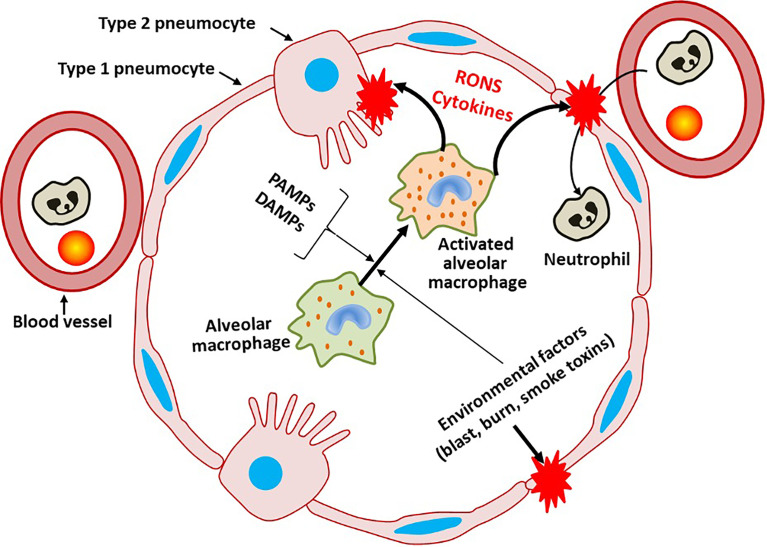
Pulmonary diseases represent four out of ten most common causes for worldwide mortality. Thus, pulmonary infections with subsequent inflammatory responses represent a major public health concern. The pulmonary barrier is a vulnerable entry site for several stress factors, including pathogens such as viruses, and bacteria, but also environmental factors e.g. toxins, air pollutants, as well as allergens. These pathogens or pathogen-associated molecular pattern and inflammatory agents e.g. damage-associated molecular pattern cause significant disturbances in the pulmonary barrier. The physiological and biological functions, as well as the architecture and homeostatic maintenance of the pulmonary barrier are highly complex. The airway epithelium, denoting the first pulmonary barrier, encompasses cells releasing a plethora of chemokines and cytokines, and is further covered with a mucus layer containing antimicrobial peptides, which are responsible for the pathogen clearance. Submucosal antigen-presenting cells and neutrophilic granulocytes are also involved in the defense mechanisms and counterregulation of pulmonary infections, and thus may directly affect the pulmonary barrier function. The detailed understanding of the pulmonary barrier including its architecture and functions is crucial for the diagnosis, prognosis, and therapeutic treatment strategies of pulmonary diseases. Thus, considering multiple side effects and limited efficacy of current therapeutic treatment strategies in patients with inflammatory diseases make experimental in vitro and in vivo models necessary to improving clinical therapy options. This review describes existing models for studyying the pulmonary barrier function under acute inflammatory conditions, which are meant to improve the translational approaches for outcome predictions, patient monitoring, and treatment decision-making.
Keywords: 2D; 3D; ALI; LOAC; PCLS; air-liquid; co-culture; organoid.
Copyright © 2022 Herminghaus, Kozlov, Szabó, Hantos, Gylstorff, Kuebart, Aghapour, Wissuwa, Walles, Walles, Coldewey and Relja.
Conflict of interest statement
The authors declare that the research was conducted in the absence of any commercial or financial relationships that could be construed as a potential conflict of interest.
Figures
References
-
- Yanagi S, Tsubouchi H, Miura A, Matsumoto N, Nakazato M. Breakdown of Epithelial Barrier Integrity and Overdrive Activation of Alveolar Epithelial Cells in the Pathogenesis of Acute Respiratory Distress Syndrome and Lung Fibrosis. BioMed Res Int (2015) 2015:573210. doi: 10.1155/2015/573210 - DOI - PMC - PubMed
-
- Nicod LP. Lung Defences: An Overview. Eur Respir Rev (2005) 14(95):45–50. doi: 10.1183/09059180.05.00009501 - DOI
Publication types
MeSH terms
Substances
LinkOut - more resources
Full Text Sources
Medical