

Paranodal Axoglial Junctions, an Essential Component in Axonal Homeostasis
- PMID: 35874818
- PMCID: PMC9299063
- DOI: 10.3389/fcell.2022.951809
Paranodal Axoglial Junctions, an Essential Component in Axonal Homeostasis
Abstract
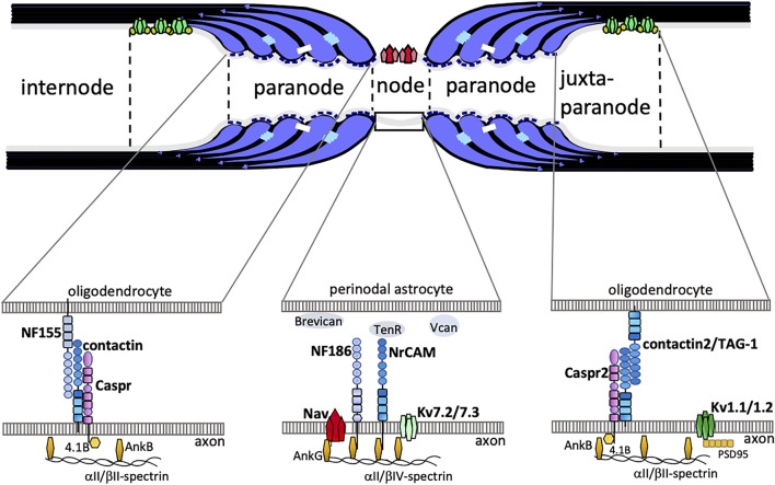
In vertebrates, a high density of voltage-gated Na+ channel at nodes of Ranvier and of voltage-gated K+ channel at juxtaparanodes is necessary for rapid propagation of action potential, that is, for saltatory conduction in myelinated axons. Myelin loops attach to the axonal membrane and form paranodal axoglial junctions (PNJs) at paranodes adjacent to nodes of Ranvier. There is growing evidence that the PNJs contribute to axonal homeostasis in addition to their roles as lateral fences that restrict the location of nodal axolemmal proteins for effective saltatory conduction. Perturbations of PNJs, as in specific PNJ protein knockouts as well as in myelin lipid deficient mice, result in internodal axonal alterations, even if their internodal myelin is preserved. Here we review studies showing that PNJs play crucial roles in the myelinated axonal homeostasis. The present evidence points to two functions in particular: 1) PNJs facilitate axonal transport of membranous organelles as well as cytoskeletal proteins; and 2) they regulate the axonal distribution of type 1 inositol 1,4,5-trisphosphate receptor (IP3R1) in cerebellar Purkinje axons. Myelinated axonal homeostasis depends among others on the state of PNJs, and consequently, a better understanding of this dependency may contribute to the clarification of CNS disease mechanisms and the development of novel therapies.
Keywords: IP3R1; Purkinje; calcium; myelin; paranodal junction.
Copyright © 2022 Ishibashi and Baba.
Conflict of interest statement
The authors declare that the research was conducted in the absence of any commercial or financial relationships that could be construed as a potential conflict of interest.
Figures
Similar articles
-
Disruption of paranodal axo-glial interaction and/or absence of sulfatide causes irregular type I inositol 1,4,5-trisphosphate receptor deposition in cerebellar Purkinje neuron axons.J Neurosci Res. 2015 Jan;93(1):19-27. doi: 10.1002/jnr.23465. Epub 2014 Aug 5. J Neurosci Res. 2015. PMID: 25093737
-
BK Channels Localize to the Paranodal Junction and Regulate Action Potentials in Myelinated Axons of Cerebellar Purkinje Cells.J Neurosci. 2015 May 6;35(18):7082-94. doi: 10.1523/JNEUROSCI.3778-14.2015. J Neurosci. 2015. PMID: 25948259 Free PMC article.
-
Accumulation of Neurofascin at Nodes of Ranvier Is Regulated by a Paranodal Switch.J Neurosci. 2020 Jul 22;40(30):5709-5723. doi: 10.1523/JNEUROSCI.0830-19.2020. Epub 2020 Jun 17. J Neurosci. 2020. PMID: 32554548 Free PMC article.
-
Multiple functions of the paranodal junction of myelinated nerve fibers.J Neurosci Res. 2009 Nov 15;87(15):3250-8. doi: 10.1002/jnr.22013. J Neurosci Res. 2009. PMID: 19224642 Review.
-
Molecular architecture of myelinated nerve fibers: leaky paranodal junctions and paranodal dysmyelination.Neuroscientist. 2013 Dec;19(6):629-41. doi: 10.1177/1073858413504627. Epub 2013 Oct 10. Neuroscientist. 2013. PMID: 24122820 Review.
Cited by
-
Is age-related myelinodegenerative change an initial risk factor of neurodegenerative diseases?Neural Regen Res. 2026 Feb 1;21(2):648-658. doi: 10.4103/NRR.NRR-D-24-00848. Epub 2025 Jan 13. Neural Regen Res. 2026. PMID: 40326982 Free PMC article.
-
Functional myelin in cognition and neurodevelopmental disorders.Cell Mol Life Sci. 2024 Apr 13;81(1):181. doi: 10.1007/s00018-024-05222-2. Cell Mol Life Sci. 2024. PMID: 38615095 Free PMC article. Review.
-
Altered plasma membrane abundance of the sulfatide-binding protein NF155 links glycosphingolipid imbalances to demyelination.Proc Natl Acad Sci U S A. 2023 Apr 4;120(14):e2218823120. doi: 10.1073/pnas.2218823120. Epub 2023 Mar 30. Proc Natl Acad Sci U S A. 2023. PMID: 36996106 Free PMC article.
References
-
- Barsukova A. G., Forte M., Bourdette D. J. (2012). Focal Increases of Axoplasmic Ca2+, Aggregation of Sodium-Calcium Exchanger, N-type Ca2+ Channel, and Actin Define the Sites of Spheroids in Axons Undergoing Oxidative Stress. J. Neurosci. 32 (35), 12028–12037. 10.1523/JNEUROSCI.0408-12.2012 - DOI - PMC - PubMed
Publication types
LinkOut - more resources
Full Text Sources