. 2022 Jul 7:10:886569.
doi: 10.3389/fcell.2022.886569. eCollection 2022.

Loss of α6β4 Integrin-Mediated Hemidesmosomes Promotes Prostate Epithelial Cell Migration by Stimulating Focal Adhesion Dynamics

Affiliations

Loss of α6β4 Integrin-Mediated Hemidesmosomes Promotes Prostate Epithelial Cell Migration by Stimulating Focal Adhesion Dynamics

Anette Schmidt et al. Front Cell Dev Biol. .

Abstract

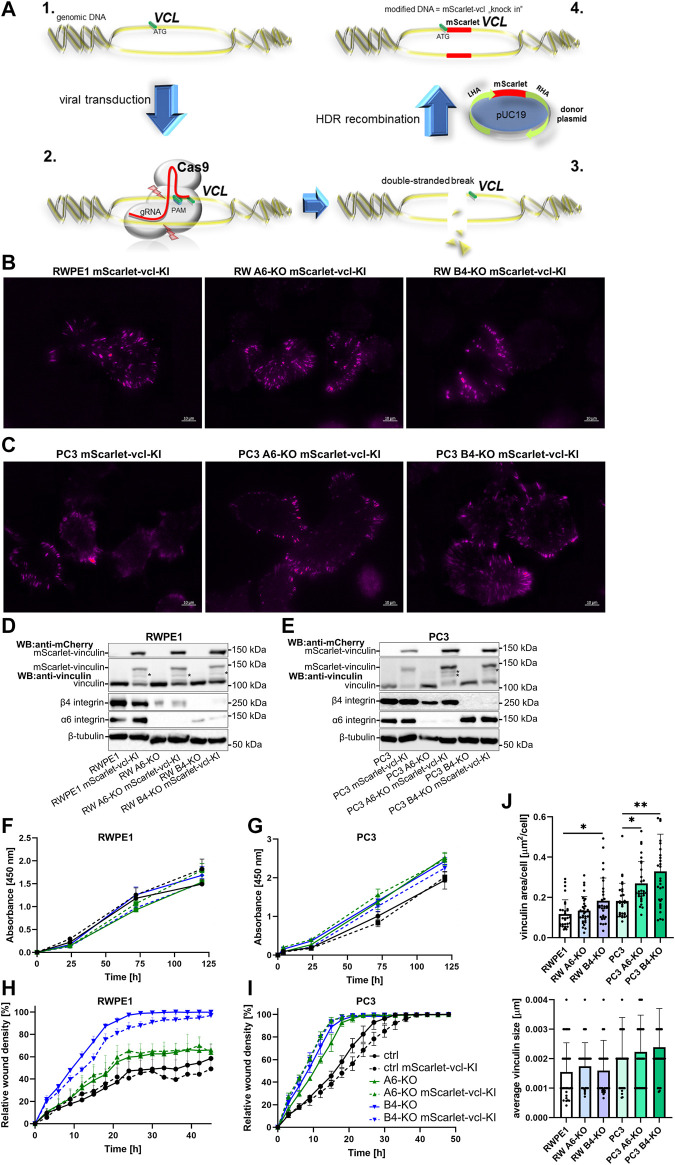
Epithelial cell adhesion is mediated by actin cytoskeleton-linked focal adhesions (FAs) and intermediate filament-associated hemidesmosomes (HDs). HDs are formed by α6β4-integrins and mediate stable anchoring to the extracellular matrix (ECM) while FAs containing β1-integrins regulate cell migration. Loss of HDs has been reported in various cancers such as prostate cancer where it correlates with increased invasive migration. Here we have studied cell migration properties and FA dynamics in genetically engineered prostate epithelial cell lines with intact or disrupted HDs. Disruption of HDs by depleting α6- or β4-integrin expression promoted collective cell migration and modulated migratory activity. Dynamic analysis of fluorescent protein-tagged FA marker proteins revealed faster FA assembly and disassembly kinetics in HD-depleted cells. FRAP analysis showed that loss of HDs correlated with faster diffusion rates of focal adhesion kinase (FAK) and vinculin in and out of FAs. These data suggest that loss of α6β4-mediated HDs promote cell migration and FA assembly dynamics by influencing the molecular diffusion rates of FAK.

Keywords: CRISPR/Cas9 knock-in; focal adhesion regulation; hemidesmosome; prostate cancer; α6β4-integrins.


Conflict of interest statement

The authors declare that the research was conducted in the absence of any commercial or financial relationships that could be construed as a potential conflict of interest.

Figures

FIGURE 1
FIGURE 1
Loss of HDs modulates migratory properties of the cells. Single-cell tracking of sparsely seeded cells was applied to analyze the cell velocity (A), directionality (B) and migration distance (C). The graphs show the mean ± SD representing data combined from two independent experiments with at least 35 single cells analyzed in each. Statistical significance was determined using Kruskal-Wallis test followed by Dunn’s multiple comparisons test (GraphPad Prism 8 software) and p-values are indicated by asterisks; 0.05 (*), 0.01 (**), or 0.001 (***).
FIGURE 2
FIGURE 2
Disruption of HDs by depleting α6- or β4-integrin induces FAs formation. RWPE1, RWPE1 α6-KO, RWPE1 β4-KO, PC3, PC3 α6-KO and PC3 β4-KO cells were stained for FA markers: paxillin (A), FAK (B), ILK (C) or vinculin (D) and imaged using TIRF microscopy. The size of individual FAs and the total area covered by the indicated markers per cell were determined using Fiji/ImageJ software. A minimum of 150 RWPE1 or 70 PC3 cells were analyzed for each variant. Statistical significance was assessed with Kruskal-Wallis test followed by Dunn’s multiple comparisons test comparing separately RWPE1 to its HD-depleted derivatives and PC3 to PC3 α6-KOs and PC3 β4-KOs using GraphPad Prism 8 software. p-values lower than 0.05 (*), 0.01 (**), or 0.001 (***) were considered statistically significant.
FIGURE 3
FIGURE 3
Establishment and characterization of RWPE1 and PC3 cells overexpressing fluorescence protein-tagged FA marker proteins. FACS-sorted populations of (A) RWPE1 and (B) PC3 cells stably transfected with mScarlet-vinculin (vcl), GFP-vcl, GFP-ILK or GFP-FAK constructs were imaged using TIRF microscopy. The expression levels of the indicated ectopically expressed fusion proteins in (C) RWPE1 and (D) PC3 cells were compared to endogenous expression levels by western blotting using antibodies recognizing the target protein. Fusion proteins were specifically detected using antibodies for GFP and mScarlet. The asterisks (*) denote the bands of proteolytically cleaved FA proteins. The symbol (#) indicates an increased FAK level in the cells expressing exogenous vinculin. (E) Parental RWPE1 and (F) PC3 cells and their respective derivatives overexpressing GFP-FAK, GFP-ILK, GFP-vinculin or mScarlet-vinculin were subjected to XTT-assay to measure cell proliferation. The data shows mean ± SD from at least two independent analyses performed in triplicates. The migratory properties of indicated variants of (G) RWPE1 and (H) PC3 cells were determined using the scratch wound assay module of IncucyteS3. The graphs show the mean ± SD. The analyses show a representative experiment out of three independent repeats with at least four replicates per variant. Statistical significance was determined using Two-way ANOVA followed by Dunnett’s multiple comparisons test (GraphPad Prism 8 software) and p-values are indicated by asterisks; 0.05 (*), 0.01 (**), or 0.001 (***).
FIGURE 4
FIGURE 4
Generation and characterization of the RWPE1 and PC3 mScarlet-vinculin knock-in cell lines. (A) Schematic of the strategy of the in-frame mScarlet knock-in into VCL gene locus. 1—A gRNA construct targeting a sequence nearby the ATG start codon of VCL was designed and cloned into plentiCRISPRv2 vector. 2—Target cells were transfected with a donor vector containing mScarlet flanked by left (LHA) and right (RHA) homology arms followed by transduction with Cas9/gRNA-expressing lentiviral vector. 3—Targeted double-stranded DNA breaks at the VCL start site are repaired by homologous recombination with the donor construct resulting in endogenous in-frame fusion of mScarlet in front of the VCL gene (4). FACS-sorted populations of (B) RWPE1 and (C) PC3 mScarlet knock-in variants were imaged using TIRF microscopy. The expression levels of endogenous fusion proteins were determined by western blots in parental and HD-depleted variants of (D) RWPE1 and (E) PC3 cells. Fusion proteins were detected using mCherry antibodies. (F) Parental RWPE1 and (G) PC3 cells and their respective derivatives with endogenously tagged mScarlet-vinculin were subjected to XTT-assay to measure cell proliferation. The data shows mean ± SD from at least two independent analyses performed in triplicates. The migratory properties of indicated variants of (H) RWPE1-mScarlet-vinculin-KI and (I) PC3-mScarlet-vinculin-KI cells were determined using the scratch wound assay module of IncucyteS3. The graphs show the mean ± SD. The analyses show a representative experiment out of three independent repeats with at least five replicates per variant. Two-way ANOVA followed by Dunnett’s multiple comparisons test was applied for statistical analysis. (J) The size of individual FAs and the total area per cell covered by endogenous mScarlet-vinculin in the indicated cell lines were determined using Fiji/ImageJ software. A minimum of 150 RWPE1 or 70 PC3 cells were analyzed for each sample. Statistical significance was determined using Kruskal-Wallis test followed by Dunn’s multiple comparisons test (GraphPad Prism 8 software) and p-values are indicated by asterisks; 0.05 (*), 0.01 (**), or 0.001 (***).
FIGURE 5
FIGURE 5
HDs depletion induces FA dynamics. Control and HD-deficient (α6- or β4-integrin-depleted) RWPE1 and PC3 cells overexpressing fluorescence FA proteins were seeded onto fibronectin-coated glass-bottom culture dishes and imaged at 1 min intervals using TIRF microscopy. The lifetime of individual (A) GFP-FAK, (B) GFP-ILK and (C) GFP-vinculin positive FAs was determined from recorded time-lapse series. (D) Lifetimes of foci formed by endogenously expressed mScarlet-vinculin were determined in RWPE1, RWPE1 α6-KO, RWPE1 β4-KO, PC3, PC3 α6-KO and PC3 β4-KO cells lines as described above. A minimum of 60 FAs were analyzed for each variant in A-D. Statistical significance was assessed with Kruskal-Wallis test followed by Dunn’s multiple comparisons test using GraphPad Prism 8 software [0.05 (*), 0.01 (**), or 0.001 (***)]. (E) A representative color-code timelapse of endogenously expressed mScarlet-vinculin foci in the indicated cell lines.
FIGURE 6
FIGURE 6
Loss of HDs affects the diffusion coefficients of FA components. Representative pictures of bleached regions of endogenous mScarlet-vinculin foci (A) and plots of relative fluorescence recovery ratio of that protein in the function of time in PC3 cell variants (B) the analysis of half-life recovery times and ratio of mobile fractions of bleached endogenous mScarlet-vinculin foci [(C) and (D), respectively], exogenous mScarlet-vinculin [(E) and (F), respectively] and exogenous GFP-FAK [(G) and (H), respectively] in RWPE1 and PC3 cells. The data are represented as a mean ± SD from at least 50 individual FAs from at least 10 random cells. Statistical significance was analyzed by Kruskal-Wallis test followed by Dunn’s multiple comparisons test using GraphPad Prism 8 software [0.05 (*), 0.01 (**), or 0.001 (***)].

Similar articles

Cited by

References

    1. Allen M. V., Smith G. J., Juliano R., Maygarden S. J., Mohler J. L. (1998). Downregulation of the β4 Integrin Subunit in Prostatic Carcinoma and Prostatic Intraepithelial Neoplasia. Hum. Pathol. 29, 311–318. 10.1016/s0046-8177(98)90109-5 - DOI - PubMed
    1. Bays J. L., DeMali K. A. (2017). Vinculin in Cell-Cell and Cell-Matrix Adhesions. Cell. Mol. Life Sci. 74, 2999–3009. 10.1007/s00018-017-2511-3 - DOI - PMC - PubMed
    1. Bello D., Webber M. M., Kleinman H. K., Wartinger D. D., Rhim J. S. (1997). Androgen Responsive Adult Human Prostatic Epithelial Cell Lines Immortalized by Human Papillomavirus 18. Carcinogenesis 18, 1215–1223. 10.1093/carcin/18.6.1215 - DOI - PubMed
    1. Bindels D. S., Haarbosch L., van Weeren L., Postma M., Wiese K. E., Mastop M., et al. (2017). mScarlet: A Bright Monomeric Red Fluorescent Protein for Cellular Imaging. Nat. Methods 14, 53–56. 10.1038/nmeth.4074 - DOI - PubMed
    1. Colburn Z. T., Jones J. C. R. (2017). α6β4 Integrin Regulates the Collective Migration of Epithelial Cells. Am. J. Respir. Cell Mol. Biol. 56, 443–452. 10.1165/rcmb.2016-0313OC - DOI - PMC - PubMed