

Controversial Role of the Immune Checkpoint OX40L Expression on Platelets in Breast Cancer Progression
- PMID: 35875148
- PMCID: PMC9304936
- DOI: 10.3389/fonc.2022.917834
Controversial Role of the Immune Checkpoint OX40L Expression on Platelets in Breast Cancer Progression
Abstract
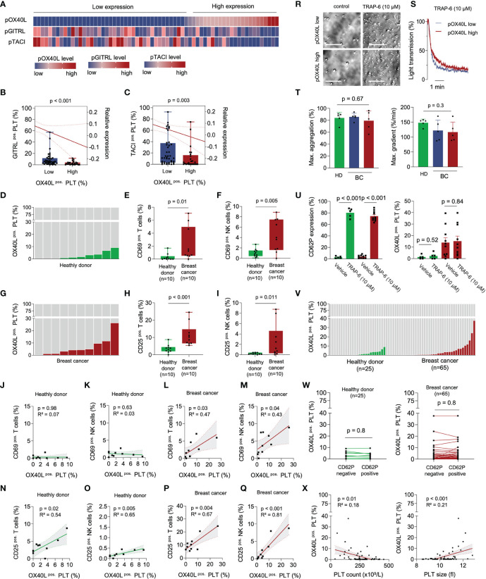
In conventional T cells, OX40 has been identified as a major costimulating receptor augmenting survival and clonal expansion of effector and memory T cell populations. In regulatory T cells, (Treg) OX40 signaling suppresses cellular activity and differentiation. However, clinical trials investigating OX40 agonists to enhance anti-tumor immunity, showed only limited success so far. Here we show that platelets from breast cancer patients express relevant levels of OX40L and platelet OX40L (pOX40L) inversely correlates with platelet-expressed immune checkpoint molecules GITRL (pGITRL) and TACI (pTACI). While high expression of pOX40L correlates with T and NK cell activation, elevated pOX40L levels identify patients with higher tumor grades, the occurrence of metastases, and shorter recurrence-free survival (RFS). Of note, OX40 mRNA levels in breast cancer correlate with enhanced expression of anti-apoptotic, immune-suppressive, and tumor-promoting mRNA gene signatures. Our data suggest that OX40L on platelets might play counteracting roles in cancer and anti-tumor immunity. Since pOX40L reflects disease relapse better than the routinely used predictive markers CA15-3, CEA, and LDH, it could serve as a novel biomarker for refractory disease in breast cancer.
Keywords: OX40L; biomarker; breast cancer; immunotherapy; platelets; prognosis.
Copyright © 2022 Rittig, Lutz, Clar, Zhou, Kropp, Koch, Hartkopf, Hinterleitner, Zender, Salih, Maurer and Hinterleitner.
Conflict of interest statement
The authors declare that the research was conducted in the absence of any commercial or financial relationships that could be construed as a potential conflict of interest.
Figures


Similar articles
-
OX40 and OX40L protein expression of tumor infiltrating lymphocytes in non-small cell lung cancer and its role in clinical outcome and relationships with other immune biomarkers.Transl Lung Cancer Res. 2019 Aug;8(4):352-366. doi: 10.21037/tlcr.2019.08.15. Transl Lung Cancer Res. 2019. PMID: 31555511 Free PMC article.
-
OX40 ligand expressed in glioblastoma modulates adaptive immunity depending on the microenvironment: a clue for successful immunotherapy.Mol Cancer. 2015 Feb 15;14:41. doi: 10.1186/s12943-015-0307-3. Mol Cancer. 2015. PMID: 25744203 Free PMC article.
-
Platelet-expressed immune checkpoint regulator GITRL in breast cancer.Cancer Immunol Immunother. 2021 Sep;70(9):2483-2496. doi: 10.1007/s00262-021-02866-y. Epub 2021 Feb 4. Cancer Immunol Immunother. 2021. PMID: 33538861 Free PMC article.
-
OX40 and OX40L Interaction in Cancer.Curr Med Chem. 2021;28(28):5659-5673. doi: 10.2174/0929867328666201229123151. Curr Med Chem. 2021. PMID: 33372866 Review.
-
OX40-OX40 ligand interaction in T-cell-mediated immunity and immunopathology.Adv Immunol. 2010;105:63-98. doi: 10.1016/S0065-2776(10)05003-0. Adv Immunol. 2010. PMID: 20510730 Review.
Cited by
-
Cross-talk between disulfidptosis and immune check point genes defines the tumor microenvironment for the prediction of prognosis and immunotherapies in glioblastoma.Sci Rep. 2024 Feb 16;14(1):3901. doi: 10.1038/s41598-024-52128-x. Sci Rep. 2024. PMID: 38365809 Free PMC article.
-
Interactions between Platelets and Tumor Microenvironment Components in Ovarian Cancer and Their Implications for Treatment and Clinical Outcomes.Cancers (Basel). 2023 Feb 17;15(4):1282. doi: 10.3390/cancers15041282. Cancers (Basel). 2023. PMID: 36831623 Free PMC article. Review.
-
Expression patterns of novel immunotherapy targets in intermediate- and high-grade lung neuroendocrine neoplasms.Cancer Immunol Immunother. 2024 May 2;73(6):114. doi: 10.1007/s00262-024-03704-7. Cancer Immunol Immunother. 2024. PMID: 38693435 Free PMC article.
-
Clinical Progress of Targeted Therapy for Breast Cancer: A Comprehensive Review.Curr Cancer Drug Targets. 2025;25(6):555-573. doi: 10.2174/0115680096289260240311062343. Curr Cancer Drug Targets. 2025. PMID: 38566384 Review.
-
OX40/OX40 ligand and its role in precision immune oncology.Cancer Metastasis Rev. 2024 Sep;43(3):1001-1013. doi: 10.1007/s10555-024-10184-9. Epub 2024 Mar 25. Cancer Metastasis Rev. 2024. PMID: 38526805 Free PMC article. Review.
References
Grants and funding
LinkOut - more resources
Full Text Sources