Review
. 2022 Jun 20;14(6):e26127.
doi: 10.7759/cureus.26127. eCollection 2022 Jun.

Recognizing Histopathological Simulators of Melanoma to Avoid Misdiagnosis

Affiliations
Review

Recognizing Histopathological Simulators of Melanoma to Avoid Misdiagnosis

Sara Waqar et al. Cureus. .

Abstract

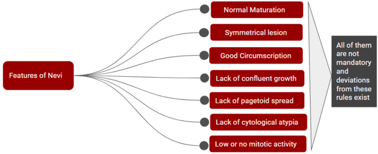
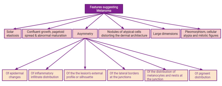
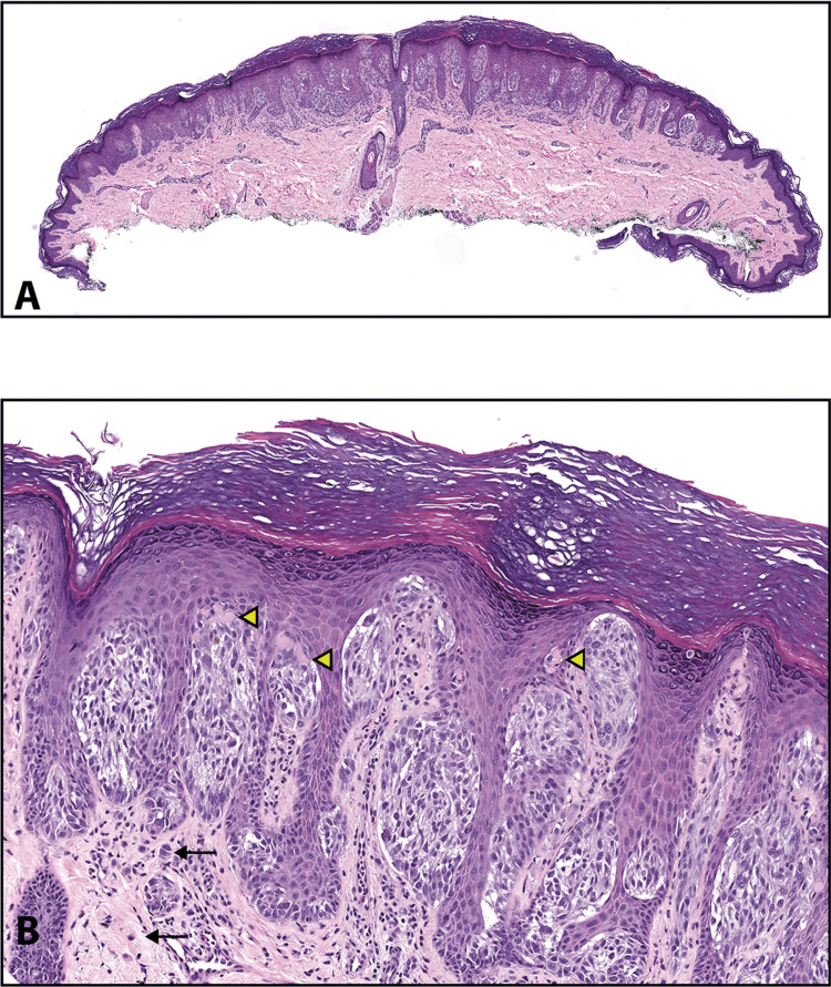
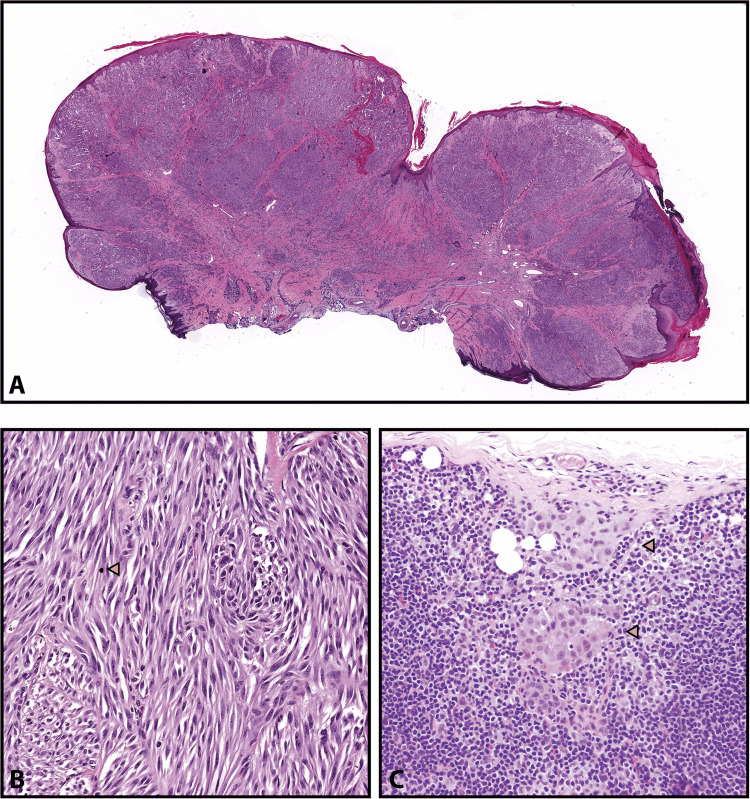
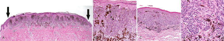
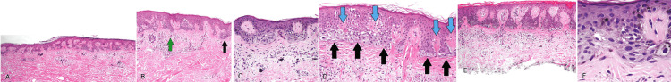
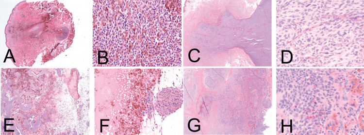
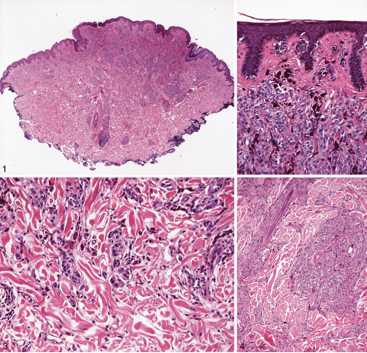
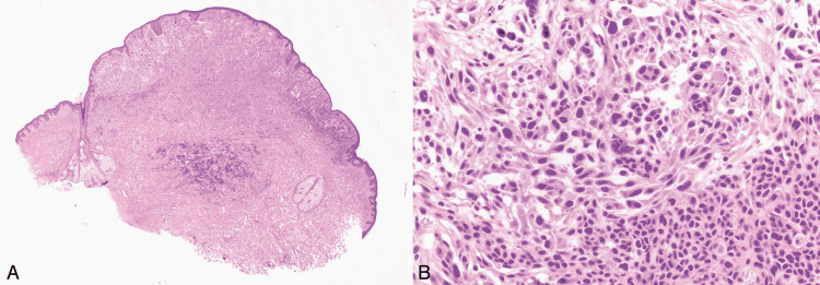
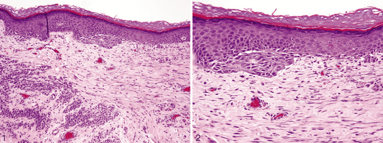
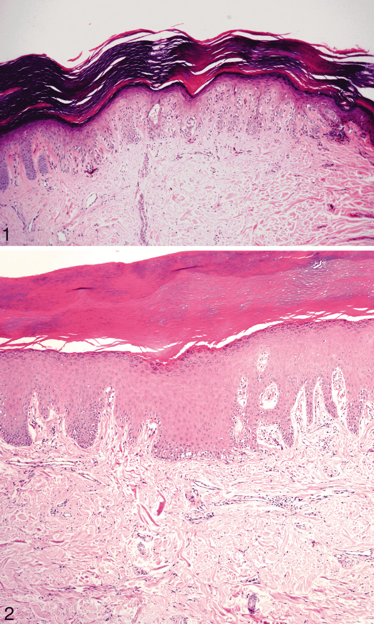
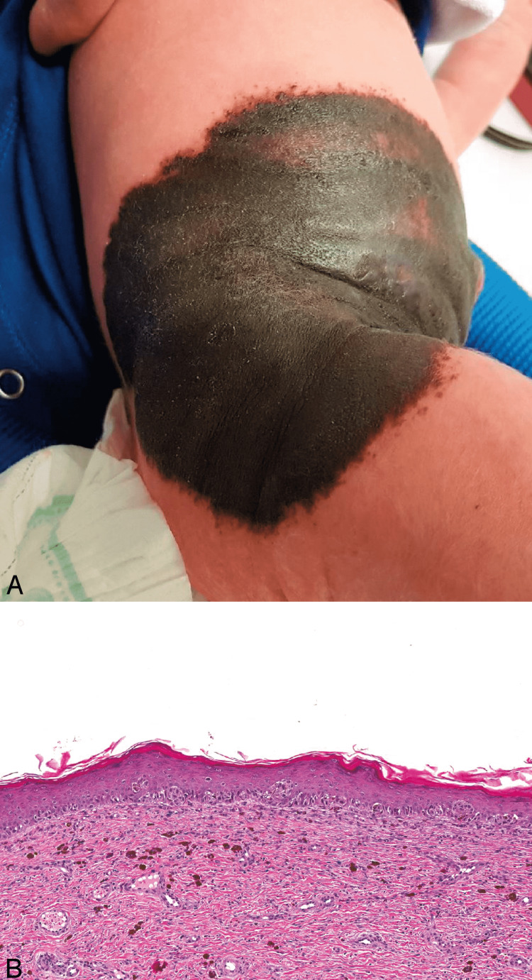
Melanocytic lesions have a wide morphological spectrum, ranging from benign nevi to malignant melanoma. In contrast to a diagnosis of a benign nevus, a diagnosis of melanoma could mean intensive treatment, lifetime monitoring, and a worse prognosis. Therefore, melanocytic tumors are notoriously challenging and associated with a high risk of litigation in surgical pathology. After describing the basic features of nevi and melanoma, this article describes the detailed clinical and histological features of those lesions that share many similar features with melanoma. The entities included are Spitz nevi and atypical Spitz tumors (AST), Reed nevus, dysplastic nevus, cellular blue nevus (CBN), deep penetrating nevus, combined nevus, recurrent nevus, irritated nevus, congenital pattern nevus, acral nevus, and nevi of special sites. Knowledge of these imitators can help pathologists distinguish between benign and malignant cases and avoid misdiagnosis.

Keywords: acral nevus; cellular blue nevus; congenital nevus; deep penetrating nevus; dysplastic nevus; melanocytic nevus; recurrent nevus; reed nevus; spitz tumors; ­melanoma and nevi.


Conflict of interest statement

This article has copyrighted material in the form of histopathological images, used with permission from © 2019 Innovative Pathology Press and Archives of Pathology & Laboratory Medicine, © 2010 College of American Pathologists.

Figures

Figure 1
Figure 1. Histological features of nevi.
This figure is created on Google Slides by Sara Waqar.
Figure 2
Figure 2. Slides showing histological features of nevi.
Reprinted from Gardner J. Survival Guide To Dermatopathology, Pathology Survival Guides Series 1. Innovative Pathology Press, Arlington Virginia; 2019 [2] with permission from Copyright 2019 Innovative Pathology Press. (A) Nests of epithelioid melanocytes in a junctional nevus, arranged at the tips of rete ridges. (B) Melanocytes at the basal layer (black arrows) are spaced out, and there are intervening keratinocytes. Mitosis in a basal keratinocyte can be seen (green arrow). (C) Small clusters, cords, or single cells of dermal melanocytes can be seen trickling down the reticular dermis. (D) Shows normal maturation. Large nests in the superficial dermis transition to smaller ones, and finally, single cells are dispersed from one another in the deep dermis. (E) Type A melanocytes are large epithelioid cells at the top, and type B are small round blue cells at the bottom in the dermis. (F) Type A cells have oval to round nuclei with abundant pale pink cytoplasm, forming nests at the junction. (G) Type B melanocytes at low power, resembling lymphocytes. They can mimic an inflamed nevus. (H) At high power, type B melanocytes (green arrows) can be differentiated from lymphocytes (yellow arrows). Lymphocytes are smaller in size than type B melanocytes with darker chromatin.
Figure 3
Figure 3. Histological features of melanoma.
This figure is created on Google Slides by Sara Waqar [18].
Figure 4
Figure 4. Slides showing histological features of melanoma.
Reprinted from Gardner J. Survival Guide To Dermatopathology, Pathology Survival Guides Series 1. Innovative Pathology Press, Arlington Virginia; 2019 [2] with permission from Copyright 2019 Innovative Pathology Press. (A) Pagetoid spread is indicated by single melanocytes going above the basal layer of the epidermis into the upper spinous and even granular layer. (B) Confluent growth is indicated by single melanocytes crowding out the basal keratinocytes. (C) Acral lentiginous melanoma. The epidermis can be seen detaching from the dermis, giving the illusion of a blister. It is called an “unzipping” artifact, which indicates confluent growth. (D) Lentigo maligna type of melanoma. The melanocytes involve the hair follicle, trickling down along the basal layer into the follicle either as nests or single cells. (E) Phenomenon of “epidermal consumption.” Nests occupy the epidermis entirely, and there is only a thin strip of keratinocytes left in the epidermis above the melanocytes. (F) Abnormal maturation is present as large nests of melanocytes can be seen from top to bottom of the lesion. (G) Cytological atypia is present as cells are hyperchromatic and pleomorphic with prominent nucleoli. (H) Invasive melanoma. Sheets of pleomorphic cells with clumped chromatin and a prominent atypical mitotic figure in the dermis can be seen.
Figure 5
Figure 5. Spitz nevus.
Reprinted from Harms KL, Lowe L, Fullen DR, Harms PW. Atypical Spitz Tumors: A Diagnostic Challenge. Arch Pathol Lab Med. 2015; 139(10): 1263-1270 [21] with permission from Archives of Pathology & Laboratory Medicine. Copyright 2010 College of American Pathologists. (A) This is the scanning magnification of a Spitz nevus. The lesion is symmetrical and predominantly nested with clefting artifacts around the nests. The epidermis is acanthotic (hematoxylin-eosin, original magnifications ×15). (B) This higher magnification shows large epithelioid melanocytes with prominent nucleoli. Kamino bodies are indicated with yellow arrowheads. Black arrows in the dermis point towards the nevomelanocytes that have retained their spitzoid morphology but have become smaller with their descent into the dermis (hematoxylin-eosin, original magnifications ×100).
Figure 6
Figure 6. Atypical Spitz tumor.
Reprinted from Harms KL, Lowe L, Fullen DR, Harms PW. Atypical Spitz Tumors: A Diagnostic Challenge. Arch Pathol Lab Med. 2015; 139(10): 1263-1270 [21] with permission from Archives of Pathology & Laboratory Medicine. Copyright 2010 College of American Pathologists. (A) This figure shows a large polypoid tumor with ulceration. (B) Spindled spitzoid cells are arranged in cellular fascicles with minimal pleomorphism and occasional mitotic figures (yellow arrowheads). (C) Yellow arrowheads show subcapsular deposits of spitzoid cells in a sentinel lymph node for an atypical Spitz tumor. These slides are stained with hematoxylin-eosin, seen in the above images at original magnifications ×5 (A), ×400 (B), and ×200 (C).
Figure 7
Figure 7. Reed nevus.
Reprinted from Gardner J. Survival Guide To Dermatopathology, Pathology Survival Guides Series 1. Innovative Pathology Press, Arlington Virginia; 2019 [2] with permission from Copyright 2019 Innovative Pathology Press. (A) Reed nevus. A small, thin, and circumscribed lesion ends in a nest abruptly at both peripheral borders. (B) Junctional nests show vertically oriented spindle cells with fine melanin pigment. The nests of these cells are hanging down from the acanthotic epidermis like “bunches of bananas.” A melanophage band can be seen underlying the lesion, giving the lesion a dark pigmentation clinically. (C) Nests of spindle cells are sometimes seen running horizontally parallel to the epidermis from rete to rete, giving a “bridging” appearance similar to dysplastic nevus. (D) The melanocytes have plump spindled nuclei that resemble spindled melanocytes of Spitz nevus; that is why it is considered a variant of Spitz nevus. The underlying papillary dermis has darkly pigmented melanophages.
Figure 8
Figure 8. Dysplastic nevus.
Reprinted from Gardner J. Survival Guide To Dermatopathology, Pathology Survival Guides Series 1. Innovative Pathology Press, Arlington Virginia; 2019 [2] with permission from Copyright 2019 Innovative Pathology Press. (A) A thin, broad lesion similar to its flat clinical appearance. The dermal component, if present, is in the papillary dermis and not in the deep dermis. (B) “Shouldering.” The green arrow points to the dermal component and the black to the junctional component. The junctional component extends to the periphery beyond the dermal component. (C) “Bridging.” The nests of melanocytes in the tips of rete ridges bridge across and are connected. Lamellar fibroplasia is seen underlying the nests and is wrapping around them. (D) Black arrows are pointing towards nests of melanocytes which are bridging across multiple adjacent retia and can be confused with confluent growth. The key here is that the inter-rete space above the papillary dermal tips (indicated by blue arrows) is uninvolved. (E) Lamellar fibroplasia. Bands of pink collagen wrap around the rete and nests of melanocytes that bridge between them. Lymphocytes can be seen scattered in the dermis. (F) Melanocytes are displaying cytological atypia. The nuclei of melanocytes are two to three times larger than the nuclei of spinous layer keratinocytes. The melanocytes have abundant grayish cytoplasm with fine dusty melanin pigment, giving it an appearance of “dirty dishwater.”
Figure 9
Figure 9. Cellular blue nevus.
Reprinted from Zembowicz A, Phadke PA. Blue nevi and variants: an update. Arch Pathol Lab Med. 2011; 135(3): 327-336 [35] with permission from Archives of Pathology & Laboratory Medicine. Copyright 2010 College of American Pathologists. (A) & (B) CBN comprises small cellular nodules of epithelioid cells. (C) & (D) Dumbbell-shaped appearance of large CBN. At high power, it shows uniform oval cells with inconspicuous nucleoli and clear cytoplasm. (E) & (F) An area of cystic degeneration can be seen, which should not be confused with tumor necrosis. (G) & (H) Areas of hemorrhage and hemosiderin deposition in large atypical CBN. Hot spots of cellular atypia and increased mitotic activity can also be seen. These slides are stained with hematoxylin-eosin, seen in the above images at original magnifications ×20 (A, C, and E), ×400 (B, D, and H), ×200 (F), and ×100 (G).
Figure 10
Figure 10. Deep penetrating nevus.
Reprinted from Luzar B, Calonje E. Deep penetrating nevus: a review. Arch Pathol Lab Med. 2011; 135(3): 321-326 [37] with permission from Archives of Pathology & Laboratory Medicine. Copyright 2010 College of American Pathologists. (1) In this figure, the nevus shows a symmetrical wedge-shaped dermal proliferation. The lesion has a plexiform appearance due to several extensions going deep into the reticular dermis along the adnexa and neurovascular bundles (hematoxylin-eosin, original magnification ×40). (2) A junctional component is seen in most deep penetrating nevi, which shows nests and minimal lentiginous proliferation of melanocytes, just like in an ordinary nevus. There is a discontinuity between the junctional and the dermal component of the lesion in the papillary dermis (hematoxylin-eosin, original magnification ×200). (3) Discohesion of melanocytes is seen in the deeper aspects (hematoxylin-eosin, original magnification ×200). (4) Melanocytes grow along the skin adnexa and neurovascular bundles (hematoxylin-eosin, original magnification ×100).
Figure 11
Figure 11. Combined nevus.
Reprinted from Harvey NT, Wood BA. A Practical Approach to the Diagnosis of Melanocytic Lesions. Arch Pathol Lab Med. 2019; 143(7): 789-810 [17] with permission from Archives of Pathology & Laboratory Medicine. Copyright 2010 College of American Pathologists. (A) The lesion displays asymmetry, raising concern for melanoma developing within a nevus. (B) On high power, the deep penetrating nevus cells are in the upper panel, and the conventional nevus cells can be seen in the lower panel. Slides are stained with hematoxylin-eosin and can be seen at original magnifications ×20 (A) and ×400 (B).
Figure 12
Figure 12. Recurrent nevus.
Reprinted from Fox JC, Reed JA, Shea CR. The recurrent nevus phenomenon: a history of challenge, controversy, and discovery. Arch Pathol Lab Med. 2011; 135(7): 842-846 [41] with permission from Archives of Pathology & Laboratory Medicine. Copyright 2010 College of American Pathologists. (1) Trizonal pattern. The first one is atypically proliferating melanocytes and increased melanin at the dermo-epidermal junction; the second is fibrous tissue in the superficial and mid-dermis; the third is the congenital nevus at the base of the lesion (hematoxylin-eosin, original magnification ×100). (2) An architecturally disordered proliferation of melanocytes represented by confluent nests and single cells can be seen at the dermo-epidermal junction (hematoxylin-eosin, original magnification ×200).
Figure 13
Figure 13. Acral junctional nevus and acral lentiginous melanoma.
Reprinted from Bravo Puccio F, Chian C. Acral junctional nevus versus acral lentiginous melanoma in situ: a differential diagnosis that should be based on clinicopathologic correlation. Arch Pathol Lab Med. 2011; 135(7): 847-852 [44] with permission from Archives of Pathology & Laboratory Medicine. Copyright 2010 College of American Pathologists. (1) Medium power field of acral junctional nevus. Most cells are in solitary units, but they form groups at the base of the rete ridges (hematoxylin-eosin, original magnification ×100). (2) Acral lentiginous melanoma. Single melanocytes proliferate along the dermo-epidermal junction (hematoxylin-eosin, original magnification ×100).
Figure 14
Figure 14. Giant congenital nevus.
Reprinted from Harvey NT, Wood BA. A Practical Approach to the Diagnosis of Melanocytic Lesions. Arch Pathol Lab Med. 2019; 143(7): 789-810 [17] with permission from Archives of Pathology & Laboratory Medicine. Copyright 2010 College of American Pathologists. (A) A giant congenital nevus. (B) The junctional component shows atypia, which could be construed as melanoma in situ in an adult patient. In a neonatal setting, these changes are invariably benign (hematoxylin-eosin, original magnification ×84).

Similar articles

Cited by

References

    1. Malignant melanoma: skin cancer-diagnosis, prevention, and treatment. Ahmed B, Qadir MI, Ghafoor S. Crit Rev Eukaryot Gene Expr. 2020;30:291–297. - PubMed
    1. Gardner J. Arlington Virginia: Innovative Pathology Press; 2019. Survival Guide To Dermatopathology, Pathology Survival Guides Series 1.
    1. Cancer stat facts: melanoma of the skin. [ May; 2022 ];https://seer.cancer.gov/statfacts/html/melan.html 2022
    1. A new understanding in the epidemiology of melanoma. Erdei E, Torres SM. Expert Rev Anticancer Ther. 2010;10:1811–1823. - PMC - PubMed
    1. Current state of melanoma diagnosis and treatment. Davis LE, Shalin SC, Tackett AJ. Cancer Biol Ther. 2019;20:1366–1379. - PMC - PubMed

LinkOut - more resources