. 2022 Jul 6:16:893030.
doi: 10.3389/fncel.2022.893030. eCollection 2022.

GPR39 Deficiency Impairs Memory and Alters Oxylipins and Inflammatory Cytokines Without Affecting Cerebral Blood Flow in a High-Fat Diet Mouse Model of Cognitive Impairment

Affiliations

GPR39 Deficiency Impairs Memory and Alters Oxylipins and Inflammatory Cytokines Without Affecting Cerebral Blood Flow in a High-Fat Diet Mouse Model of Cognitive Impairment

Thierno M Bah et al. Front Cell Neurosci. .

Abstract

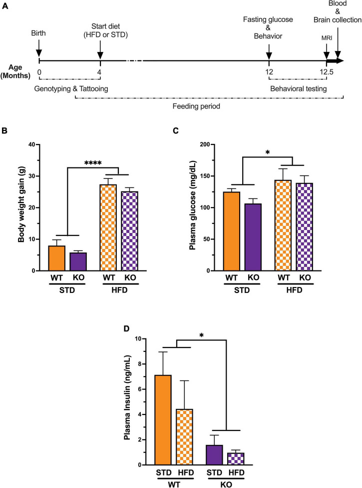
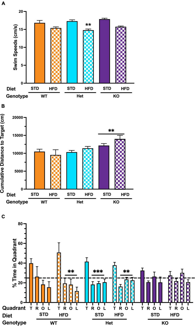
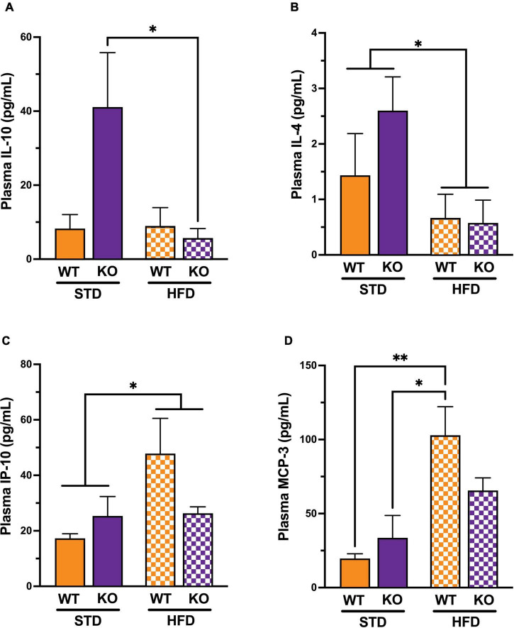
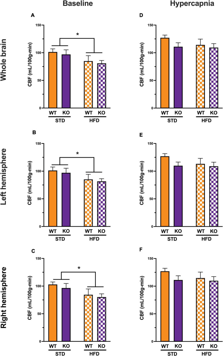
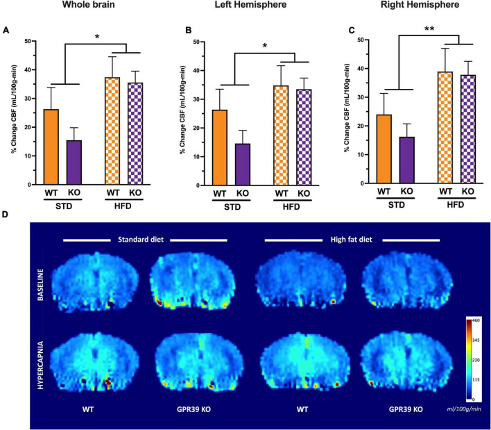
Vascular cognitive impairment (VCI) is the second most common cause of dementia. There is no treatment for VCI, in part due to a lack of understanding of the underlying mechanisms. The G-protein coupled receptor 39 (GPR39) is regulated by arachidonic acid (AA)-derived oxylipins that have been implicated in VCI. Furthermore, GPR39 is increased in microglia of post mortem human brains with VCI. Carriers of homozygous GPR39 SNPs have a higher burden of white matter hyperintensity, an MRI marker of VCI. We tested the hypothesis that GPR39 plays a protective role against high-fat diet (HFD)-induced cognitive impairment, in part mediated via oxylipins actions on cerebral blood flow (CBF) and neuroinflammation. Homozygous (KO) and heterozygous (Het) GPR39 knockout mice and wild-type (WT) littermates with and without HFD for 8 months were tested for cognitive performance using the novel object recognition (NOR) and the Morris water maze (MWM) tests, followed by CBF measurements using MRI. Brain tissue and plasma oxylipins were quantified with high-performance liquid chromatography coupled to mass spectrometry. Cytokines and chemokines were measured using a multiplex assay. KO mice, regardless of diet, swam further away from platform location in the MWM compared to WT and Het mice. In the NOR test, there were no effects of genotype or diet. Brain and plasma AA-derived oxylipins formed by 11- and 15-lipoxygenase (LOX), cyclooxygenase (COX) and non-enzymatically were increased by HFD and GPR39 deletion. Interleukin-10 (IL-10) was lower in KO mice on HFD than standard diet (STD), whereas IL-4, interferon γ-induced protein-10 (IP-10) and monocyte chemotactic protein-3 (MCP-3) were altered by diet in both WT and KO, but were not affected by genotype. Resting CBF was reduced in WT and KO mice on HFD, with no change in vasoreactivity. The deletion of GPR39 did not change CBF compared to WT mice on either STD or HFD. We conclude that GPR39 plays a role in spatial memory retention and protects against HFD-induced cognitive impairment in part by modulating inflammation and AA-derived oxylipins. The results indicate that GPR39 and oxylipin pathways play a role and may serve as therapeutic targets in VCI.

Keywords: GPR39; cerebral blood flow; dementia; high fat diet; mouse; neuroinflammation; oxylipins; vascular cognitive impairment.


Conflict of interest statement

NA is co-inventor of technologies related to GPR39 that have been licensed by OHSU to Vasocardea. This potential conflict of interest has been reviewed and managed by OHSU. The remaining authors declare that the research was conducted in the absence of any commercial or financial relationships that could be construed as a potential conflict of interest.

Figures

FIGURE 1
FIGURE 1
Experimental timeline and Impact of high fat diet on metabolic syndrome. (A) All genotypes of mice were placed on a high fat diet or standard diet at 3–5 months of age. Behavioral and cognitive performance was assessed using the water maze and the novel object recognition tests. Fasting glucose was measured just before behavior and magnetic resonance imaging (n = 6 per group, randomly selected) upon completion of the study. Plasma samples and brains were collected for biochemical assays and oxylipins analysis respectively. (B) Mice on a long-term high fat diet develop a diabetic phenotype. Prior to initiation of dietary intervention, there were no significant difference in body weight between groups. High fat diet caused a significant increase in body weight in both genotype after 8 months on the diet. (C) Plasma fasting glucose showed significant diet effect without genotype effect. (D) Plasma insulin levels showed significant genotype effect with low insulin in KO mice compared to WT littermates regardless of diet. All values represent means ± standard error of the mean, *p < 0.05, ****p < 0.0001, n = 5–10 per group.
FIGURE 2
FIGURE 2
Effects of GPR39 deficiency on memory retention in the water maze. (A) Swim speeds during the visible platform showed an effect of diet in Het mice, with lower swim speeds in Het mice fed with HFD. (B) There was no effect of diet on latency or cumulative distance to the target in any genotype during visible or hidden platform training. However, KO mice fed with HFD took longer to locate the platform than KO mice fed with STD. (C) Spatial memory retention in the water maze probe trial showed an effect of quadrant in WT mice on a STD. However, they did not spend more time in the target quadrant than any non-target quadrant. WT on HFD spent more time in the target quadrant than any other quadrant. Het mice on a STD and HFD also spent more time in the target quadrant than any other quadrant. In contrast, no effect of quadrant was seen in KO mice on STD or HFD. All values represent means ± standard error of the mean, **p < 0.001, ***p < 0.0001, n = 5–10 per group.
FIGURE 3
FIGURE 3
Effects of GPR39 deficiency on systemic inflammation. (A) IL-10 showed a significant interaction between genotype and diet and a significant diet effect. The post-hoc test revealed a significant difference between KO fed with HFD and KO fed with STD, whereas no significant difference existed between WT regardless of the diet. (B) IL-4 showed a significant diet effect between mice on STD compared to mice on HFD, whereas no significant difference existed between genotypes. (C) IP-10 showed a significant diet effect between mice on STD compared to mice on HFD, whereas no significant difference existed between genotypes. (D) MCP-3 showed a significant diet effect between mice on STD compared to mice on HFD, whereas no significant difference existed between genotypes. The post-hoc test revealed a significant difference between WT fed with HFD and WT fed with STD and a significant difference between WT fed with HFD and KO fed with STD without genotype effect. All values represent means ± standard error of the mean, *p < 0.05, **p < 0.001, n = 3–4 per group.
FIGURE 4
FIGURE 4
Effects of GPR39 deficiency on cerebral blood flow. (A) At baseline, CBF (in ml/100 g/min) showed a diet effect between STD and HFD mice with lower CBF in HFD, but without genotype effect for the whole brain. (B,C) Left and right hemispheres showed a diet effect between STD and HFD mice with lower CBF in HFD, but without genotype effect. (D–F) With hypercapnia (CO2 challenge), the absolute mean CBF increased in all groups with no diet or genotype effect for the whole brain and for both brain hemispheres. All values represent means ± standard error of the mean, *p < 0.05, n = 4–7 per group.
FIGURE 5
FIGURE 5
Effects of GPR39 deficiency on cerebrovascular reactivity. (A–C) Cerebrovascular reactivity to CO2 challenge showed a diet effect without genotype effect for the whole brain and for both brain hemispheres with a trend toward higher CBF in HFD fed mice compared to STD mice. (D) Grouped color perfusion maps of cerebral blood flow (mL/100g/min) showing reduced CBF associated with HFD. All values represent means ± standard error of the mean, *p < 0.05, **p < 0.001, n = 4–7 per group.

Similar articles

Cited by

References

    1. Alkayed N. J., Cao Z., Qian Z. Y., Nagarajan S., Liu X., Nelson J. W., et al. (2022). Control of Coronary Vascular Resistance by Eicosanoids via a Novel GPCR. Am. J. Physiol. Cell Physiol. 322 C1011–C1021. 10.1152/ajpcell.00454.2021 - DOI - PMC - PubMed
    1. Benice T. S., Raber J. (2008). Object recognition analysis in mice using nose-point digital video tracking. J. Neurosci. Methods 168 422–430. 10.1016/j.jneumeth.2007.11.002 - DOI - PubMed
    1. Bocarsly M. E., Fasolino M., Kane G. A., LaMarca E. A., Kirschen G. W., Karatsoreos I. N., et al. (2015). Obesity diminishes synaptic markers, alters microglial morphology, and impairs cognitive function. Proc. Natl. Acad. Sci. U. S. A. 112 15731–15736. 10.1073/pnas.1511593112 - DOI - PMC - PubMed
    1. Bracko O., Vinarcsik L. K., Cruz Hernandez J. C., Ruiz-Uribe N. E., Haft-Javaherian M., Falkenhain K., et al. (2020). High fat diet worsens Alzheimer’s disease-related behavioral abnormalities and neuropathology in APP/PS1 mice, but not by synergistically decreasing cerebral blood flow. Sci. Rep. 10:9884. 10.1038/s41598-020-65908-y - DOI - PMC - PubMed
    1. Buckman L. B., Hasty A. H., Flaherty D. K., Buckman C. T., Thompson M. M., Matlock B. K., et al. (2014). Obesity induced by a high-fat diet is associated with increased immune cell entry into the central nervous system. Brain Behav. Immun. 35 33–42. 10.1016/j.bbi.2013.06.007 - DOI - PMC - PubMed