

Cytomorphological Analysis and Interpretation of Nitric Oxide-Mediated Neurotoxicity in Sleep-Deprived Mice Model
- PMID: 35875423
- PMCID: PMC9305911
- DOI: 10.1177/09727531211059925
Cytomorphological Analysis and Interpretation of Nitric Oxide-Mediated Neurotoxicity in Sleep-Deprived Mice Model
Abstract
Background: Sleep deprivation (SD) is a biological stress condition for the brain, and the pathogenesis of SD is closely related to elevated oxidative stress, mitochondrial dysfunction, a major cause of neurodegeneration. This oxidative stress-mediated cell death is attributed to rise in calcium ion influx which further excites or alters the neurotransmitters level by activating neuronal nitric oxide (NO) synthase (nNOS) release of NO in mouse SD model. This study indicates that the nitrergic neurons are possible therapeutic targets for the amelioration of SD-induced cognitive dysfunction and behavioral alterations.
Purpose: SD is considered as a risk factor for various neurodegenerative diseases. SD leads to biochemical, behavioral, and neurochemical alterations in animals. This study was designed to explore the possible involvement of a nitrergic neuron system in six days SD-induced morphological and neurodegenerative changes in mice.
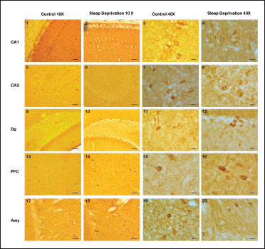
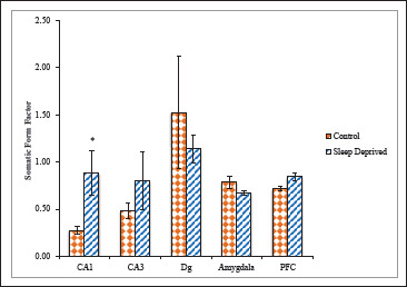
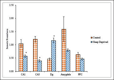
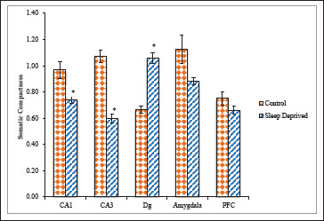
Methods: Using nNOS immunohistochemistry, we have investigated the effects of SD on nNOS positive neurons. Immunohistochemical study for the distribution of nNOS positive neuronal cell bodies was carried out in the hippocampus, prefrontal cortex (PFC), and amygdaloid nuclei of mice brain.
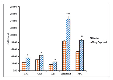
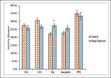
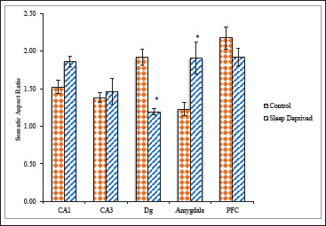
Results: Sleep-deprived animals showed a significantly increased number of nNOS positive neurons and altered neuronal cytomorphology as compared with the control group.
Conclusion: These results indicate that total SD may induce morphological changes in nNOS positive neurons in the brain, thus increasing NO synthesis, which is implicated in SD-induced neuronal cell death.
Keywords: Neurodegeneration; Neuronal nitric oxide synthase; Neurotoxicity; Nitric oxide; Sleep deprivation.
© 2022 Indian Academy of Neurosciences (IAN).
Conflict of interest statement
Declaration of Conflicting Interests: The authors declared no potential conflicts of interest with respect to the research, authorship, and/or publication of this article.
Figures

Similar articles
-
Increased nitric oxide-mediated neurotransmission in the medial prefrontal cortex is associated with the long lasting anxiogenic-like effect of predator exposure.Behav Brain Res. 2013 Nov 1;256:391-7. doi: 10.1016/j.bbr.2013.08.006. Epub 2013 Aug 12. Behav Brain Res. 2013. PMID: 23948217
-
Effect of repeated restraint on homotypic stress-induced nitric oxide synthases expression in brain structures regulating HPA axis.Pharmacol Rep. 2012;64(6):1381-90. doi: 10.1016/s1734-1140(12)70935-0. Pharmacol Rep. 2012. PMID: 23406748
-
Melatonin ameliorates cognitive impairment induced by sleep deprivation in rats: role of oxidative stress, BDNF and CaMKII.Behav Brain Res. 2013 Nov 1;256:72-81. doi: 10.1016/j.bbr.2013.07.051. Epub 2013 Aug 6. Behav Brain Res. 2013. PMID: 23933144
-
Neuronal nitric oxide synthase and affective disorders.IBRO Rep. 2018 Nov 17;5:116-132. doi: 10.1016/j.ibror.2018.11.004. eCollection 2018 Dec. IBRO Rep. 2018. PMID: 30591953 Free PMC article. Review.
-
Point of NO return for nitrergic nerves in diabetes: a new insight into diabetic complications.Curr Pharm Des. 2004;10(29):3683-95. doi: 10.2174/1381612043382792. Curr Pharm Des. 2004. PMID: 15579064 Review.
Cited by
-
Cerebrospinal Fluid Nitric Oxide Synthase is a Potential Mediator Between Cigarette Smoke Exposure and Sleep Disorders.Nat Sci Sleep. 2024 Jul 1;16:897-906. doi: 10.2147/NSS.S458294. eCollection 2024. Nat Sci Sleep. 2024. PMID: 38974692 Free PMC article.
-
Sleep deprivation induces late deleterious effects in a pharmacological model of Parkinsonism.Exp Brain Res. 2024 May;242(5):1175-1190. doi: 10.1007/s00221-024-06811-0. Epub 2024 Mar 18. Exp Brain Res. 2024. PMID: 38499659
References
-
- Wang GP, Huang LQ, Wu HJ, et al.. Calcineurin contributes to spatial memory impairment induced by rapid eye movement sleep deprivation. Neuroreport 2009; 20(13): 1172–1176. - PubMed
-
- Alhaider IA, Aleisa AM, Tran TT, et al.. Sleep deprivation prevents stimulation-induced increases of levels of P-CREB and BDNF: Protection by caffeine. Mol Cell Neurosci 2011; 46(4): 742–751. - PubMed
-
- Kiss JP. Role of nitric oxide in the regulation of monoaminergic neurotransmission. Brain Res Bull 2000; 52(6): 459–466. - PubMed
LinkOut - more resources
Full Text Sources
Miscellaneous
