. 2022 Jul 6:10:914137.
doi: 10.3389/fbioe.2022.914137. eCollection 2022.

Subject-Specific 3D Models to Investigate the Influence of Rehabilitation Exercises and the Twisted Structure on Achilles Tendon Strains

Affiliations

Subject-Specific 3D Models to Investigate the Influence of Rehabilitation Exercises and the Twisted Structure on Achilles Tendon Strains

Alessia Funaro et al. Front Bioeng Biotechnol. .

Abstract

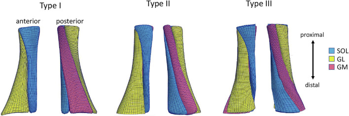
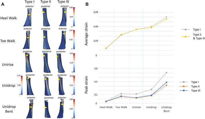
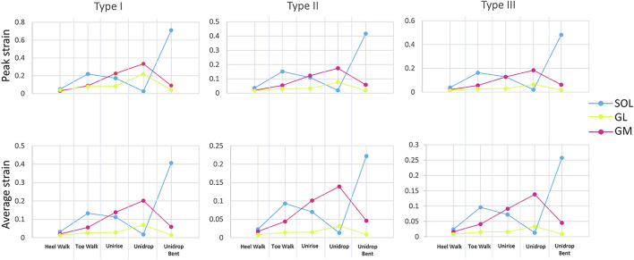
The Achilles tendon (AT) is the largest tendon of the human body and has a primary role in locomotor activities. The complex structure of the AT includes twisting of three sub-tendons, non-uniform tissue deformations and differential triceps surae muscle forces. The main aim of this study was to investigate the impact of commonly used rehabilitation exercises (walking on heels, walking on toes, unilateral heel rise, heel drop with extended knee and heel drop with the knee bent) and different twists on AT strains. 3D freehand ultrasound based subject-specific geometry and subject-specific muscle forces during different types of rehabilitation exercises were used to determine tendon strains magnitudes and differences in strains between the sub-tendons. In addition, three Finite Element models were developed to investigate the impact of AT twist. While walking on heels developed the lowest average strain, heel drop with knee bent exhibited the highest average strain. The eccentric heel drop resulted in higher peak and average strain, compared to concentric heel rise for all the three models. The isolated exercises (heel rise and heel drop) presented higher average strains compared to the functional exercises (walking tasks). The amount of twist influences the peak strains but not the average. Type I consistently showed highest peak strains among the five rehabilitation exercises. The ranking of the exercises based on the AT strains was independent of AT twist. These findings might help clinicians to prescribe rehabilitation exercises for Achilles tendinopathy based on their impact on the AT strains.

Keywords: achilles tendon; fascicle twist; finite element modeling; rehabilitation exercises; sub-tendon morphology; tendon strain.


Conflict of interest statement

The authors declare that the research was conducted in the absence of any commercial or financial relationships that could be construed as a potential conflict of interest.

Figures

FIGURE 1
FIGURE 1
Anterior and posterior view of the Achilles free tendon geometries representing the three types of twist: Type I (least), Type II (moderate), Type III (extreme). Each 3D tendon model was divided in three sub-tendons, each one arising from one of the three triceps surae muscles: the soleus (SOL) and the two heads (medialis, GM and lateralis, GL) of the gastrocnemius muscle.
FIGURE 2
FIGURE 2
(A). View of the distribution of the maximum Lagrange strain for the three types of twist and for all the five rehabilitation exercises. The yellow dots indicate the location of the peak strain. (B) Trend of the peak and average of the maximum Lagrange strain for all the types of twist and all the rehabilitation exercises, in the mid-portion of the tendon models. In average strain, Type I and Type II strain are described with the same line, since the values are the same.
FIGURE 3
FIGURE 3
Peak and average of the maximum Lagrange strain at the mid-portion of each sub-tendon, soleus (SOL), gastrocnemius medialis (GM) and gastrocnemius lateralis (GL), for all the five rehabilitation exercises and twist.

Similar articles

Cited by

References

    1. Albers I. S., Zwerver J., Diercks R. L., Dekker J. H., Van den Akker-Scheek I. (2016). Incidence and Prevalence of Lower Extremity Tendinopathy in a Dutch General Practice Population: A Cross Sectional Study. BMC Musculoskelet. Disord. 17. 10.1186/s12891-016-0885-2 - DOI - PMC - PubMed
    1. Alfredson H., Pietilä T., Jonsson P., Lorentzon R. (1998). Heavy-Load Eccentric Calf Muscle Training for the Treatment of Chronic Achilles Tendinosis. Am. J. Sports Med. 26 (3), 360–366. 10.1177/03635465980260030301 - DOI - PubMed
    1. Arndt A., Bengtsson A.-S., Peolsson M., Thorstensson A., Movin T. (2012). Non-uniform Displacement within the Achilles Tendon during Passive Ankle Joint Motion. Knee Surg. Sports Traumatol. Arthrosc. 20 (9), 1868–1874. 10.1007/s00167-011-1801-9 - DOI - PubMed
    1. Baxter J. R., Corrigan P., Hullfish T. J., O’Rourke P., Silbernagel K. G. (2021). Exercise Progression to Incrementally Load the Achilles Tendon. Med. Sci. Sports Exerc. 53 (1), 124–130. 10.1249/MSS.0000000000002459 - DOI - PubMed
    1. Bogaerts S., De Brito Carvalho C., Scheys L., Desloovere K., D’hooge J., Maes F., et al. (2017). Evaluation of Tissue Displacement and Regional Strain in the Achilles Tendon Using Quantitative High-Frequency Ultrasound. Plos One 12 (7), e0181364. 10.1371/journal.pone.0181364 - DOI - PMC - PubMed

LinkOut - more resources