

Bioinspired Protein/Peptide Loaded 3D Printed PLGA Scaffold Promotes Bone Regeneration
- PMID: 35875498
- PMCID: PMC9300829
- DOI: 10.3389/fbioe.2022.832727
Bioinspired Protein/Peptide Loaded 3D Printed PLGA Scaffold Promotes Bone Regeneration
Abstract
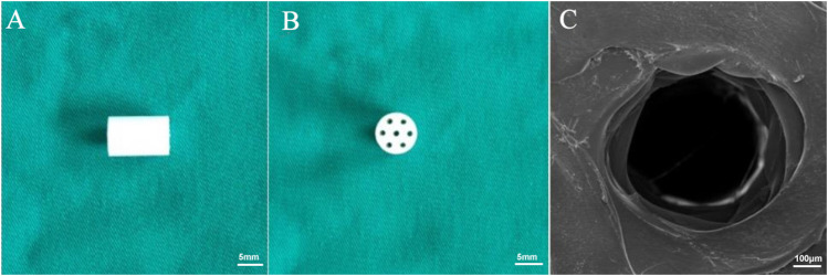
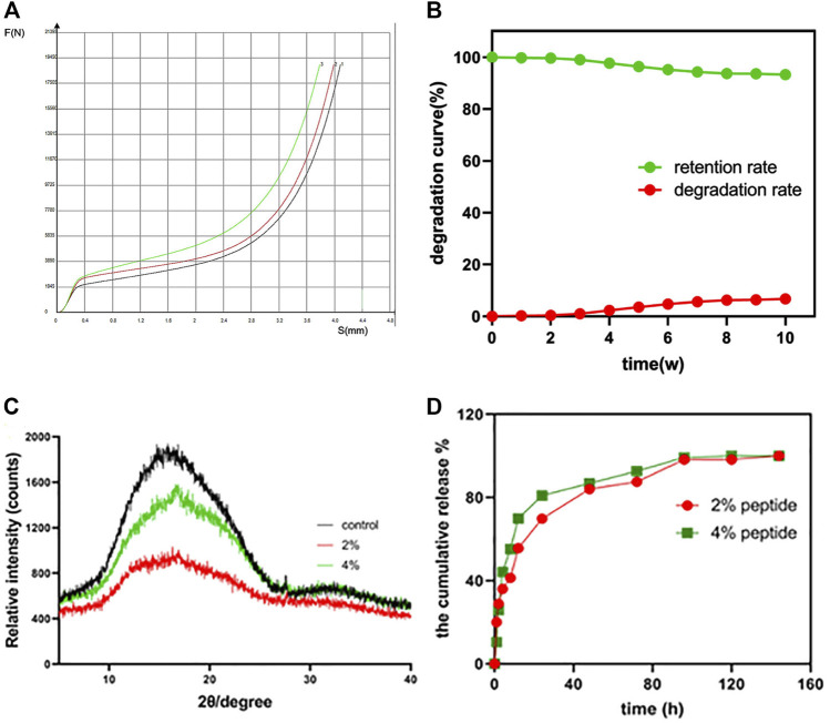
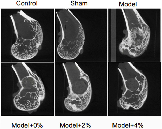
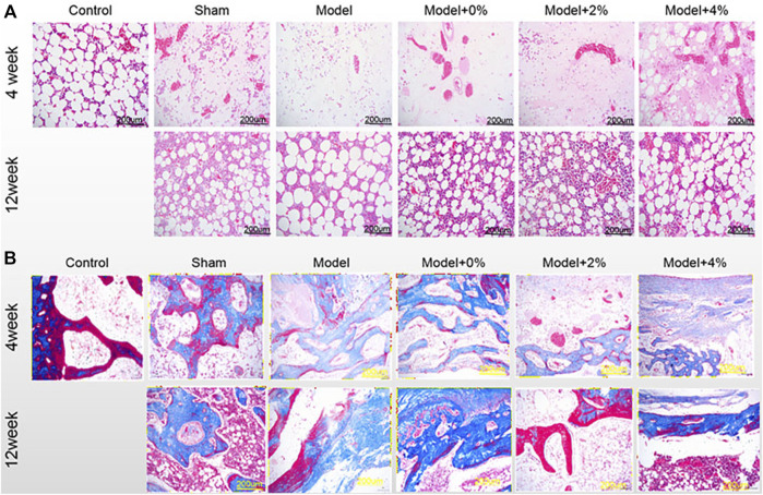
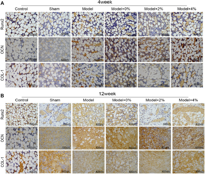
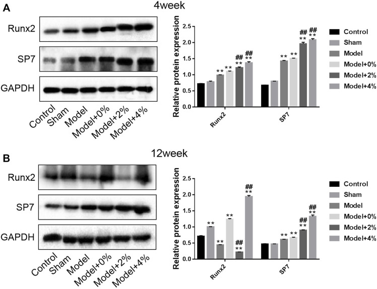
Background: This study was aimed to investigate the effect of three dimensional (3D)printed poly lactide-co-glycolide (PLGA) scaffolds combined with Gly-Phe-Hyp-Gly-Arg (GFOGER) and bone morphogenetic protein 9 (BMP-9) on the repair of large bone defects. Methods: 3D printing method was used to produce PLGA scaffolds, and the sample was viewed by both optical microscopy and SEM, XRD analysis, water absorption and compressive strength analysis, etc. The rabbits were divided into six groups randomly and bone defect models were constructed (6 mm in diameter and 9 mm in depth): control group (n = 2), sham group (n = 4), model group (n = 4) and model + scaffold group (n = 4 rabbits for each group, 0%,2% and 4%). The rabbits were sacrificed at the 4th and 12th weeks after surgery, and the samples were collected for quantitative analysis of new bone mineral density by micro-CT, histopathological observation, immunohistochemistry and Western blot to detect the protein expression of osteoblast-related genes. Results: This scaffold presented acceptable mechanical properties and slower degradation rates. After surface modification with GFOGER peptide and BMP-9, the scaffold demonstrated enhanced new bone mineral deposition and density over the course of a 12 week in vivo study. Histological analysis and WB confirmed that this scaffold up-regulated the expression of Runx7, OCN, COL-1 and SP7, contributing to the noted uniform trabeculae formation and new bone regeneration. Conclusions: The application of this strategy in the manufacture of composite scaffolds provided extensive guidance for the application of bone tissue engineering.
Keywords: 3D printing; PLGA scaffold; bio-inspired; bone defect; protein/peptide decoration.
Copyright © 2022 Song, Li, Wang, Wang, Lv, Xie, Zhang and Shao.
Conflict of interest statement
The authors declare that the research was conducted in the absence of any commercial or financial relationships that could be construed as a potential conflict of interest.
Figures
Similar articles
-
Preparation of 3D Printing PLGA Scaffold with BMP-9 and P-15 Peptide Hydrogel and Its Application in the Treatment of Bone Defects in Rabbits.Contrast Media Mol Imaging. 2022 Jul 31;2022:1081957. doi: 10.1155/2022/1081957. eCollection 2022. Contrast Media Mol Imaging. 2022. PMID: 35965616 Free PMC article.
-
Dual-functional 3D-printed composite scaffold for inhibiting bacterial infection and promoting bone regeneration in infected bone defect models.Acta Biomater. 2018 Oct 1;79:265-275. doi: 10.1016/j.actbio.2018.08.015. Epub 2018 Aug 18. Acta Biomater. 2018. PMID: 30125670
-
3D printed scaffold for repairing bone defects in apical periodontitis.BMC Oral Health. 2022 Aug 8;22(1):327. doi: 10.1186/s12903-022-02362-4. BMC Oral Health. 2022. PMID: 35941678 Free PMC article.
-
Enhancement in sustained release of antimicrobial peptide and BMP-2 from degradable three dimensional-printed PLGA scaffold for bone regeneration.RSC Adv. 2019 Apr 4;9(19):10494-10507. doi: 10.1039/c8ra08788a. eCollection 2019 Apr 3. RSC Adv. 2019. PMID: 35515290 Free PMC article.
-
Tissue-engineered composite scaffold of poly(lactide-co-glycolide) and hydroxyapatite nanoparticles seeded with autologous mesenchymal stem cells for bone regeneration.J Zhejiang Univ Sci B. 2017 Nov.;18(11):963-976. doi: 10.1631/jzus.B1600412. J Zhejiang Univ Sci B. 2017. PMID: 29119734 Free PMC article.
Cited by
-
Scaffold Adhering to Peptide-Based Biomimetic Extracellular Matrix Composite Nanobioglass Promotes the Proliferation and Migration of Skin Fibroblasts Through the GSK-3β/β-Catenin Signaling Axis.Int J Nanomedicine. 2024 Mar 23;19:2957-2972. doi: 10.2147/IJN.S449385. eCollection 2024. Int J Nanomedicine. 2024. PMID: 38549840 Free PMC article.
-
Hydrogel promotes bone regeneration through various mechanisms: a review.Biomed Tech (Berl). 2024 Nov 22;70(2):103-114. doi: 10.1515/bmt-2024-0391. Print 2025 Apr 28. Biomed Tech (Berl). 2024. PMID: 39571066 Review.
-
Silk-Based Biomaterials for Designing Bioinspired Microarchitecture for Various Biomedical Applications.Biomimetics (Basel). 2023 Jan 28;8(1):55. doi: 10.3390/biomimetics8010055. Biomimetics (Basel). 2023. PMID: 36810386 Free PMC article. Review.
-
3D Printing Technologies in Personalized Medicine, Nanomedicines, and Biopharmaceuticals.Pharmaceutics. 2023 Jan 17;15(2):313. doi: 10.3390/pharmaceutics15020313. Pharmaceutics. 2023. PMID: 36839636 Free PMC article. Review.
-
A bifunctional bortezomib-loaded porous nano-hydroxyapatite/alginate scaffold for simultaneous tumor inhibition and bone regeneration.J Nanobiotechnology. 2023 Jun 1;21(1):174. doi: 10.1186/s12951-023-01940-0. J Nanobiotechnology. 2023. PMID: 37264410 Free PMC article.
References
LinkOut - more resources
Full Text Sources