

Deep Learning-Based Recognition of Different Thyroid Cancer Categories Using Whole Frozen-Slide Images
- PMID: 35875502
- PMCID: PMC9298848
- DOI: 10.3389/fbioe.2022.857377
Deep Learning-Based Recognition of Different Thyroid Cancer Categories Using Whole Frozen-Slide Images
Abstract
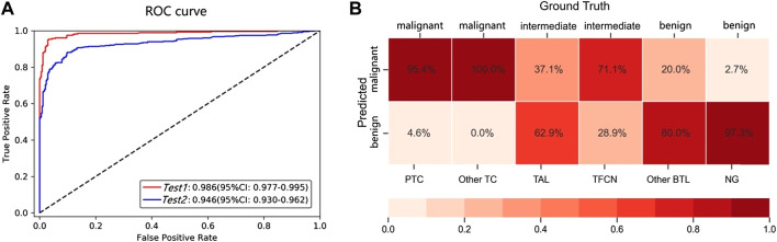
Introduction: The pathological rare category of thyroid is a type of lesion with a low incidence rate and is easily misdiagnosed in clinical practice, which directly affects a patient's treatment decision. However, it has not been adequately investigated to recognize the rare, benign, and malignant categories of thyroid using the deep learning method and recommend the rare to pathologists. Methods: We present an empirical decision tree based on the binary classification results of the patch-based UNet model to predict rare categories and recommend annotated lesion areas to be rereviewed by pathologists. Results: Applying this framework to 1,374 whole-slide images (WSIs) of frozen sections from thyroid lesions, we obtained an area under a curve of 0.946 and 0.986 for the test datasets with and without WSIs, respectively, of rare types. However, the recognition error rate for the rare categories was significantly higher than that for the benign and malignant categories (p < 0.00001). For rare WSIs, the addition of the empirical decision tree obtained a recall rate and precision of 0.882 and 0.498, respectively; the rare types (only 33.4% of all WSIs) were further recommended to be rereviewed by pathologists. Additionally, we demonstrated that the performance of our framework was comparable to that of pathologists in clinical practice for the predicted benign and malignant sections. Conclusion: Our study provides a baseline for the recommendation of the uncertain predicted rare category to pathologists, offering potential feasibility for the improvement of pathologists' work efficiency.
Keywords: WSI; deep learning model; pathology; rare category; thyroid cancer.
Copyright © 2022 Zhu, Chen, Guo, Ma, Sun and Lu.
Conflict of interest statement
CC and FS were employed by the company Digital Health China Technologies Corporation Limited. The remaining authors declare that the research was conducted in the absence of any commercial or financial relationships that could be construed as a potential conflict of interest.
Figures


Similar articles
-
Identification of misdiagnosis by deep neural networks on a histopathologic review of breast cancer lymph node metastases.Sci Rep. 2022 Aug 5;12(1):13482. doi: 10.1038/s41598-022-17606-0. Sci Rep. 2022. PMID: 35931718 Free PMC article.
-
Deep learning-based six-type classifier for lung cancer and mimics from histopathological whole slide images: a retrospective study.BMC Med. 2021 Mar 29;19(1):80. doi: 10.1186/s12916-021-01953-2. BMC Med. 2021. PMID: 33775248 Free PMC article.
-
Rule-based automatic diagnosis of thyroid nodules from intraoperative frozen sections using deep learning.Artif Intell Med. 2020 Aug;108:101918. doi: 10.1016/j.artmed.2020.101918. Epub 2020 Aug 9. Artif Intell Med. 2020. PMID: 32972671 Free PMC article.
-
Deep learning-based fully automated diagnosis of melanocytic lesions by using whole slide images.J Dermatolog Treat. 2022 Aug;33(5):2571-2577. doi: 10.1080/09546634.2022.2038772. Epub 2022 Feb 10. J Dermatolog Treat. 2022. PMID: 35112978
-
Clinical-grade endometrial cancer detection system via whole-slide images using deep learning.Front Oncol. 2022 Nov 2;12:1040238. doi: 10.3389/fonc.2022.1040238. eCollection 2022. Front Oncol. 2022. PMID: 36408137 Free PMC article.
Cited by
-
Quantitative analysis of studies that use artificial intelligence on thyroid cancer: a 20-year bibliometric analysis.Front Oncol. 2025 Mar 18;15:1525650. doi: 10.3389/fonc.2025.1525650. eCollection 2025. Front Oncol. 2025. PMID: 40171256 Free PMC article. Review.
-
A study of machine learning models for rapid intraoperative diagnosis of thyroid nodules for clinical practice in China.Cancer Med. 2024 Feb;13(3):e6854. doi: 10.1002/cam4.6854. Epub 2024 Jan 8. Cancer Med. 2024. PMID: 38189547 Free PMC article.
-
Prediction of TNFRSF9 expression and molecular pathological features in thyroid cancer using machine learning to construct Pathomics models.Endocrine. 2024 Oct;86(1):324-332. doi: 10.1007/s12020-024-03862-9. Epub 2024 May 16. Endocrine. 2024. PMID: 38753243
-
The Use of Artificial Intelligence in the Diagnosis and Classification of Thyroid Nodules: An Update.Cancers (Basel). 2023 Jan 24;15(3):708. doi: 10.3390/cancers15030708. Cancers (Basel). 2023. PMID: 36765671 Free PMC article. Review.
-
Trends in AI-powered Classification of Thyroid Neoplasms Based on Histopathology Images - a Systematic Review.Acta Inform Med. 2023;31(4):280-286. doi: 10.5455/aim.2023.31.280-286. Acta Inform Med. 2023. PMID: 38379694 Free PMC article.
References
-
- Böhland M., Tharun L., Scherr T., Mikut R., Hagenmeyer V., Thompson L. D. R., et al. (2021). Machine Learning Methods for Automated Classification of Tumors with Papillary Thyroid Carcinoma-Like Nuclei: A Quantitative Analysis. PLoS One 16 (9), e0257635. 10.1371/journal.pone.0257635 - DOI - PMC - PubMed
LinkOut - more resources
Full Text Sources