

A retrospective study of 3D deep learning approach incorporating coordinate information to improve the segmentation of pre- and post-operative abdominal aortic aneurysm
- PMID: 35875647
- PMCID: PMC9299237
- DOI: 10.7717/peerj-cs.1033
A retrospective study of 3D deep learning approach incorporating coordinate information to improve the segmentation of pre- and post-operative abdominal aortic aneurysm
Abstract
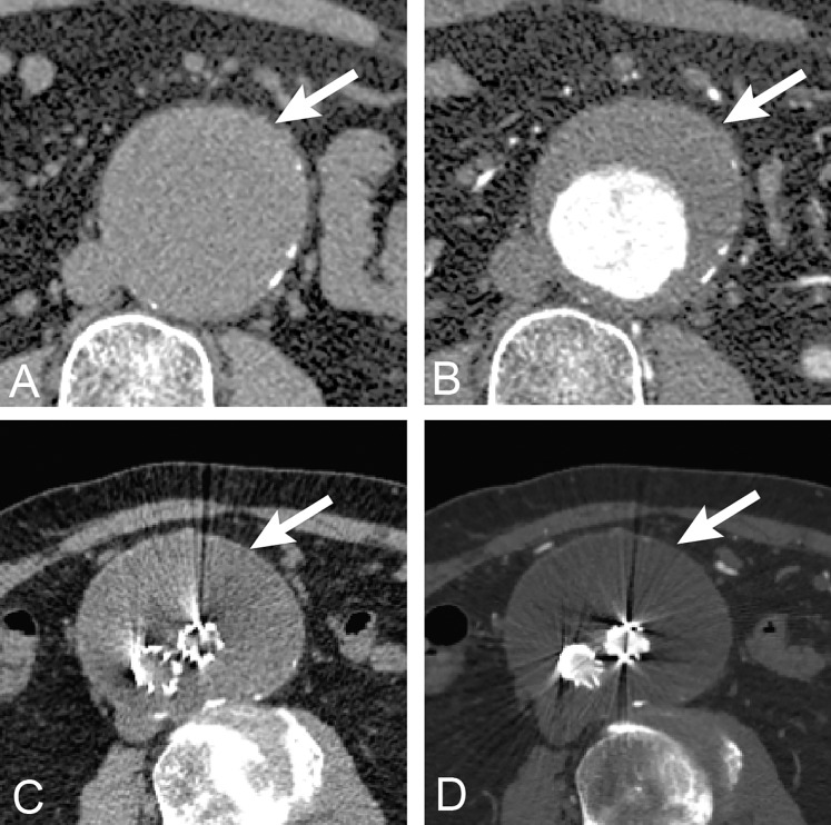
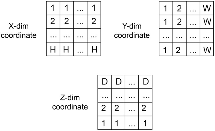
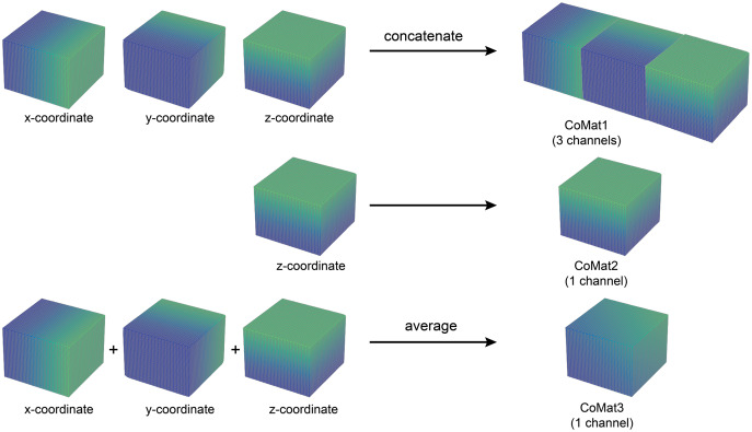
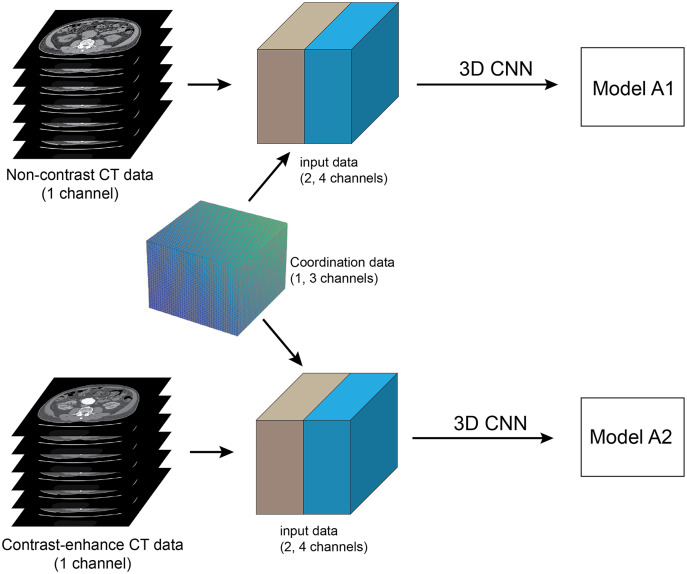
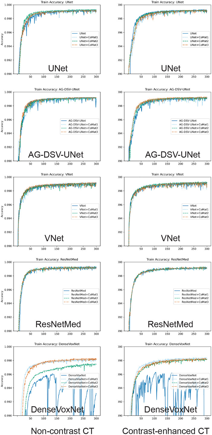
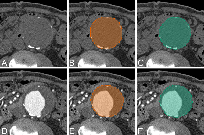
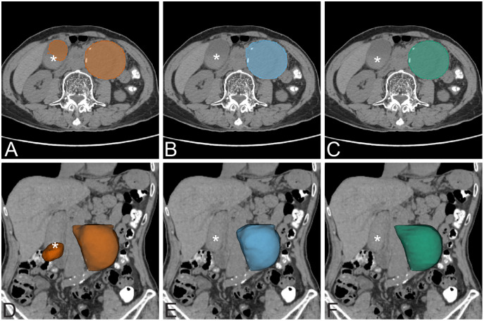
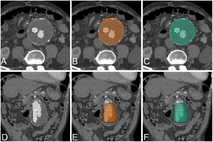
Abdominal aortic aneurysm (AAA) is one of the most common diseases worldwide. 3D segmentation of AAA provides useful information for surgical decisions and follow-up treatment. However, existing segmentation methods are time consuming and not practical in routine use. In this article, the segmentation task will be addressed automatically using a deep learning based approach which has been proved to successfully solve several medical imaging problems with excellent performances. This article therefore proposes a new solution of AAA segmentation using deep learning in a type of 3D convolutional neural network (CNN) architecture that also incorporates coordinate information. The tested CNNs are UNet, AG-DSV-UNet, VNet, ResNetMed and DenseVoxNet. The 3D-CNNs are trained with a dataset of high resolution (256 × 256) non-contrast and post-contrast CT images containing 64 slices from each of 200 patients. The dataset consists of contiguous CT slices without augmentation and no post-processing step. The experiments show that incorporation of coordinate information improves the segmentation results. The best accuracies on non-contrast and contrast-enhanced images have average dice scores of 97.13% and 96.74%, respectively. Transfer learning from a pre-trained network of a pre-operative dataset to post-operative endovascular aneurysm repair (EVAR) was also performed. The segmentation accuracy of post-operative EVAR using transfer learning on non-contrast and contrast-enhanced CT datasets achieved the best dice scores of 94.90% and 95.66%, respectively.
Keywords: 3D segmentation; Abdominal aortic aneurysm; Computed tomography; Coordinate information; Deep learning; Transfer learning.
© 2022 Siriapisith et al.
Conflict of interest statement
The authors declare that they have no competing interests.
Figures
Similar articles
-
A benchmark study of convolutional neural networks in fully automatic segmentation of aortic root.Front Bioeng Biotechnol. 2023 Jun 15;11:1171868. doi: 10.3389/fbioe.2023.1171868. eCollection 2023. Front Bioeng Biotechnol. 2023. PMID: 37397959 Free PMC article.
-
Deep learning-based recognition and segmentation of intracranial aneurysms under small sample size.Front Physiol. 2022 Dec 19;13:1084202. doi: 10.3389/fphys.2022.1084202. eCollection 2022. Front Physiol. 2022. PMID: 36601346 Free PMC article.
-
Machine deep learning accurately detects endoleak after endovascular abdominal aortic aneurysm repair.JVS Vasc Sci. 2020 Jan 7;1:5-12. doi: 10.1016/j.jvssci.2019.12.003. eCollection 2020. JVS Vasc Sci. 2020. PMID: 34617036 Free PMC article.
-
3D segmentation of exterior wall surface of abdominal aortic aneurysm from CT images using variable neighborhood search.Comput Biol Med. 2019 Apr;107:73-85. doi: 10.1016/j.compbiomed.2019.01.027. Epub 2019 Feb 6. Comput Biol Med. 2019. PMID: 30782525
-
Automatic Segmentation of Multiple Organs on 3D CT Images by Using Deep Learning Approaches.Adv Exp Med Biol. 2020;1213:135-147. doi: 10.1007/978-3-030-33128-3_9. Adv Exp Med Biol. 2020. PMID: 32030668 Review.
Cited by
-
Automatic CTA analysis for blood vessels and aneurysm features extraction in EVAR planning.Sci Rep. 2025 May 12;15(1):16431. doi: 10.1038/s41598-025-00484-7. Sci Rep. 2025. PMID: 40355533 Free PMC article.
-
Deep learning techniques for imaging diagnosis and treatment of aortic aneurysm.Front Cardiovasc Med. 2024 Feb 28;11:1354517. doi: 10.3389/fcvm.2024.1354517. eCollection 2024. Front Cardiovasc Med. 2024. PMID: 38481955 Free PMC article. Review.
References
-
- Chen S, Ma K, Zheng Y. Med3D: transfer learning for 3D medical image analysis. ArXiv preprint. 1904. - DOI
-
- Cheng J-Z, Ni D, Chou Y-H, Qin J, Tiu C-M, Chang Y-C, Huang C-S, Shen D, Chen C-M. Computer-aided diagnosis with deep learning architecture: applications to breast lesions in US images and pulmonary nodules in CT scans. Scientific Reports. 2016;6(1):24454. doi: 10.1038/srep24454. - DOI - PMC - PubMed
-
- Dziubich T, Białas P, Znaniecki Ł, Halman J. Abdominal aortic aneurysm segmentation from contrast-enhanced computed tomography angiography using deep convolutional networks. ADBIS, TPDL and EDA 2020 Common Workshops and Doctoral Consortium; Cham: Springer International Publishing; 2020.
-
- Fedorov A, Beichel R, Kalpathy-Cramer J, Finet J, Fillion-Robin JC, Pujol S, Bauer C, Jennings D, Fennessy F, Sonka M, Buatti J, Aylward S, Miller JV, Pieper S, Kikinis R. 3D Slicer as an image computing platform for the Quantitative Imaging Network. Magnetic Resonance Imaging. 2012;30(9):1323–1341. doi: 10.1016/j.mri.2012.05.001. - DOI - PMC - PubMed
Associated data
LinkOut - more resources
Full Text Sources