

Structural Brain Network Abnormalities in Parkinson's Disease With Freezing of Gait
- PMID: 35875794
- PMCID: PMC9304752
- DOI: 10.3389/fnagi.2022.944925
Structural Brain Network Abnormalities in Parkinson's Disease With Freezing of Gait
Abstract
Objective: Diffusion tensor imaging (DTI) studies have investigated white matter (WM) integrity abnormalities in Parkinson's disease (PD). However, little is known about the topological changes in the brain network. This study aims to reveal these changes by comparing PD without freezing of gait (FOG) (PD FOG-), PD with FOG (PD FOG+), and healthy control (HC).
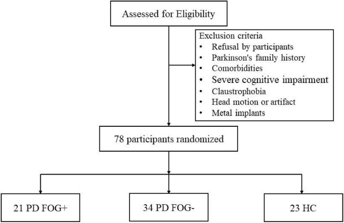
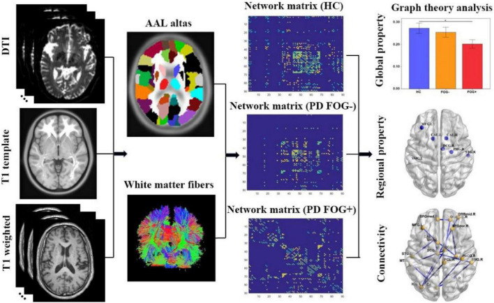
Methods: 21 PD FOG+, 34 PD FOG-, and 23 HC were recruited, and DTI images were acquired. The graph theoretical analysis and network-based statistical method were used to calculate the topological parameters and assess connections.
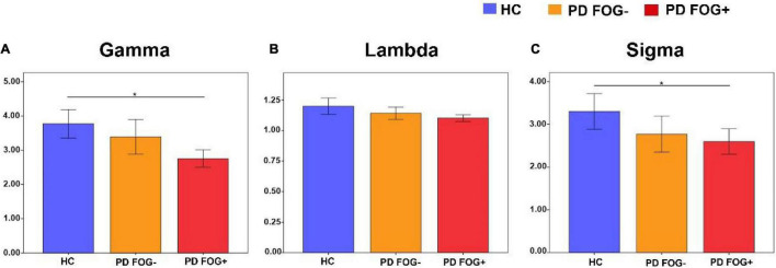
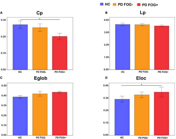
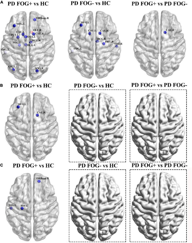
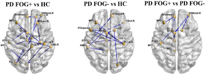
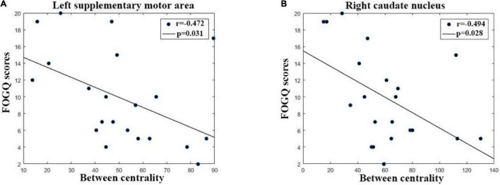
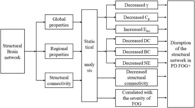
Results: PD FOG+ showed a decreased normalized clustering coefficient, small-worldness, clustering coefficient, and increased local network efficiency compared with HCs. PD FOG+ showed decreased centrality, degree centrality, and nodal efficiency in the striatum, frontal gyrus, and supplementary motor area (SMA). PD FOG+ showed decreased connections in the frontal gyrus, cingulate gyrus, and caudate nucleus (CAU). The between centrality of the left SMA and left CAU was negatively correlated with FOG questionnaire scores.
Conclusion: This study demonstrates that PD FOG+ exhibits disruption of global and local topological organization in structural brain networks, and the disrupted topological organization can be potential biomarkers in PD FOG+. These new findings may provide increasing insight into the pathophysiological mechanism of PD FOG+.
Keywords: Parkinson’s disease; diffusion tensor imaging; freezing of gait; graph theory analysis; network-based statistic.
Copyright © 2022 Jin, Yang, Qi, Teng, Li, Yao, Ruan and Wei.
Conflict of interest statement
The authors declare that the research was conducted in the absence of any commercial or financial relationships that could be construed as a potential conflict of interest.
Figures
Similar articles
-
Impaired Topographical Organization of Functional Brain Networks in Parkinson's Disease Patients With Freezing of Gait.Front Aging Neurosci. 2020 Oct 21;12:580564. doi: 10.3389/fnagi.2020.580564. eCollection 2020. Front Aging Neurosci. 2020. PMID: 33192473 Free PMC article.
-
Impaired structural and reserved functional topological organizations of brain networks in Parkinson's disease with freezing of gait.Quant Imaging Med Surg. 2023 Jan 1;13(1):66-79. doi: 10.21037/qims-22-351. Epub 2022 Sep 28. Quant Imaging Med Surg. 2023. PMID: 36620158 Free PMC article.
-
Brain network topology and future development of freezing of gait in Parkinson's disease: a longitudinal study.J Neurol. 2022 May;269(5):2503-2512. doi: 10.1007/s00415-021-10817-x. Epub 2021 Oct 7. J Neurol. 2022. PMID: 34618223
-
The Relationship of Rapid Eye Movement Sleep Behavior Disorder and Freezing of Gait in Parkinson's Disease.Cureus. 2020 Dec 30;12(12):e12385. doi: 10.7759/cureus.12385. Cureus. 2020. PMID: 33532150 Free PMC article. Review.
-
Oscillation-specific nodal alterations in early to middle stages Parkinson's disease.Transl Neurodegener. 2019 Nov 15;8:36. doi: 10.1186/s40035-019-0177-5. eCollection 2019. Transl Neurodegener. 2019. PMID: 31807287 Free PMC article. Review.
Cited by
-
Abnormal topological structure of structural covariance networks based on fractal dimension in noise induced hearing loss.Sci Rep. 2024 Nov 28;14(1):29644. doi: 10.1038/s41598-024-80731-5. Sci Rep. 2024. PMID: 39609512 Free PMC article.
-
Topological disruption of low- and high-order functional networks in presbycusis.Brain Commun. 2024 Apr 6;6(2):fcae119. doi: 10.1093/braincomms/fcae119. eCollection 2024. Brain Commun. 2024. PMID: 38638149 Free PMC article.
-
Reduced white matter integrity and disrupted brain network in children with type 2 and 3 spinal muscular atrophy.J Neurodev Disord. 2025 Jan 24;17(1):3. doi: 10.1186/s11689-025-09592-x. J Neurodev Disord. 2025. PMID: 39856544 Free PMC article.
-
Abnormal Alterations of the White Matter Structural Network in Patients with Herpes Zoster and Postherpetic Neuralgia.Brain Topogr. 2025 Feb 6;38(2):28. doi: 10.1007/s10548-025-01104-3. Brain Topogr. 2025. PMID: 39912964
References
LinkOut - more resources
Full Text Sources