. 2022 Aug 3;144(30):13851-13864.
doi: 10.1021/jacs.2c05349. Epub 2022 Jul 24.

Discovery of a Transferrin Receptor 1-Binding Aptamer and Its Application in Cancer Cell Depletion for Adoptive T-Cell Therapy Manufacturing

Affiliations

Discovery of a Transferrin Receptor 1-Binding Aptamer and Its Application in Cancer Cell Depletion for Adoptive T-Cell Therapy Manufacturing

Emmeline L Cheng et al. J Am Chem Soc. .

Abstract

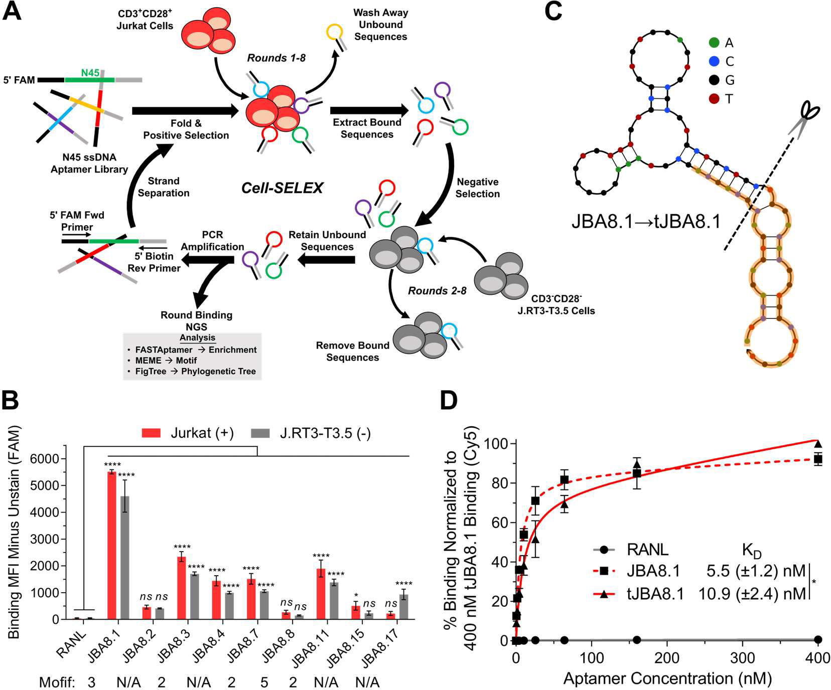
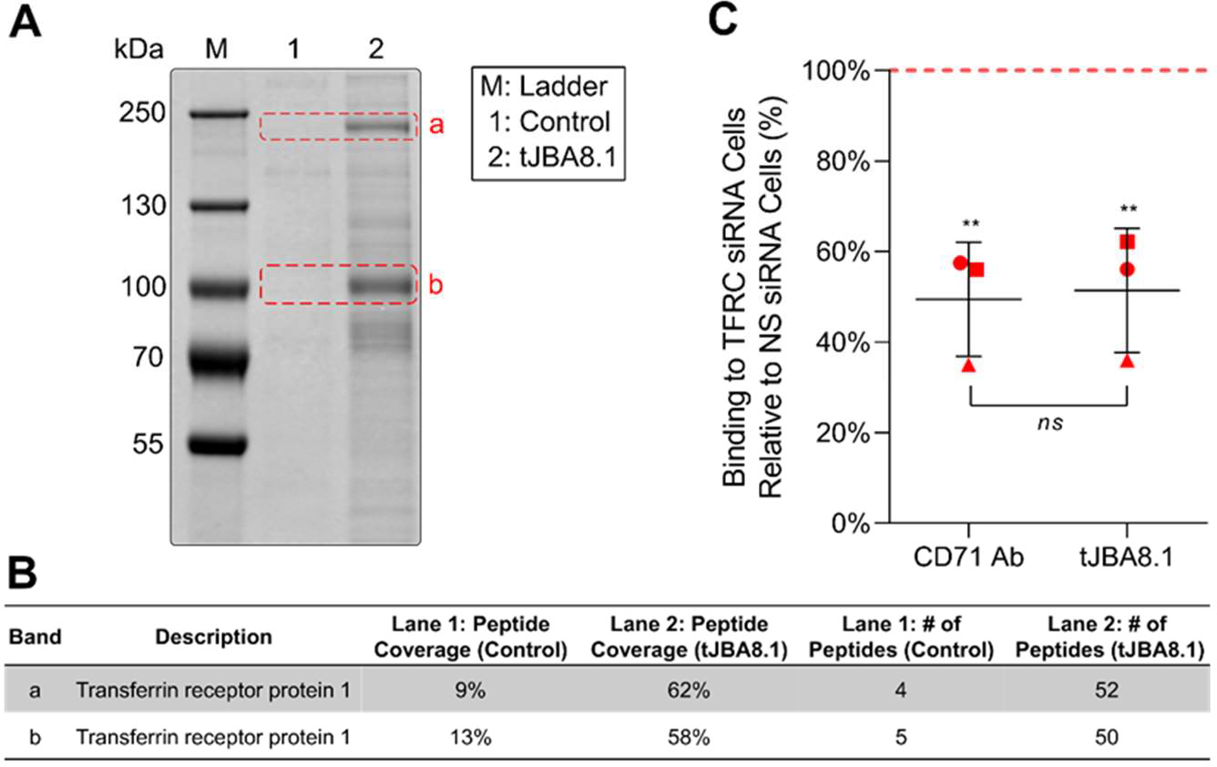
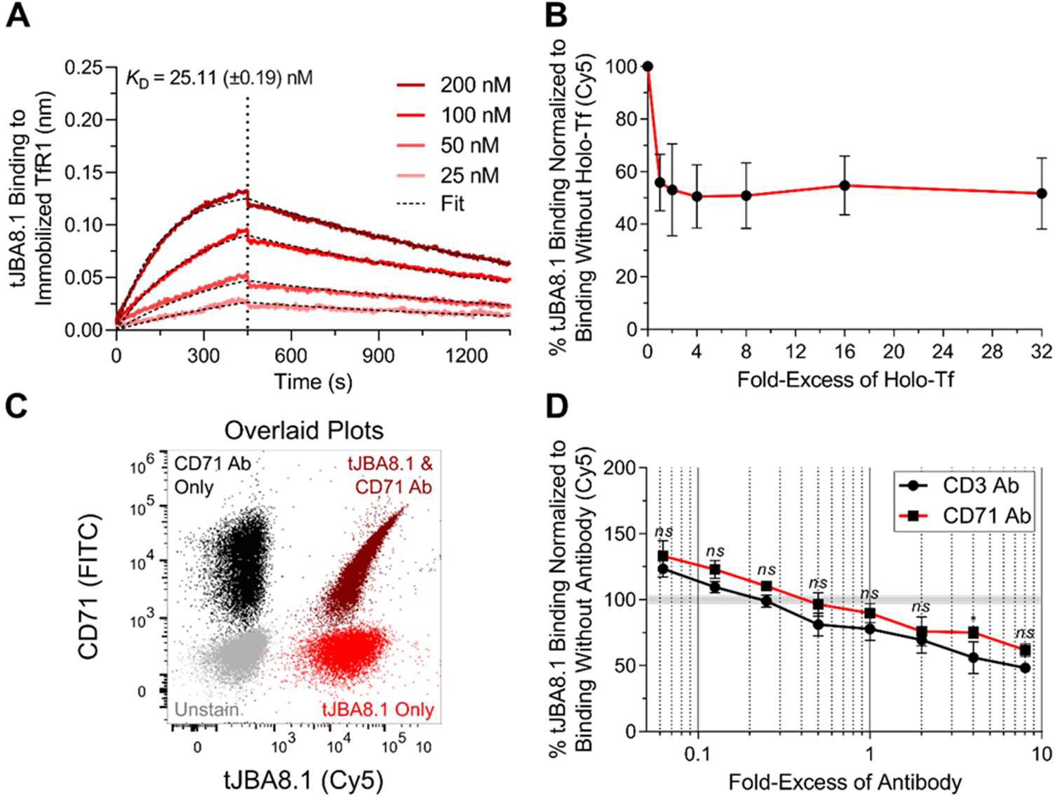
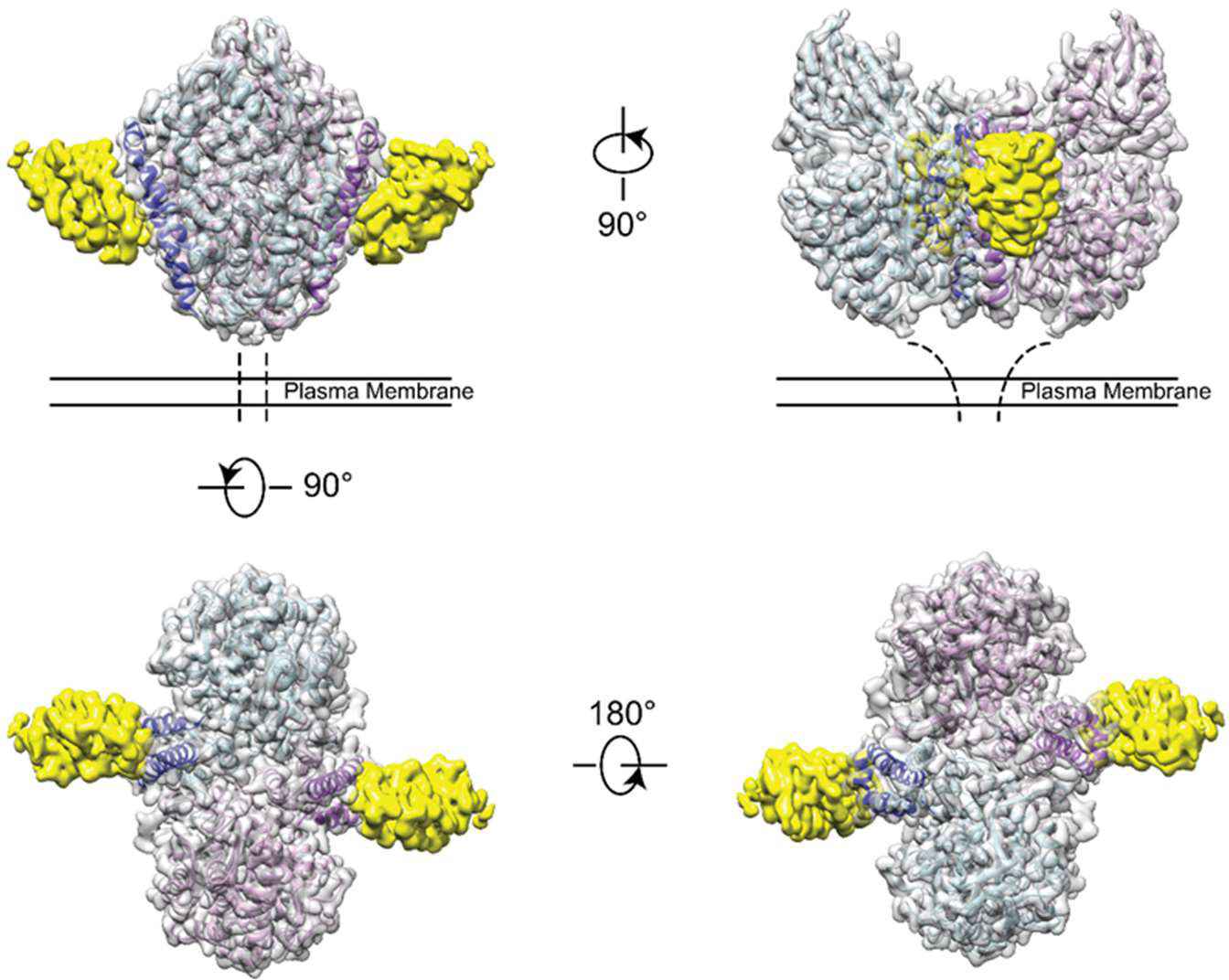
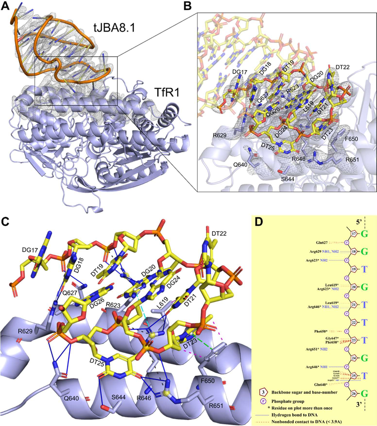
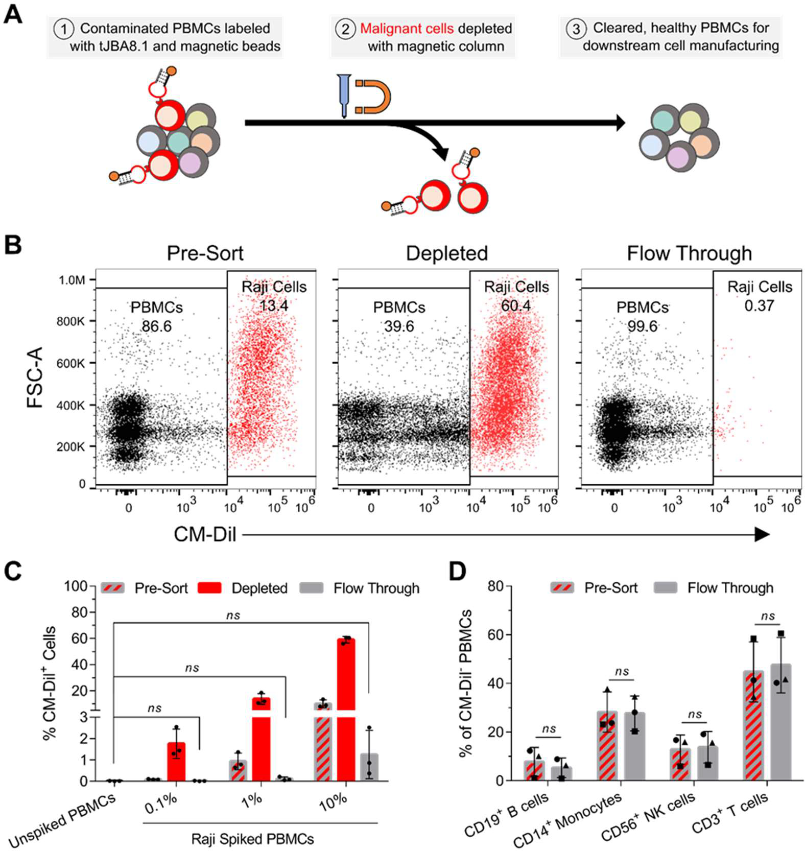
The clinical manufacturing of chimeric antigen receptor (CAR) T cells includes cell selection, activation, gene transduction, and expansion. While the method of T-cell selection varies across companies, current methods do not actively eliminate the cancer cells in the patient's apheresis product from the healthy immune cells. Alarmingly, it has been found that transduction of a single leukemic B cell with the CAR gene can confer resistance to CAR T-cell therapy and lead to treatment failure. In this study, we report the identification of a novel high-affinity DNA aptamer, termed tJBA8.1, that binds transferrin receptor 1 (TfR1), a receptor broadly upregulated by cancer cells. Using competition assays, high resolution cryo-EM, and de novo model building of the aptamer into the resulting electron density, we reveal that tJBA8.1 shares a binding site on TfR1 with holo-transferrin, the natural ligand of TfR1. We use tJBA8.1 to effectively deplete B lymphoma cells spiked into peripheral blood mononuclear cells with minimal impact on the healthy immune cell composition. Lastly, we present opportunities for affinity improvement of tJBA8.1. As TfR1 expression is broadly upregulated in many cancers, including difficult-to-treat T-cell leukemias and lymphomas, our work provides a facile, universal, and inexpensive approach for comprehensively removing cancerous cells from patient apheresis products for safe manufacturing of adoptive T-cell therapies.


Conflict of interest statement

The authors declare the following competing financial interest(s): Michael Jensen has interests in Umoja Biopharma and Juno Therapeutics, a Bristol-Myers Squibb company. Michael Jensen is a seed investor and holds ownership equity in Umoja, serves as a member of the Umoja Joint Steering Committee, and is a Board Observer of the Umoja Board of Directors. Michael Jensen holds patents, some of which are licensed to Umoja Biopharma and Juno Therapeutics. Suzie Pun, Nataly Kacherovsky, Emmeline Cheng, Ian Cardle, and Michael Jensen are co-inventors on a provisional patent application for the aptamers and cell depletion strategy described in this article.

Figures

Figure 1.
Figure 1.
Cell-SELEX and post-SELEX truncation lead to the development of the tJBA8.1 aptamer. (A) Schematic of cell-SELEX using CD3+CD28+ Jurkat cells for positive selection and CD3CD28 J.RT3-T3.5 cells for negative selection. (B) Binding median fluorescence intensity (MFI) of 100 nM RANL and individual aptamers identified from round 8 of cell-SELEX to Jurkat cells and J.RT3-T3.5 cells by flow cytometry. Aptamers belonging to predicted motifs are indicated. Graph bars and error bars represent mean ± standard deviation; n = 3 independent experiments. ns > 0.05, *P < 0.05, ****P < 0.0001 (ordinary two-way ANOVA with Šídák correction). (C) MFE secondary structures of JBA8.1 and its truncation (tJBA8.1), predicted using NUPACK (temperature = 4 °C; Na+ = 137 mM; Mg2+ = 5.5 mM). The dashed line indicates the site of truncation, whereas the orange highlighting denotes the 18-nt flanking constant regions. (D) Flow cytometry binding curves of RANL, JBA8.1, and tJBA8.1 to Jurkat cells, normalized to 400 nM tJBA8.1 binding. The curves represent a nonlinear regression assuming one-site total binding. KD values were calculated by averaging the individual regression values of the independent experiments. Data points and error bars, and KD values, represent mean ± standard deviation; n = 3 independent experiments with technical duplicates. *P < 0.05 (two-sided unpaired t-test). FAM, 6-carboxyfluorescein; Cy5, cyanine 5.
Figure 2.
Figure 2.
TfR1 is identified as the target of tJBA8.1. (A) Colloidal blue-stained 8% SDS-PAGE gel of Jurkat cell membrane proteins pulled down by tJBA8.1. The control lane represents proteins captured by biotin-saturated magnetic beads only. Bands a and b (dashed red boxes) from both lanes were excised for mass spectrometry analysis. (B) Summary of the protein with the highest peptide coverage and number of peptides identified in each excised band by mass spectrometry. (C) Flow cytometry analysis of FITC-labeled CD71 Ab and 25 nM Cy5-labeled tJBA8.1 binding to Jurkat cells 24 h after nucleofection with TFRC siRNA duplexes. Red, dashed horizontal line represents binding to non-specific (NS) siRNA-treated controls to which the TFRC siRNA data points were normalized. Horizontal lines and error bars represent mean ± standard deviation; n = 3 independent experiments. **P < 0.01 (significance between ligand staining on TFRC siRNA- and NS siRNA-treated cells; one-way ANOVA with Bonferroni correction). Ns > 0.05 (significance between the relative CD71 Ab and tJBA8.1 staining in pairwise experiments; two-sided paired t-test). Cy5, cyanine 5; FITC, fluorescein isothiocyanate.
Figure 3.
Figure 3.
tJBA8.1 competes with holo-Tf but not antibody clone CY1G4 for binding to TfR1. (A) Association and dissociation kinetics of serially diluted FAM-labeled tJBA8.1 binding to biotinylated TfR1 immobilized on streptavidin biosensors by BLI. The association phase is illustrated from 0–450 s, whereas dissociation is shown from 450–1350 s (separated by the vertical dotted line). KD values were calculated by performing a global fit of the multi-concentration kinetic data to a 1:1 binding model. KD values represent mean ± standard deviation; n = 4 individual concentrations of aptamers. (B) Competitive binding of 25 nM Cy5-labeled tJBA8.1 with varying fold-excess of holo-Tf to Jurkat cells by flow cytometry. Binding was normalized to aptamer-stained controls without holo-Tf. Data points and error bars represent mean ± standard deviation; n = 3 independent experiments. ns > 0.05, *P < 0.05 (ordinary two-way ANOVA with Šídák correction). (C) Overlaid flow cytometry plots of unstained (grey), FITC-labeled CD71 Ab single-stained (black), 25 nM Cy5-labeled tJBA8.1 single-stained (red), and antibody and aptamer co-stained (dark red) Jurkat cells. Plots are representative of n = 2 independent experiments. (D) Competitive binding of 25 nM Cy5-labeled tJBA8.1 with varying fold-excess of CD3 or CD71 Ab to Jurkat cells by flow cytometry. Binding was normalized to aptamer-stained controls without antibody. Data points and error bars represent mean ± standard deviation; n = 3 independent experiments. ns > 0.05, *P < 0.05 (ordinary two-way ANOVA with Šídák correction). FITC, fluorescein isothiocyanate; Cy5, cyanine 5; FAM, 6-carboxyfluorescein.
Figure 4.
Figure 4.
tJBA8.1 binds the helical domain of TfR1. Cryo-EM density map and refined structure of tJBA8.1-bound TfR1. Four views are shown with color coding (yellow: tJBA8.1 density; gray: TfR1 homodimer density; light blue and light purple: individual TfR1 monomer structures; dark blue and dark purple: α helices 1, 2, and 3 of each TfR1 monomer helical domain).
Figure 5.
Figure 5.
De novo model of tJBA8.1 built into the cryo-EM map enables detailed characterization of the interactions at the tJBA8.1-TfR1 interface. (A) Cartoon representation of tJBA8.1-TfR1 complex (yellow: tJBA8.1; lavender blue: TfR1) modeled in the cryo-EM density maps (overlayed grey mesh). For clarity, only one monomer of the tJBA8.1-TfR1 complex is shown, and the overlaid cryo-EM density maps are restricted to tJBA8.1 and the binding interface on TfR1. (B) Zoomed-in view of the modeled tJBA8.1-TfR1 interface within the cryo-EM density maps showing the overall fit of the model. tJBA8.1 nucleotides and TfR1 protein residues in close contact at the binding interface are labeled and shown as sticks with atom color coding (yellow: nucleotide carbon; lavender blue: protein carbon; blue: nitrogen; red: oxygen; orange: phosphate). (C) Molecular interactions at the modeled tJBA8.1-TfR1 interface, calculated using PDBePISA and PLIP. Different lines and color coding are used to denote the different interactions (blue solid: hydrogen bonds; black dashed: salt bridges; magenta dashed: hydrophobic interactions; green dashed with ring centroids: pistacking; cyan dashed with centroids: cation-pi). The list of detected interactions at the tJBA8.1-TfR1 interface can be found in Table S8. (D) Schematic diagram of hydrogen bonds and nonbonded contacts at the modeled tJBA8.1-TfR1 interface, generated using NUCPLOT.
Figure 6.
Figure 6.
tJBA8.1 thoroughly depletes Raji B-lymphoma cells from PBMCs without altering healthy immune cell composition. (A) Schematic of malignant cell depletion from PBMCs using tJBA8.1-mediated MACS. (B) Flow cytometry plots of CM-Dil+ Raji cell depletion from high (10%) Raji spiked PBMCs. The different cell fractions from the depletion process are shown. Plots for low (0.1%) and medium (1%) Raji spiked PBMCs can be found in Figure S11B,C. Plots are representative of n = 3 independent experiments with different PBMC donors. (C) Flow cytometry analysis of the percentage of CM-Dil+ Raji cells in each cell fraction of the depletion process using low (0.1%), medium (1%), and high (10%) Raji spiked PBMCs. Unspiked PBMCs were included as a benchmark of complete depletion. Graph bars and error bars represent mean ± standard deviation; n = 3 independent experiments with different PBMC donors. ns > 0.05 (ordinary one-way ANOVA with Dunnett’s correction). (D) Flow cytometry analysis of the healthy immune cell composition within CM-Dil PBMCs before (pre-sort) and after (flow through) Raji depletion from high (10%) Raji spiked PBMCs. Analysis for low (0.1%) and medium (1%) Raji spiked PBMCs can be found in Figure S12A,B. The circles, squares and triangles represent different PBMC donors from separate depletion studies. Graph bars and error bars represent mean ± standard deviation; n = 3 independent experiments with different PBMC donors. ns > 0.05 (paired two-way ANOVA with Šídák correction). CM-Dil, chloromethylbenzamido-1,1’-dioctadecyl-3,3,3’,3’-tetramethylindocarbocyanine perchlorate.

References

    1. Maude SL; Laetsch TW; Buechner J; Rives S; Boyer M; Bittencourt H; Bader P; Verneris MR; Stefanski HE; Myers GD; Qayed M; De Moerloose B; Hiramatsu H; Schlis K; Davis KL; Martin PL; Nemecek ER; Yanik GA; Peters C; Baruchel A; Boissel N; Mechinaud F; Balduzzi A; Krueger J; June CH; Levine BL; Wood P; Taran T; Leung M; Mueller KT; Zhang Y; Sen K; Lebwohl D; Pulsipher MA; Grupp SA Tisagenlecleucel in Children and Young Adults with B-Cell Lymphoblastic Leukemia. New England Journal of Medicine 2018, 378 (5), 439–448. - PMC - PubMed
    1. Schuster SJ; Bishop MR; Tam CS; Waller EK; Borchmann P; McGuirk JP; Jäger U; Jaglowski S; Andreadis C; Westin JR; Fleury I; Bachanova V; Foley SR; Ho PJ; Mielke S; Magenau JM; Holte H; Pantano S; Pacaud LB; Awasthi R; Chu J; Anak Ö; Salles G; Maziarz RT Tisagenlecleucel in Adult Relapsed or Refractory Diffuse Large B-Cell Lymphoma. New England Journal of Medicine 2018, 380 (1), 45–56. - PubMed
    1. Neelapu SS; Locke FL; Bartlett NL; Lekakis LJ; Miklos DB; Jacobson CA; Braunschweig I; Oluwole OO; Siddiqi T; Lin Y; Timmerman JM; Stiff PJ; Friedberg JW; Flinn IW; Goy A; Hill BT; Smith MR; Deol A; Farooq U; McSweeney P; Munoz J; Avivi I; Castro JE; Westin JR; Chavez JC; Ghobadi A; Komanduri KV; Levy R; Jacobsen ED; Witzig TE; Reagan P; Bot A; Rossi J; Navale L; Jiang Y; Aycock J; Elias M; Chang D; Wiezorek J; Go WY Axicabtagene Ciloleucel CAR T-Cell Therapy in Refractory Large B-Cell Lymphoma. New England Journal of Medicine 2017, 377 (26), 2531–2544. - PMC - PubMed
    1. Locke FL; Ghobadi A; Jacobson CA; Miklos DB; Lekakis LJ; Oluwole OO; Lin Y; Braunschweig I; Hill BT; Timmerman JM; Deol A; Reagan PM; Stiff P; Flinn IW; Farooq U; Goy A; McSweeney PA; Munoz J; Siddiqi T; Chavez JC; Herrera AF; Bartlett NL; Wiezorek JS; Navale L; Xue A; Jiang Y; Bot A; Rossi JM; Kim JJ; Go WY; Neelapu SS Long-Term Safety and Activity of Axicabtagene Ciloleucel in Refractory Large B-Cell Lymphoma (ZUMA-1): A Single-Arm, Multicentre, Phase 1–2 Trial. The Lancet Oncology 2019, 20 (1), 31–42. - PMC - PubMed
    1. Wang M; Munoz J; Goy A; Locke FL; Jacobson CA; Hill BT; Timmerman JM; Holmes H; Jaglowski S; Flinn IW; McSweeney PA; Miklos DB; Pagel JM; Kersten M-J; Milpied N; Fung H; Topp MS; Houot R; Beitinjaneh A; Peng W; Zheng L; Rossi JM; Jain RK; Rao AV; Reagan PM KTE-X19 CAR T-Cell Therapy in Relapsed or Refractory Mantle-Cell Lymphoma. New England Journal of Medicine 2020, 382 (14), 1331–1342. - PMC - PubMed

Publication types

MeSH terms

Substances