Review
. 2023 Jul 3;11(3):2104085.
doi: 10.1080/21688370.2022.2104085. Epub 2022 Jul 24.

Interplay between EGFR, E-cadherin, and PTP1B in epidermal homeostasis

Affiliations
Review

Interplay between EGFR, E-cadherin, and PTP1B in epidermal homeostasis

Tessa Arnaud et al. Tissue Barriers. .

Abstract

Maintaining epithelial homeostasis is crucial to allow embryo development but also the protective barrier which is ensured by the epidermis. This homeostasis is regulated through the expression of several molecules among which EGFR and E-cadherin which are of major importance. Indeed, defects in the regulation of these proteins lead to abnormalities in cell adhesion, proliferation, differentiation, and migration. Hence, regulation of these two proteins is of the utmost importance as they are involved in numerous skin pathologies and cancers. In the last decades it has been described several pathways of regulation of these two proteins and notably several mechanisms of cross-regulation between these partners. In this review, we aimed to describe the current understanding of the regulation of EGFR and interactions between EGFR and E-cadherin and, in particular, the implication of these cross-regulations in epithelium homeostasis. We pay particular attention to PTP1B, a phosphatase involved in the regulation of EGFR.

Keywords: E-cadherin; EGFR; Epithelium; PTP1B; differentiation.


Conflict of interest statement

No potential conflict of interest was reported by the author(s).

Figures
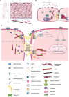
Figure 1.
Figure 1.
A Schematic representation of human epidermis. At the outermost side of the skin, the epidermis is composed of several layers of cells: basal, replicative cells, and suprabasal layers that undergo a regulated program of terminal differentiation as cells move outward to the stratum corneum (dead cells). On the dermis interface, in the basal layer of keratinocytes, EGFR signaling is highly activated and cells are proliferative. From the base and up in the epidermis, cells reduce their proliferation and start to differentiate and form more cell-cell junctions, such as tight, adherens and gap junctions; a precise regulation and balance between EGFR and E-Cadherin (E-Cad) are keystone in maintaining structure and integrity of the epidermis. B Regulation of EGFR intracellular trafficking and activation. EGFR activated in the presence of various concentrations of EGF is driven in two distinct endocytic pathways. 1: At low concentrations of EGF, the receptor undergoes the “recycling pathway”, through early and recycling endosomes, before being recycled back to the plasma membrane. 2: At high concentrations of EGF, it undergoes the “degradation pathway”. Signaling is down-regulated by two major mechanisms: PTP1B-dependent dephosphorylation at the RE membrane contact, and degradation inside the endolysosomes upon control by ubiquitination. C Overview of EGFR, E-Cadherin and PTP1B interactions, and their roles in the maintenance of AJ and intracellular signaling. 1: By dephosphorylating beta-catenin, PTP1B promotes the proper assembly of AJ. Their integrity is also conserved by the direct interaction of PTP1B with E-Cadherin (still at AJ location). 2: When PTP1B is not phosphorylated, it cannot dephosphorylate beta-catenin, and fails to maintain AJ integrity. 3: EGFR promotes proliferation, differentiation, cell growth and survival by activating several signaling pathways such as MAPK, STAT, PI3K/Akt and Erk. The latters are also regulated by E-Cadherin. EGFR interaction with E-Cadherin through galectin-7 partially impairs EGFR phosphorylation, contributing to the E-Cadherin/EGFR balance. 4: p120 direct interaction with EGFR enhances Akt and Erk signaling. 5: PTP1B inhibits EGFR by dephosphorylation, both at the plasma membrane and near the RE (Figure 1b), therefore down-regulating the corresponding signaling pathways. 6: Under tension, vinculin binds to unfolded alpha-catenin, after phosphorylation by Abl. This complex binds myosin II and F-actin, forming an organized contractile actin network.

References

    1. Kulukian A, Fuchs E.. Spindle orientation and epidermal morphogenesis. Philos Trans R Soc Lond B Biol Sci. 2013;368(1629):211. doi:10.1098/rstb.2013.0016. - DOI - PMC - PubMed
    1. Fuchs E. Epithelial skin biology: three decades of developmental biology, a hundred questions answered and a thousand new ones to address. Curr Top Dev Biol. 2016;116:357–221. - PMC - PubMed
    1. Tinkle CL, Lechler T, Pasolli HA, Fuchs E. Conditional targeting of E-cadherin in skin: insights into hyperproliferative and degenerative responses. Proc Natl Acad Sci U S A. 2004;101(2):552–557. doi:10.1073/pnas.0307437100. - DOI - PMC - PubMed
    1. Tunggal JA, Helfrich I, Schmitz A, Schwarz H, Günzel D, Fromm M, Kemler R, Krieg T, Niessen CM. E-cadherin is essential for in vivo epidermal barrier function by regulating tight junctions. EMBO J. 2005;24(6):1146–1156. doi:10.1038/sj.emboj.7600605. - DOI - PMC - PubMed
    1. Young P, Boussadia O, Halfter H, Grose R, Berger P, Leone DP, Robenek H, Charnay P, Kemler R, Suter U. E-cadherin controls adherens junctions in the epidermis and the renewal of hair follicles. EMBO J. 2003;22(21):5723–5733. doi:10.1093/emboj/cdg560. - DOI - PMC - PubMed

Publication types