. 2022 Aug 3;14(30):34365-34376.
doi: 10.1021/acsami.2c06503. Epub 2022 Jul 24.

Multifunctional Magnetic CuS/Gd2O3 Nanoparticles for Fluorescence/Magnetic Resonance Bimodal Imaging-Guided Photothermal-Intensified Chemodynamic Synergetic Therapy of Targeted Tumors

Affiliations

Multifunctional Magnetic CuS/Gd2O3 Nanoparticles for Fluorescence/Magnetic Resonance Bimodal Imaging-Guided Photothermal-Intensified Chemodynamic Synergetic Therapy of Targeted Tumors

Minchuan Luo et al. ACS Appl Mater Interfaces. .

Abstract

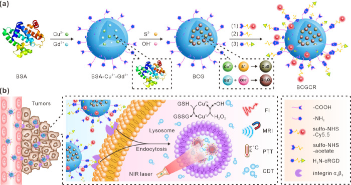
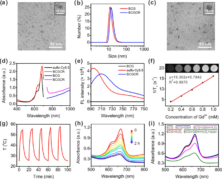
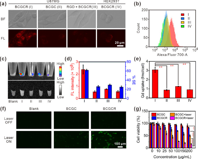
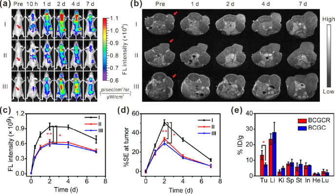
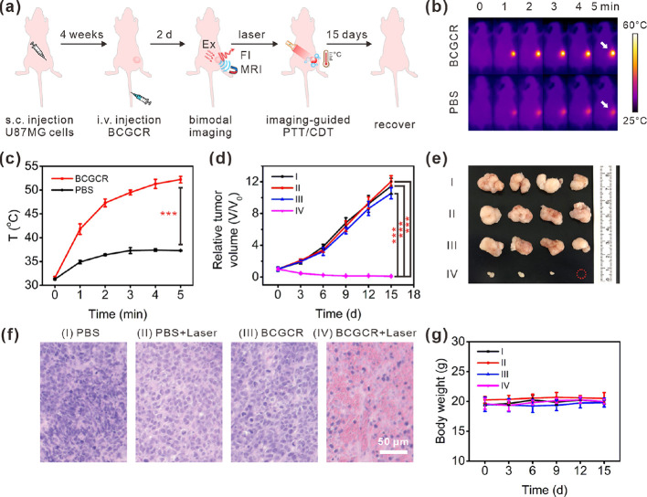
Chemodynamic therapy (CDT), which consumes endogenous hydrogen peroxide (H2O2) to generate reactive oxygen species (ROS) and causes oxidative damage to tumor cells, shows tremendous promise for advanced cancer treatment. However, the rate of ROS generation based on the Fenton reaction is prone to being restricted by inadequate H2O2 and unattainable acidity in the hypoxic tumor microenvironment. We herein report a multifunctional nanoprobe (BCGCR) integrating bimodal imaging and photothermal-enhanced CDT of the targeted tumor, which is produced by covalent conjugation of bovine serum albumin-stabilized CuS/Gd2O3 nanoparticles (NPs) with the Cy5.5 fluorophore and the tumor-targeting ligand RGD. BCGCR exhibits intense near-infrared (NIR) fluorescence and acceptable r1 relaxivity (∼15.3 mM-1 s-1) for both sensitive fluorescence imaging and high-spatial-resolution magnetic resonance imaging of tumors in living mice. Moreover, owing to the strong NIR absorbance from the internal CuS NPs, BCGCR can generate localized heat and displays a high photothermal conversion efficiency (30.3%) under 980 nm laser irradiation, which enables photothermal therapy and further intensifies ROS generation arising from the Cu-induced Fenton-like reaction for enhanced CDT. This synergetic effect shows such an excellent therapeutic efficacy that it can ablate xenografted tumors in vivo. We believe that this strategy will be beneficial to exploring other advanced nanomaterials for the clinical application of multimodal imaging-guided synergetic cancer therapies.

Keywords: copper sulfide; enhanced chemodynamic therapy; fluorescence imaging; magnetic resonance imaging; photothermal therapy.


Conflict of interest statement

The authors declare no competing financial interest.

Figures

Figure 1
Figure 1
Schematic illustration of BCGCR. (a) Design. (b) Mechanisms of fluorescence/MR bimodal imaging-guided PTT and intensified CDT of targeted tumors.
Figure 2
Figure 2
Characterization of BCG and BCGCR. (a) TEM image of BCG. (b) DLS analysis of BCG and BCGCR. (c) TEM image of BCGCR. (d) Absorption spectra of sulfo-Cy5.5 (0.1 mg/mL, black), BCGCR (2 mg/mL, red), BCG (20 mg/mL, blue), and BCGCR (25 mg/mL, pink). (e) Fluorescence (FL) spectra of BCG (10 mg/mL), sulfo-Cy5.5 (2 μg/mL), and BCGCR (1 mg/mL). λex = 660 nm. (f) Plots of 1/T1 of BCGCR versus different Gd3+ concentrations from 0.2 to 1.0 mM. Inset: T1-weighted MR images (1.5 T) of aqueous BCGCR with Gd3+ concentrations of 0, 0.2, 0.4, 0.6, 0.8, and 1.0 mM. TR/TE, 500/9.0 ms. (g) Photothermal stability of aqueous BCGCR (1 mM Cu2+) for five cycles of heating by irradiation (980 nm, 0.8 W/cm2) and natural cooling. (h) Absorption spectra of MB (10 μg/mL) incubated with BCG-GSH (0.5 mM Cu2+) and H2O2 (20 μM) at pH 5.4 for 0, 3, 5, 10, 15, 20, 30, 60, 90, and 120 min. (i) Absorption spectra of (10 μg/mL) MB under different conditions at pH 5.4 with or without laser irradiation (980 nm, 0.8 W/cm2) for 5 min.
Figure 3
Figure 3
In vitro bimodal imaging of cells, CDT performance, and cytotoxicity studies. (a) Fluorescence images of (I) U87MG cells incubated with BCGCR (100 μg/mL, 2 h), (II) U87MG cells incubated with BCGC (100 μg/mL, 2 h), (III) U87MG cells incubated with RGD (10 μM, 1 h) followed by BCGCR (100 μg/mL, 2 h), or (IV) HEK293T cells incubated with BCGCR (100 μg/mL, 2 h) (ex: 650 ± 22 nm, em: 720 ± 30 nm). (b) Flow cytometry assays of fluorescence intensity in cells after similar treatments with BCGC or BCGCR (10 μg/mL) for 2 h (ex: 633 nm, em: 710 ± 25 nm). (c) Fluorescence (top) (ex/em. = 675/720 nm) images of cell pellets after similar treatments with BCGC or BCGCR (10 μg/mL) for 2 h and T1-weighted MR (bottom) (TR/TE = 500/9.0 ms at 1.5 T) images of cell pellets after similar treatments with BCGC or BCGCR (10 μM Gd3+) for 24 h. (d) Statistics of total FL intensities (red) and average % MR signal enhancements (% SE, blue) of cell pellets in (c). (e) ICP-AES analysis of the average Gd uptake in each cell after indicated treatments in (c, bottom). (f) Fluorescence images of U87MG cells (untreated, incubated with 100 μg/mL BCGC or BCGCR, 6 h) followed by 10 μM DCFH-DA staining with or without laser irradiation (980 nm, 0.8 W/cm2) for 5 min (ex: 470 ± 20 nm, em: 525 ± 25 nm). (g) U87MG cell viability after incubation with different concentrations of BCGC or BCGCR for 24 h followed by exposure (or nonexposure) to laser irradiation (980 nm, 0.8 W/cm2) for 5 min. Values denote the mean ± SD (n = 5 for cell viability and n = 3 for others; **P < 0.01, ***P < 0.001).
Figure 4
Figure 4
Bimodal imaging of U87MG tumors in living mice as well as biodistribution analysis. (a) FL images of mice receiving (I) i.v. injection of BCGCR (5 mg/kg, 100 μL), (II) i.t. injection of free cRGDfk (2 mM, 100 μL) followed by i.v. injection of BCGCR (5 mg/kg, 100 μL) 1 h later, and (III) i.v. injection of BCGC (5 mg/kg, 100 μL) at 0 h, 10 h, 1 day, 2 days, 4 days, and 7 days (ex: 675 nm, em: 720 nm). (b) T1-weighted MR images of mice receiving (I) i.v. injection of BCGCR (20 μmol/kg Gd3+, 200 μL), (II) i.t. injection of free cRGDfk (2 mM, 100 μL) followed by i.v. injection of BCGCR (20 μmol/kg Gd3+, 200 μL) 1 h later, and (III) i.v. injection of BCGC (20 μmol/kg Gd3+, 200 μL) at 0 h, 1 day, 2 days, 4 days, and 7 days (TR/TE = 500/9.0 ms at 1.5 T). Red arrows indicate the tumor locations in mice. (c) Variations of average FL intensity at the tumor with time in (a). (d) Variations of average % SE at the tumor with time in (b). (e) Biodistribution (% ID/g) of BCGCR (red) or BCGC (blue) in the main organs and the U87MG tumor (Tu: tumor; Li: liver; Ki: kidneys; Sp: spleen; St: stomach; In: intestines; He: heart; Lu: lungs) at 2 days after i.v. injection of BCGC (10 mg/kg Cu2+) or BCGCR (10 mg/kg Cu2+) determined via a quantification analysis of the amount of Cu2+ by ICP-AES. Values denote the mean ± SD (n = 3, *P < 0.05, and **P < 0.01).
Figure 5
Figure 5
Synergetic PTT and intensified CDT of tumors in vivo. (a) Schematic illustration of the process of fluorescence/MR bimodal imaging-guided synergetic PTT/CDT of U87MG tumors in living mice. (b) IR thermal images of U87MG tumors in living mice during exposure to 980 nm laser irradiation (0.8 W/cm2) for 0–5 min at 2 days after i.v. injection of PBS (200 μL) or BCGCR (5 mg/kg Cu2+, 200 μL). White arrows indicate the tumor locations in mice. (c) Average temperature variations of the tumor with time in (b). (d) Relative tumor volume variation in living mice with time after treatments with (I) PBS (200 μL), (II) PBS (200 μL) + laser (0.8 W/cm2, 10 min), (III) BCGCR (5 mg/kg Cu2+, 200 μL), and (IV) BCGCR (5 mg/kg Cu2+, 200 μL) + laser (0.8 W/cm2, 10 min). (e) Photograph of tumors resected from mice on the 15th day after the indicated treatments. (f) H&E staining of U87MG tumor slices resected from mice on the second day after indicated treatments. (g) Average body weight variation of mice with time after the indicated treatments. Values denote the mean ± SD (n = 4, ***P < 0.001).

References

    1. Aggarwal V.; Tuli H. S.; Varol A.; Thakral F.; Yerer M. B.; Sak K.; Varol M.; Jain A.; Khan M. A.; Sethi G. Role of Reactive Oxygen Species in Cancer Progression: Molecular Mechanisms and Recent Advancements. Biomolecules 2019, 9, 735.10.3390/biom9110735. - DOI - PMC - PubMed
    1. Yang B.; Chen Y.; Shi J. Reactive Oxygen Species (ROS)-Based Nanomedicine. Chem. Rev. 2019, 119, 4881–4985. 10.1021/acs.chemrev.8b00626. - DOI - PubMed
    1. Jin H.; Zhu T.; Huang X.; Sun M.; Li H.; Zhu X.; Liu M.; Xie Y.; Huang W.; Yan D. ROS-responsive Nanoparticles Based on Amphiphilic Hyperbranched Polyphosphoester for Drug Delivery: Light-triggered Size-reducing and Enhanced Tumor Penetration. Biomaterials 2019, 211, 68–80. 10.1016/j.biomaterials.2019.04.029. - DOI - PubMed
    1. Hu P.; Wang R.; Zhou L.; Chen L.; Wu Q.; Han M.; El-Toni A. M.; Zhao D.; Zhang F. Near-Infrared-Activated Upconversion Nanoprobes for Sensitive Endogenous Zn2+ Detection and Selective On-Demand Photodynamic Therapy. Anal. Chem. 2017, 89, 3492–3500. 10.1021/acs.analchem.6b04548. - DOI - PubMed
    1. Lan M.; Zhao S.; Liu W.; Lee C.; Zhang W.; Wang P. Photosensitizers for Photodynamic Therapy. Adv. Healthcare Mater. 2019, 8, 1900132.10.1002/adhm.201900132. - DOI - PubMed