. 2022 Aug;70(8):557-569.
doi: 10.1369/00221554221115767. Epub 2022 Jul 25.

Quantitative Assessment of the Apical and Basolateral Membrane Expression of VEGFR2 and NRP2 in VEGF-A-stimulated Cultured Human Umbilical Vein Endothelial Cells

Affiliations

Quantitative Assessment of the Apical and Basolateral Membrane Expression of VEGFR2 and NRP2 in VEGF-A-stimulated Cultured Human Umbilical Vein Endothelial Cells

Esmeralda K Bosma et al. J Histochem Cytochem. 2022 Aug.

Abstract

Endothelial cells (ECs) form a precisely regulated polarized monolayer in capillary walls. Vascular endothelial growth factor-A (VEGF-A) induces endothelial hyperpermeability, and VEGF-A applied to the basolateral side, but not the apical side, has been shown to be a strong barrier disruptor in blood-retinal barrier ECs. We show here that VEGF-A presented to the basolateral side of human umbilical vein ECs (HUVECs) induces higher permeability than apical stimulation, which is similar to results obtained with bovine retinal ECs. We investigated with immunocytochemistry and confocal imaging the distribution of VEGF receptor-2 (VEGFR2) and neuropilin-2 (NRP2) in perinuclear apical and basolateral membrane domains. Orthogonal z-sections of cultured HUVECs were obtained, and the fluorescence intensity at the apical and basolateral membrane compartments was measured. We found that VEGFR2 and NRP2 are evenly distributed throughout perinuclear apical and basolateral membrane compartments in unstimulated HUVECs grown on Transwell inserts, whereas basolateral VEGF-A stimulation induces a shift toward basolateral VEGFR2 and NRP2 localization. When HUVECs were grown on coverslips, the distribution of VEGFR2 and NRP2 across the perinuclear apical and basolateral membrane domains was different. Our findings demonstrate that HUVECs dynamically regulate VEGFR2 and NRP2 localization on membrane microdomains, depending on growth conditions and the polarity of VEGF-A stimulation.

Keywords: apicobasal; endothelial barrier; membrane microdomains; receptors; vascular endothelial growth factor A; vascular endothelial growth factor receptor.


Conflict of interest statement

Competing Interests: The author(s) declared no potential conflicts of interest with respect to the research, authorship, and/or publication of this article.

Figures

Figure 1.
Figure 1.
Basolateral VEGF-A induces permeability more effectively than apical VEGF-A in BRECs. Relative permeability to dextran-FITC is shown in BRECs after apical and/or basolateral stimulation with VEGF-A (25 ng/ml or 100 ng/ml) for 48 hr. Data are normalized to the untreated condition. n=8–12 Transwell inserts from three independent experiments. One-way ANOVA followed by Dunnett’s multiple comparisons test was used to compare with untreated controls. *P < 0.05, ***P < 0.001. Abbreviations: VEGF-A, vascular endothelial growth factor-A; BRECs, bovine retinal endothelial cells.
Figure 2.
Figure 2.
Basolateral VEGF-A induces permeability more effectively than apical VEGF-A in HUVECs. Relative permeability to dextran-FITC (A) and 766 Da Cy3 tracer (B) is shown in HUVECs after apical and/or basolateral stimulation with VEGF-A (25 ng/ml or 100 ng/ml) for 48 hr. Data are normalized to the untreated condition. n=9–11 Transwell inserts from three independent experiments. One-way ANOVA followed by Dunnett’s multiple comparisons test (A) or Kruskal–Wallis test, followed by Dunn’s multiple comparisons test (B) was used to compare with untreated controls. *P < 0.05, **P < 0.01, ***P < 0.001. Abbreviations: VEGF-A, vascular endothelial growth factor-A; HUVECs, human umbilical vein endothelial cells.
Figure 3.
Figure 3.
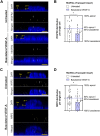
VEGFR2 and NRP2 distribution in HUVECs cultured on Transwell inserts. Representative orthogonal images of VEGFR2 (A) and NRP2 (C) staining in HUVECs grown on Transwell inserts after stimulation with VEGF-A (25 ng/ml for 48 hr). DAPI staining was used to identify the cell nucleus, but the translucent Transwell inserts showed positivity for DAPI as well. Labeling in the images: a marks the apical membrane, b marks the basolateral membrane, and tw marks the Transwell insert. Quantification of the subcellular signal distribution is shown in (B) and (D). n=17–26 cells from three different donor experiments. Welch’s t-test was used to test whether there is a preference to apical or basolateral localization within one condition. Student’s t-test was used to investigate differences between conditions. *P < 0.05. Scale bars, 5 µm. Abbreviations: HUVECs, human umbilical vein endothelial cells; VEGF-A, vascular endothelial growth factor-A.
Figure 4.
Figure 4.
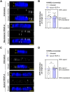
VEGFR2 and NRP2 distribution in HUVECs cultured on coverslips. Representative orthogonal images of VEGFR2 (A) and NRP2 (C) staining in HUVECs grown on coverslips after stimulation with VEGF-A (25 ng/ml for 48 hr). DAPI staining was used to identify the cell nucleus. Labeling in the images: a marks the apical membrane and b marks the basolateral membrane. Quantification of the subcellular signal distribution is shown in (B) and (D). n=11–18 cells from three different donor experiments. Welch’s t-test was used to test whether there is a preference to apical or basolateral localization within one condition. Student’s t-test was used to investigate differences between conditions. *P < 0.05, **P < 0.01. Scale bars, 5 µm. Abbreviations: HUVECs, human umbilical vein endothelial cells; VEGF-A, vascular endothelial growth factor-A.
Figure 5.
Figure 5.
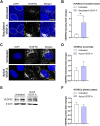
Total VEGFR2 protein expression in HUVECs. (A and C) Representative merged projection images of VEGFR2 staining in HUVECs grown on Transwell inserts (A) and coverslips (C) after stimulation with VEGF-A (25 ng/ml for 48 hr). DAPI staining was used to identify the cell nucleus, but the translucent Transwell inserts showed positivity for DAPI as well. A yellow dotted line demonstrates boundaries of cell nuclei in the DAPI channel. Quantification of the fluorescence signal is shown in (B) and (D). Representative Western blots of VEGFR2 and actin (loading control) protein expression after stimulation with VEGF-A (25 ng/ml for 48 hr) are shown in (E). Quantification of Western blot data is shown in (F). (B) n=7–10 images of three different donor experiments. (D) n=10–13 images of three different donors experiments. (F) n=6 wells of three independent donor experiments. (B, D) Mann–Whitney U -test. (F) Student’s t-test. *P < 0.05. Scale bars, 10 µm. Abbreviations: HUVECs, human umbilical vein endothelial cells; VEGF-A, vascular endothelial growth factor-A.
Figure 6.
Figure 6.
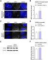
Total NRP2 protein expression in HUVECs. (A and C) Representative merged projection images of NRP2 staining in HUVECs grown on Transwell inserts (A) and coverslips (C) after stimulation with VEGF-A (25 ng/ml for 48 hr). DAPI staining was used to identify the cell nucleus, but the translucent Transwell inserts showed positivity for DAPI as well. A yellow dotted line demonstrates boundaries of cell nuclei in the DAPI channel. Quantification of the fluorescence signal is shown in (B) and (D). Representative Western blots of NRP2 and actin (loading control) protein expression after stimulation with VEGF-A (25 ng/ml for 48 hr) are shown (E) and quantified (F). (B) n=12 images of three different donor experiments. (D) n=8–9 images of three different donor experiments. (F) n=6 wells of three different donor experiments. (B, D, F) Mann–Whitney U-test. Scale bars, 10 µm. Abbreviations: HUVECs, human umbilical vein endothelial cells; VEGF-A, vascular endothelial growth factor-A.

Similar articles

Cited by

References

    1. Worzfeld T, Schwaninger M. Apicobasal polarity of brain endothelial cells. J Cereb Blood Flow Metab. 2016;36(2):340–62. - PMC - PubMed
    1. Witmer AN, Vrensen GFJM, van Noorden CJF, Schlingemann RO. Vascular endothelial growth factors and angiogenesis in eye disease. Prog Retin Eye Res. 2003;22(1):1–29. - PubMed
    1. Dvorak HF, Brown LF, Detmar M, Dvorak AM. Vascular permeability factor/vascular endothelial growth factor, microvascular hyperpermeability, and angiogenesis. Am J Pathol. 1995;146(5):1029–39. - PMC - PubMed
    1. Hudson N, Powner MB, Sarker MH, Burgoyne T, Campbell M, Ockrim ZK, Martinelli R, Futter CE, Grant MB, Fraser PA, Shima DT, Greenwood J, Turowski P. Differential apicobasal VEGF signaling at vascular blood-neural barriers. Dev Cell. 2014;30(5):541–52. - PMC - PubMed
    1. Klaassen I, van Noorden CJF, Schlingemann RO. Molecular basis of the inner blood-retinal barrier and its breakdown in diabetic macular edema and other pathological conditions. Prog Retin Eye Res. 2013;34:19–48. - PubMed

MeSH terms

Substances