. 2022 Sep 28;190(2):1117-1133.
doi: 10.1093/plphys/kiac333.

Pyruvate, phosphate dikinase regulatory protein impacts light response of C4 photosynthesis in Setaria viridis

Affiliations

Pyruvate, phosphate dikinase regulatory protein impacts light response of C4 photosynthesis in Setaria viridis

Kuenzang Om et al. Plant Physiol. .

Abstract

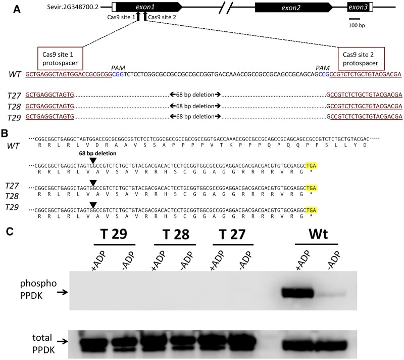
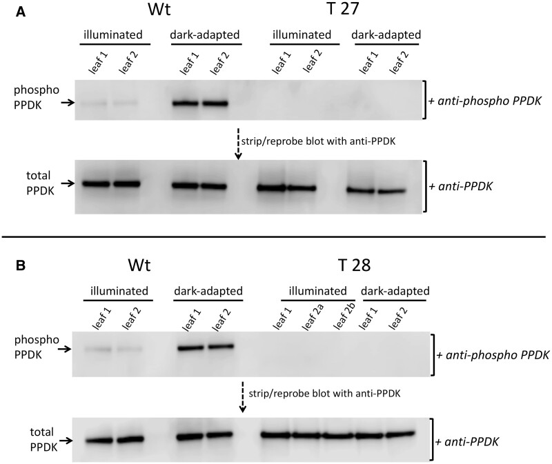
In C4 plants, the pyruvate (Pyr), phosphate dikinase regulatory protein (PDRP) regulates the activity of the C4 pathway enzyme Pyr, phosphate dikinase (PPDK) in a light-/dark-dependent manner. The importance of this regulatory action to C4 pathway function and overall C4 photosynthesis is unknown. To resolve this question, we assessed in vivo PPDK phospho-regulation and whole leaf photophysiology in a CRISPR-Cas9 PDRP knockout (KO) mutant of the NADP-ME C4 grass green millet (Setaria viridis). PDRP enzyme activity was undetectable in leaf extracts from PDRP KO lines. Likewise, PPDK phosphorylated at the PDRP-regulatory Thr residue was immunologically undetectable in leaf extracts. PPDK enzyme activity in rapid leaf extracts was constitutively high in the PDRP KO lines, irrespective of light or dark pretreatment of leaves. Gas exchange analysis of net CO2 assimilation revealed PDRP KO leaves had markedly slower light induction kinetics when leaves transition from dark to high-light or low-light to high-light. In the initial 30 min of the light induction phase, KO leaves had an ∼15% lower net CO2 assimilation rate versus the wild-type (WT). Despite the impaired slower induction kinetics, we found growth and vigor of the KO lines to be visibly indistinguishable from the WT when grown in normal air and under standard growth chamber conditions. However, the PDRP KO plants grown under a fluctuating light regime exhibited a gradual multi-day decline in Fv/Fm, indicative of progressive photosystem II damage due to the absence of PDRP. Collectively, our results demonstrate that one of PDRP's functions in C4 photosynthesis is to ensure optimal photosynthetic light induction kinetics during dynamic changes in incident light.


Figures

Figure 1
Figure 1
Overview of the NADP-ME C4 metabolic cycle with emphasis on the roles of PPDK and PDRP. A, Context view of reversible phosphorylation of PPDK by PDRP in the C4 cycle. In the light, PPDK fulfills a cardinal role in the C4 metabolic pathway by regenerating the primary CO2 fixation substrate PEP from Pyr. The linked action of stromal pyrophosphatase (PPi-ase) and adenyl kinase (AK) exert a substantial influence on the direction of catalysis of both PDRP and PPDK activities. CA, carbonic anhydrase; PEPc, PEP carboxylase; NADP-MDH, NADP malate dehydrogenase; NADP-ME, NADP malic enzyme;OAA, oxaloacetic acid; Mal, malic acid; C/B, Calvin Benson cycle. B, PDRP regulates PPDK activity by reversible phosphorylation of an active-site Thr located one residue removed from the central catalytic His. Only the His-P PPDK catalytic intermediate is amenable to phosphorylation/inactivation by PDRPs protein kinase function.
Figure 2
Figure 2
CRISPR–Cas9 edited S. viridis PDRP gene knockout mutants lack PDRP enzyme activity. A, Schematic diagram of the S. viridis PDRP gene model (Sevir.2G348700.2) showing Cas9 target sites within the first exon. Shown directly below is an alignment of exon 1 WT sequence with the corresponding CRISPR–Cas9 edited DNA sequence of KO lines T27, T28, and T29 with the resultant 68-bp deletion. CRISPR–Cas9 target protospacer sequences are underlined and highlighted in red. PAM sequences are highlighted in blue. B, CRISPR–Cas9 deleted PDRP exon 1 sequence shifts the reading frame of exon 1 in the KO lines to encode missense codons until a stop codon (highlighted) is encountered 66-bp downstream from the deletion site. C, In vitro PDRP protein kinase assays of desalted leaf extracts. PDRP knockout lines T27, T28, T29, and WT were assayed for ADP-dependent PDRP protein kinase (PPDK phosphorylating) activity using an immuno-based assay with recombinant maize C4 PPDK as substrate. Upper part shows immunoblots of terminated reaction aliquots probed with anti-phospho-PPDK antibody. Control assays (-ADP) omitted ADP in the reaction mix. Lower part serves as a lane load control and is the same blot stripped and reprobed with anti-PPDK polyclonal antibody. The lower molecular mass band evident in T27, T28, T29 lanes (lower, C) is most likely attributable to the presence of a higher proteolytic background in the leaf extract used in the assays and is apparently unrelated to genotype. Additional assay sets also show the occurrence of this minor band in WT lanes (see Supplemental Figure S2). Results identical to those of the single experiment shown in the upper panel were obtained in three independent assays.
Figure 3
Figure 3
Immunoblot detection of PPDK and phospho-PPDK in leaf extracts isolated from dark-adapted or illuminated leaves of PDRP knockout lines T27, T28, and WT. Leaves were dark-adapted for 2 h and then illuminated for 30 min at 1,300 µmol quanta m−2s−1 as described in “Materials and methods.” Shown are separate immunoblots for WT versus T27 (A) and WT versus T28 (B). Upper parts are immunoblots of duplicate leaf extracts probed with anti-phospho-PPDK antibody. The T28 immunoblot (B) contained three separate leaf samples. Lower parts show the same immunoblots stripped and reprobed with anti-PPDK antibody. Each lane contained 2-µg total soluble protein.
Figure 4
Figure 4
PPDK enzyme activity in desalted leaf extracts isolated from dark adapted and illuminated leaves. T27, T28, T29, and WT. Leaves were dark adapted for 2 h and then illuminated for 30 min at 1,300 µmol quanta m−2s−1 as described in “Materials and methods”. PPDK activity is shown as percentage of illuminated WT leaves (WT PPDK activity = 1.04 µmol min−1mg prot−1). Values shown are the means ± se, n = 3.
Figure 5
Figure 5
WT and PDRP knockout plants at the 3-to 4-week stage after germination and growth under standard growth chamber conditions. Plants were germinated and grown under standard growth chamber conditions (normal air conditions, ∼700-μmol quanta m−2 s−1, 16-h/8-h day/night, 31°C/22°C photoperiod). Additional, details provided in “Materials and methods.”
Figure 6
Figure 6
Leaf PPDK activation state in WT and T27 leaves as a function of increasing irradiance. PPDK enzyme activity was measured in leaf extracts from leaves that had first been dark adapted for 30 min and then illuminated for 15 min at the designated irradiance level. Data points for T27 leaves at 100 and 1,200 μmol quanta m−2s−1 were omitted due to insufficient number of replicates. Values shown are the means ± se, n = 3. Differences between genotypes for data points 600, 800, 1,000, and 1,600 μmol quanta m−2 s−1 are not significant (P > 0.05, two-tailed Student’s t test).
Figure 7
Figure 7
Light activation and dark deactivation of photosynthetic CO2 assimilation and PPDK activity in WT and T27 leaves. A, Net CO2 assimilation (Anet) in WT and T27 leaves during a dark → high light → low light transition. Anet values are the means ± se, n = 11, WT; n = 10, T27; differences between WT and T27 for measurements at 40–59 min are statistically significant (P < 0.05, two-tailed Student’s t test). B, PPDK light activation/dark deactivation in WT and T27 leaves during dark→light→dark transitions. Leaves used for PPDK light activation curve were first dark adapted for 2 h prior to illumination at 1,300 µmol quanta m−2s−1. Anet measurements and PPDK enzyme activity were conducted separately using leaves grown under identical conditions. Values shown for PPDK enzyme activity are the means ± se, n = 3.
Figure 8
Figure 8
Simultaneous measurement of net CO2 assimilation, gs, and intercellular CO2 in WT and T27 leaves. A, Net CO2 assimilation (Anet), (B) gs, and (C) intercellular CO2 (Ci) from 30 min of dark into 90 min of light. Anet, gs, Ci values are the means ± se, n = 7, WT; n = 8, T27; differences in Anet between WT and T27 for measurements at 45–60 min are statistically significant (P < 0.05, two-tailed Student’s t test). Differences in gs between WT and T27 for measurements at 48–60 min are statistically significant (P < 0.05, two-tailed Student’s t test).
Figure 9
Figure 9
Leaf Pyr content of dark adapted and 3 min illuminated WT and T27 leaves. Leaves were dark adapted for 2 h prior to illumination for 3 min at 1,300 µmol quanta m−2s−1. Values are the means ± se, n = 8 except for dark adapted T27 where n = 6. Asterisks indicate statistically significant differences by two-tailed Student’s t test: *P < 0.05; **P < 0.01; ****P < 0.0001.
Figure 10
Figure 10
Effect of a fluctuating light photoperiod on Fv/Fm. Shown are daily measurements of Fv/Fm in WT and T27 KO leaves during 7-day growth under a fluctuating light photoperiod versus a control steady high light or low light photoperiod. The fluctuating light regime consisted of repeating cycles of 1 min at 1,200 and 4 min at 300 µmol quanta m−2 s−1 per 16-h/8-h light/dark photoperiod. For the steady high light and steady low light controls, plants were grown under a 16-h/8-h light/dark photoperiod of 850 µmol quanta m−2 s−1 and 90 µmol quanta m−2 s−1, respectively. Time points are the means ± se, n = 6–8. A three-way repeated measure ANOVA for Genotype × Light treatment × Day (see Supplemental Table S2) and post-hoc Tukey’s test was used to examine differences in Fv/Fm between genotypes within growth Light treatment across 7 days (Supplemental Table S3). Where P-values ns, not significant, * ≤0.05, ** ≤0.01, and *** ≤0.001

Similar articles

Cited by

References

    1. Acevedo-Siaca LG, Coe R, Wang Y, Kromdijk J, Quick WP, Long SP (2020) Variation in photosynthetic induction between rice accessions and its potential for improving productivity. New Phytologist 227: 1097–1108 - PMC - PubMed
    1. Ashton AR, Burnell JN, Furbank RT, Jenkins CLD, Hatch MD (1990) Enzymes of C4 photosynthesis. InLea PJ, ed, Methods in Plant Biochemistry, Vol 3, Academic Press, San Diego, CA, pp 39–72
    1. Brutnell TP, Wang L, Swartwood K, Goldschmidt A, Jackson D, Zhu XG, Kellogg E, Van Eck J (2010) Setaria viridis: a model for C4 photosynthesis. Plant Cell 22: 2537–2544 - PMC - PubMed
    1. Burnell JN, Hatch MD (1985) Regulation of C4 photosynthesis: purification and properties of the protein catalyzing ADP-mediated inactivation and Pi-mediated activation of pyruvate, Pi dikinase. Arch Biochem Biophys 237: 490–503 - PubMed
    1. Cermák T, Curtin SJ, Gil-Humanes J, Cegan R, Kono TJY, Konecná E, Belanto JJ, Starker CG, Mathre JW, Greenstein RL, et al. (2017) A multipurpose toolkit to enable advanced genome engineering in plants. Plant Cell 29: 119–1217 - PMC - PubMed

Publication types

MeSH terms