. 2022 Jul 25;8(1):55.
doi: 10.1186/s40981-022-00545-0.

One-lung ventilation and 3D image analysis in a case of tracheal bronchus with steeply angled branching of left main bronchus: a case report

Affiliations

One-lung ventilation and 3D image analysis in a case of tracheal bronchus with steeply angled branching of left main bronchus: a case report

Taichi Onimaru et al. JA Clin Rep. .

Abstract

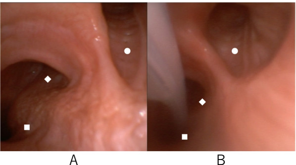
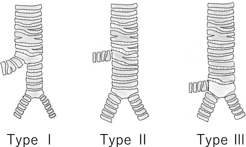
Background: Establishing one-lung ventilation (OLV) in patients with tracheal bronchus (TB) may be challenging due to its unusual bronchial anatomy. We present a case of difficult OLV in a patient with right TB and steeply angled bifurcation of the left main bronchus.

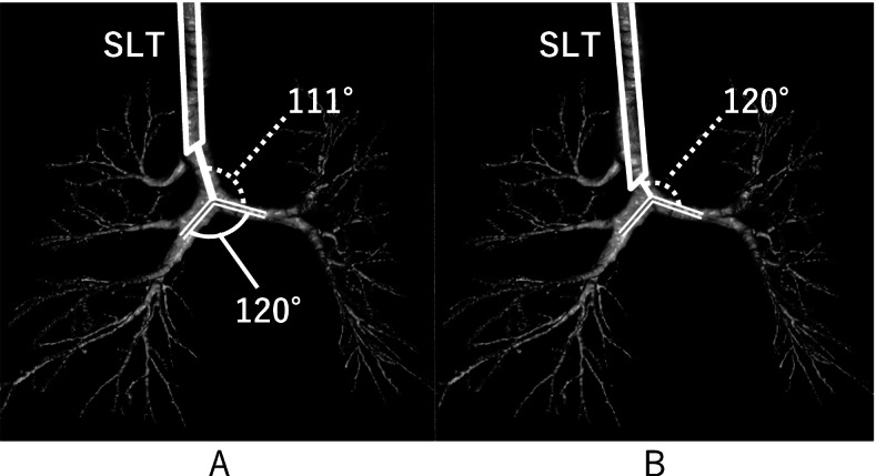
Case presentation: A 79-year-old woman was scheduled to undergo video-assisted thoracic surgery left upper lobectomy. We planned right OLV with a bronchial blocker; however, it was difficult to place the blocker in the left main bronchus due to a steep bifurcation angle. Therefore, we changed the entry angle of the lumen tip by advancing the tracheal tube to just above the tracheal bifurcation, allowing successful placement of the bronchial blocker into the bronchus.

Conclusion: For airway management in patients with TB, especially for OLV, it is essential to understand the anatomy of the trachea, bronchus, and TB and to select the appropriate device for each case.

Keywords: Anesthesia; Bronchial blocker; Double-lumen tracheal tube; Main bronchus angle; One-lung ventilation; Tracheal bronchus.


Conflict of interest statement

The authors declare that they have no competing interests.

Figures

Fig. 1
Fig. 1
Bronchoscopy before (A) and after (B) bronchial blocker placement. ■: Left main bronchus ◆: right main bronchus ●: tracheal bronchus
Fig. 2
Fig. 2
Three-dimensional (3D) image evaluation of the airway. Entry angles from the SLT tip were 111° and 120° when the tip was placed above the TB entry (A) and tracheal bifurcation (B), respectively. Main bronchus angle was 120° (A). SLT: single-lumen endotracheal tube, TB: tracheal bronchus
Fig. 3
Fig. 3
Modified schematic diagram of right TB classification (Conacher’s classification) [9]. Types I and II: the right TB is distant (> 2 cm) from the tracheal bifurcation. The distal trachea is either a narrow bronchial structure (type I) or a normal tracheal structure (type II). Type III: the right TB is close (< 2 cm) to the tracheal bifurcation and appears as a three-branching structure. TB: tracheal bronchus
Fig. 4
Fig. 4
Strategy for OLV in patients with a right TB. *a: Using a right-sided DLT with a shortened bronchial side tube design. *b: Dual BB technique; BB (e.g., Arndt endotracheal blocker or uninvent bronchial blocker) and Fogarty catheters are inserted inside and outside the SLT, respectively, to occlude the right main bronchus and right TB [6, 11]. *c: Selective BB; since the right upper lobe is not deflated by this technique, this technique is dependent on the type of surgical procedure being performed. BB, bronchial blocker; DLT, double-lumen endotracheal tube; OLV, one-lung ventilation; SLT, single-lumen endotracheal tube; TB, tracheal bronchus

References

    1. Ghaye B, Szapiro D, Fanchamps JM, Dondelinger RF. Congenital bronchial abnormalities revisited. Radiographics. 2001;21:105–119. doi: 10.1148/radiographics.21.1.g01ja06105. - DOI - PubMed
    1. Suzuki M, Matsui O, Kawashima H, Takemura A, Matsubara K, Hayashi N, et al. Radioanatomical study of a true tracheal bronchus using multidetector computed tomography. Jpn J Radiol. 2010;28:188–192. doi: 10.1007/s11604-009-0405-5. - DOI - PubMed
    1. Siegel MJ, Shackelford GD, Francis RS, McAllister WA. Tracheal bronchus. Radiology. 1979;130:353–355. doi: 10.1148/130.2.353. - DOI - PubMed
    1. Moon YJ, Kim SH, Park SW, Lee YM. The implications of a tracheal bronchus on one-lung ventilation and fibreoptic bronchoscopy in a patient undergoing thoracic surgery: a case report. Can J Anaesth. 2015;62:399–402. doi: 10.1007/s12630-014-0293-8. - DOI - PubMed
    1. Kawagoe I, Hayashida M, Suzuki K, Kitamura Y, Oh S, Satoh D, et al. Anesthetic management of patients undergoing right lung surgery after left upper lobectomy: selection of tubes for one-lung ventilation (OLV) and oxygenation during OLV. J Cardiothorac Vasc Anesth. 2016;30:961–966. doi: 10.1053/j.jvca.2015.10.004. - DOI - PubMed

LinkOut - more resources