. 2022 Jul 26;6(14):4310-4319.
doi: 10.1182/bloodadvances.2022007453.

Cerebral vascular injury in transplant-associated thrombotic microangiopathy

Affiliations

Cerebral vascular injury in transplant-associated thrombotic microangiopathy

Anthony Sabulski et al. Blood Adv. .

Abstract

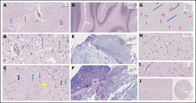
Transplant-associated thrombotic microangiopathy (TA-TMA) and atypical hemolytic uremic syndrome (aHUS) are complement-mediated TMAs. The central nervous system (CNS) is the most common extrarenal organ affected by aHUS, and, despite mechanistic overlap between aHUS and TA-TMA, CNS involvement is rarely reported in TA-TMA, suggesting that CNS involvement in TA-TMA may be underdiagnosed and that these patients may benefit from complement blockers. In addition, there are no widely used histologic or radiologic criteria for the diagnosis of TMA in the brain. Thirteen recipients of pediatric hematopoietic cell transplants (HCTs) who had TA-TMA and who underwent autopsy were studied. Seven of 13 brains had vascular injury, and 2 had severe vascular injury. Neurologic symptoms correlated with severe vascular injury. Classic TMA histology was present and most often observed in the cerebellum, brainstem, and cerebral white matter. Abnormalities in similar anatomic regions were seen on imaging. Brain imaging findings related to TMA included hemorrhages, siderosis, and posterior reversible encephalopathy syndrome. We then studied 100 consecutive HCT recipients to identify differences in neurologic complications between patients with and those without TA-TMA. Patients with TA-TMA were significantly more likely to have a clinical concern for seizure, have an electroencephalogram performed, and develop altered mental status. In summary, our study confirms that TA-TMA involves the brains of recipients of HCT and is associated with an increased incidence of neurologic symptoms. Based on these findings, we propose that patients with low- or moderate-risk TA-TMA who develop neurologic complications should be considered for TA-TMA-directed therapy.


Figures

None
Graphical abstract
Figure 1.
Figure 1.
Severe histologic sequelae in the CNS of patients with TA-TMA. Cerebral (A-B; original magnification ×8 (A), original magnification ×40 (B)) and cerebellar (C-D; original magnification x20.5 (C), original magnification ×2.5 (D)) white matter show extensive demyelination, with scattered macrophages (C, yellow arrow, original magnification ×20.5), though not to confluence and not an infarct microscopically or in vascular distribution. Many vessels are affected by chronic changes in other organs that are related to TA-TMA. (C) Capillaries with thickened rigid walls shown at low power, appearing “wire-like” with a ratio of wall to lumen to wall occasionally 1:1:1 (red arrow) are present. (A-C) Capillary basement membrane layering and splitting and pericapillary clearing are also seen (light blue arrows and most pronounced, dark blue arrow). A range of capillaries and small arteries show similar features. Pigment is seen in small capillaries (A, orange arrows) and macrophages are present in the periarteriolar stroma (A, green arrow). (E-F; original magnification ×1.8 (E), original magnification ×8.5 (F)) Loss of myelination is readily evident on Luxol fast blue stain with periodic acid–Schiff highlighting vascular features. (G-J) These findings were compared with the histology in a patient without TA-TMA. There were normal delicate capillaries (red arrows) and tracks (blue arrows), as well as preserved parenchyma in cerebellar (G-H; original magnification x13 (G), original magnification ×40 (H)) and cerebral white (I, original magnification ×40) matter and spinal cord (J, interface of white and gray matter, original magnification ×14/×0.4).
Figure 2.
Figure 2.
CNS radiographic abnormalities in TA-TMA. An 18-year-old man (patient 1 in Table 1) experienced progressive neurologic decline after HCT and underwent magnetic resonance imaging (A-G) on day 124 after HCT. He was diagnosed with TA-TMA on day 39. (A-B) Axial fluid-attenuated inversion recovery (FLAIR) images demonstrate multifocal areas of signal abnormality (black arrows) in the cerebellum, brainstem, periventricular white matter, and posterior limb of the left internal capsule and adjacent basal ganglia. (C) A T2-trace diffusion–weighted image demonstrates corresponding diffusion restriction predominantly within areas of signal in the cerebellum (white arrows). (D-E) Progressive hyperintense signal abnormality on FLAIR throughout the cerebellum, brainstem, cerebral white matter, and deep gray nuclei (white arrowheads). (F-G) Diffusion-weighted imaging demonstrates corresponding progressive diffusion restriction (black arrowheads). A 4-year-old patient with neuroblastoma (patient 3 in Table 1) experienced altered mental status on day 10 after autologous HCT and underwent a head CT (H-I). TA-TMA was diagnosed on the same day. Axial computed tomographic images demonstrate a new, ovoid, high-attenuation hemorrhage in the genu of the corpus callosum (arrowhead), as well as a more subtle hemorrhage along the cortical surface of the posterior right frontal lobe (arrow).
Figure 3.
Figure 3.
A modified TA-TMA management algorithm that incorporates CNS manifestations of TA-TMA in treatment decisions. As previously published, low risk TA-TMA is limited to laboratory evidence of intravascular hemolysis without end organ injury or increased terminal complement activation. Moderate risk TA-TMA is defined as the presence of either a random urine protein creatinine ratio >2 mg/mg or an elevated plasma sC5b-9 level (terminal complement, normal range <244 ng/mL)., High-risk TA-TMA includes patients with both of these laboratory abnormalities as well as patients with TA-TMA diagnosed on tissue biopsy. We propose the inclusion of neurologic symptoms or brain imaging findings potentially attributable to TA-TMA into the treatment decision process, as these patients may have CNS TA-TMA and early initiation of complement inhibitors may limit morbidity from TA-TMA–mediated brain injury.

References

    1. Jodele S, Sabulski A. Transplant-associated thrombotic microangiopathy: elucidating prevention strategies and identifying high-risk patients. Expert Rev Hematol. 2021;14(8):751-763. - PubMed
    1. Hofer J, Rosales A, Fischer C, Giner T. Extra-renal manifestations of complement-mediated thrombotic microangiopathies. Front Pediatr. 2014;2:97. - PMC - PubMed
    1. Loirat C, Frémeaux-Bacchi V. Atypical hemolytic uremic syndrome. Orphanet J Rare Dis. 2011;6(1):60. - PMC - PubMed
    1. Riedl M, Fakhouri F, Le Quintrec M, et al. . Spectrum of complement-mediated thrombotic microangiopathies: pathogenetic insights identifying novel treatment approaches. Semin Thromb Hemost. 2014;40(4):444-464. - PubMed
    1. Noris M, Remuzzi G. Cardiovascular complications in atypical haemolytic uraemic syndrome. Nat Rev Nephrol. 2014;10(3):174-180. - PubMed

Publication types

MeSH terms

Substances