

Genomically Silent Refractory Gastric Cancer in a Young Patient Exhibits Overexpression of CXCL5
- PMID: 35877235
- PMCID: PMC9320515
- DOI: 10.3390/curroncol29070375
Genomically Silent Refractory Gastric Cancer in a Young Patient Exhibits Overexpression of CXCL5
Abstract
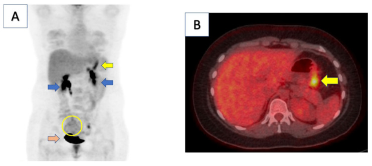
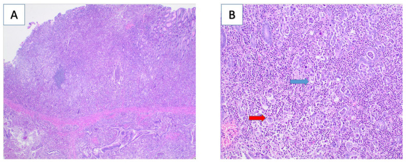
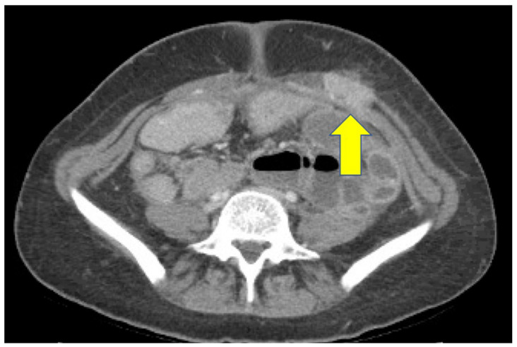
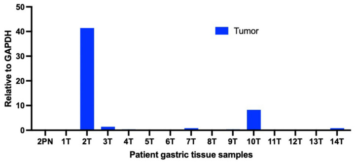
Gastric cancer is the third leading cause of cancer-related deaths, with more than one million new cases and approximately 841,000 deaths annually worldwide. We report a case of a young patient (25 years old) with an aggressive form of gastric cancer. The patient had previously been treated for Helicobacter pylori (H. pylori), which is a main risk factor for developing gastric cancer. Genetic testing showed an E-cadherin (CDH1) germline mutation of unknown significance. After eight cycles of chemotherapy, a positron emission tomography (PET) scan showed disease progression with an enlarging hypermetabolic right adnexal mass suspicious for metastatic disease. Tumor pathology demonstrated invasive and poorly differentiated gastric carcinoma. The analysis of the tumor biopsy indicated the very high expression of a chemokine, C-X-C motif chemokine 5 (CXCL5). The combination of H. pylori infection with an existence of a rare CDH1 mutation could have contributed to this aggressive gastric cancer.
Keywords: CDH1; CXCL5; Helicobacter pylori; advanced gastric cancer; young patient.
Conflict of interest statement
The authors state they have no conflicts of interest to declare.
Figures
Similar articles
-
Gastric cancer: new genetic developments.J Surg Oncol. 2005 Jun 1;90(3):114-33; discussion 133. doi: 10.1002/jso.20214. J Surg Oncol. 2005. PMID: 15895459
-
Genetic analysis of a case of Helicobacter pylori-uninfected intramucosal gastric cancer in a family with hereditary diffuse gastric cancer.Gastric Cancer. 2019 Jul;22(4):892-898. doi: 10.1007/s10120-018-00912-w. Epub 2018 Dec 12. Gastric Cancer. 2019. PMID: 30542785
-
Trefoil factor 1 expression suppresses Helicobacter pylori-induced inflammation in gastric carcinogenesis.Cancer. 2015 Dec 15;121(24):4348-58. doi: 10.1002/cncr.29644. Epub 2015 Sep 15. Cancer. 2015. PMID: 26372254 Free PMC article.
-
New aspects of Helicobacter pylori infection involvement in gastric oncogenesis.J Surg Res. 2008 May 1;146(1):149-58. doi: 10.1016/j.jss.2007.06.011. Epub 2007 Jul 27. J Surg Res. 2008. PMID: 17720195 Review.
-
Helicobacter pylori infection and molecular changes in gastric carcinogenesis.J Gastroenterol. 2002;37 Suppl 13:45-9. doi: 10.1007/BF02990099. J Gastroenterol. 2002. PMID: 12109665 Review.
Cited by
-
Repurposed Drugs in Gastric Cancer.Molecules. 2022 Dec 30;28(1):319. doi: 10.3390/molecules28010319. Molecules. 2022. PMID: 36615513 Free PMC article. Review.
-
TRIF-IFN-I pathway in Helicobacter-induced gastric cancer in an accelerated murine disease model and patient biopsies.iScience. 2024 Mar 8;27(4):109457. doi: 10.1016/j.isci.2024.109457. eCollection 2024 Apr 19. iScience. 2024. PMID: 38558931 Free PMC article.
-
Gastric cancer and mesenchymal stem cell-derived exosomes: from pro-tumorigenic effects to anti-cancer vehicles.Arch Pharm Res. 2024 Jan;47(1):1-19. doi: 10.1007/s12272-023-01477-8. Epub 2023 Dec 27. Arch Pharm Res. 2024. PMID: 38151649 Review.
References
-
- SEER Cancer Statistics Review (CSR): Stomach Cancer. [(accessed on 6 January 2022)]; Available online: https://seer.cancer.gov/statfacts/html/stomach.html.
Publication types
MeSH terms
Substances
Grants and funding
LinkOut - more resources
Full Text Sources
Medical
Miscellaneous
