. 2022 Jul 4;9(7):297.
doi: 10.3390/bioengineering9070297.

Microcarrier-Based Culture of Human Pluripotent Stem-Cell-Derived Retinal Pigmented Epithelium

Affiliations

Microcarrier-Based Culture of Human Pluripotent Stem-Cell-Derived Retinal Pigmented Epithelium

Mohamed A Faynus et al. Bioengineering (Basel). .

Abstract

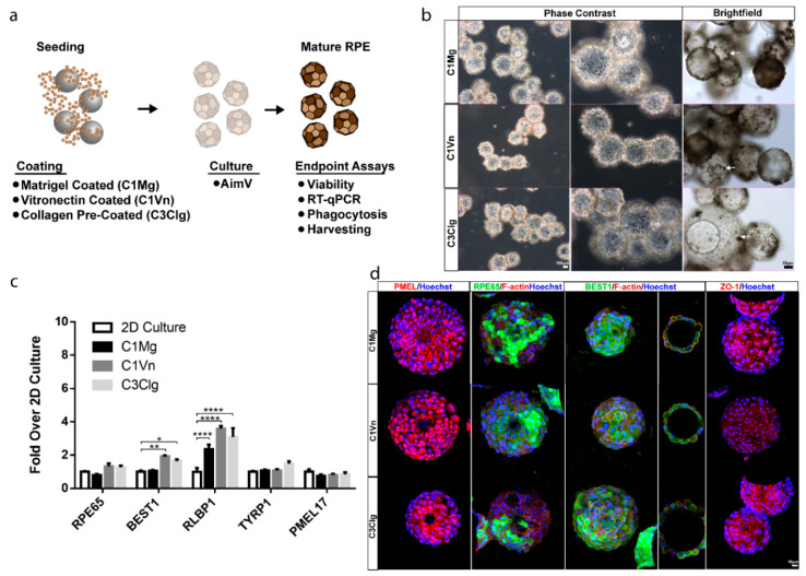
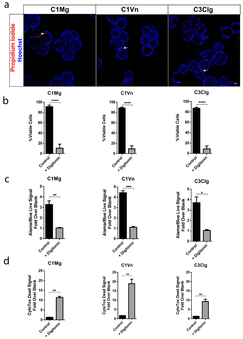
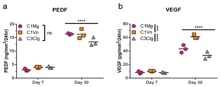
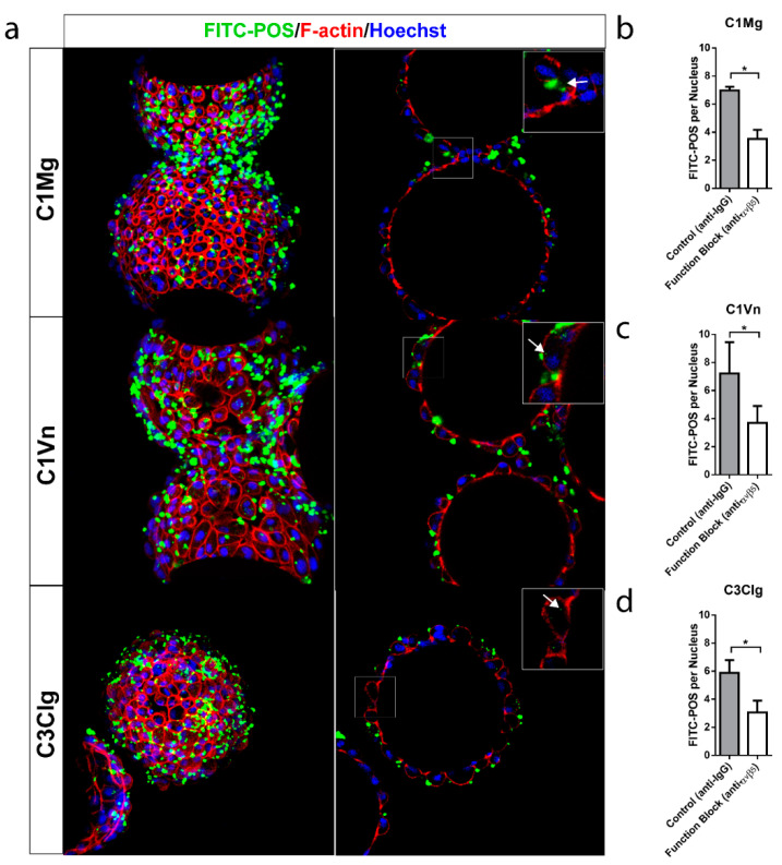
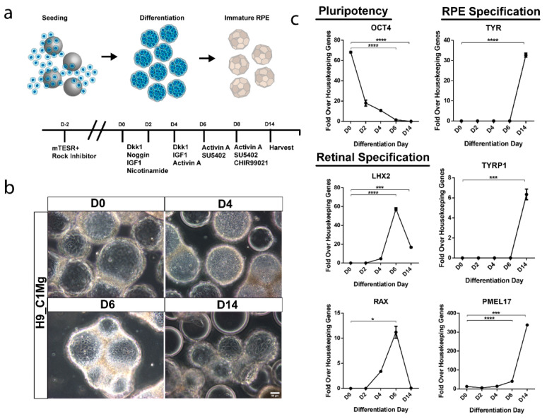
Dry age-related macular degeneration (AMD) is estimated to impact nearly 300 million individuals globally by 2040. While no treatment options are currently available, multiple clinical trials investigating retinal pigmented epithelial cells derived from human pluripotent stem cells (hPSC-RPE) as a cellular replacement therapeutic are currently underway. It has been estimated that a production capacity of >109 RPE cells annually would be required to treat the afflicted population, but current manufacturing protocols are limited, being labor-intensive and time-consuming. Microcarrier technology has enabled high-density propagation of many adherent mammalian cell types via monolayer culture on surfaces of uM-diameter matrix spheres; however, few studies have explored microcarrier-based culture of RPE cells. Here, we provide an approach to the growth, maturation, and differentiation of hPSC-RPE cells on Cytodex 1 (C1) and Cytodex 3 (C3) microcarriers. We demonstrate that hPSC-RPE cells adhere to microcarriers coated with Matrigel, vitronectin or collagen, and mature in vitro to exhibit characteristic epithelial cell morphology and pigmentation. Microcarrier-grown hPSC-RPE cells (mcRPE) are viable; metabolically active; express RPE signature genes including BEST1, RPE65, TYRP1, and PMEL17; secrete the trophic factors PEDF and VEGF; and demonstrate phagocytosis of photoreceptor outer segments. Furthermore, we show that undifferentiated hESCs also adhere to Matrigel-coated microcarriers and are amenable to directed RPE differentiation. The capacity to support hPSC-RPE cell cultures using microcarriers enables efficient large-scale production of therapeutic RPE cells sufficient to meet the treatment demands of a large AMD patient population.

Keywords: microcarriers; retinal pigment epithelium; stem cells.


Conflict of interest statement

The authors declare no conflict of interest.

Figures

Figure 1
Figure 1
Microcarrier-seeded RPE (mcRPE) mature and express RPE markers (a) Outline of experimental approach. Cytodex 1 microcarriers were coated with either Matrigel (C1Mg) or human recombinant vitronectin (C1Vn) and Cytodex 3 microcarriers precoated with denatured porcine-skin collagen (C3Clg) were seeded with hPSC-RPE cells and cultured for 4 weeks before analysis. (b) mcRPE mature to develop phase-bright borders, form polygonal morphology and produce pigment (brightfield images, white arrow). (c) Expression of RPE marker genes RPE65, BEST1, RLBP1, TYRP1 and PMEL17 as detected by RT-qPCR. mcRPE demonstrate similar gene expression profiles compared to 2D controls except for significant differences in C1Vn and C3Clg BEST1 (* p = 0.0309 and ** p = 0.0012) and C1Mg, C1Vn and C3Clg RLBP1 (**** p < 0.0001) (d) mcRPE express immunocytochemically detectable markers of RPE maturity and polarization including: premelanosome (PMEL17), retinal pigment epithelium-specific 65 kDa (RPE65), bestrophin-1 (BEST1), zonular occludens-1 (ZO-1) and co-stain with F-actin. Statistical analysis, two-way ANOVA, Sidak’s correction. Data represented as means normalized to 2D controls with error bars indicating standard error of means.
Figure 2
Figure 2
mcRPE are viable and metabolically active. (a) mcRPE exhibits high viability on all three microcarrier types with few non-viable cells (red) as demonstrated by propidium iodide nuclear staining. (b) Quantification of the propidium iodide staining data revealed an average >88% cell viability for all three microcarriers with a significant decrease when exposed to Digitonin (**** p < 0.0001, two-way ANOVA, Sidak’s correction). (c) mcRPE demonstrates active metabolism with a decrease when exposed to Digitonin as measured through alamarBlue reagent (** p = 0.006, *** p = 0.001, * p = 0.0105, paired, two-tailed t test. (d) Cells’ viability was further measured using CytoTox-Fluor assay for dead-cell protease activity. All three microcarrier conditions exhibit minimal protease activity with a significant increase when exposed to digitonin (** p < 0.05, paired, two-tailed t test). Scale bar 50 µm, data represented as means with error bars indicating standard error of mean.
Figure 3
Figure 3
mcRPE secretion of PEDF and VEGF increases between days 7 and 30 post seeding. (a) PEDF secretion by mcRPE after 7DPS and 30DPS was quantified by sandwich ELISA for three microcarrier types. mcRPE secreted more PEDF at 30DPS compared to 7DPS, but no significant differences among microcarrier types were observed (ns, not significant). (b) VEGF secretion by mcRPE was measured in the same samples by sandwich ELISA. Day 30 mcRPE also secreted more VEGF compared to day 7, and mcRPE on C1Vn secreted higher levels of VEGF than those on C1Mg or C3Clg (*** p < 0.001, **** p < 0.000, respectively; two-way ANOVA, Sidak’s correction). Horizontal bars indicate mean of three biological replicates (2 × 10⁵ cells per replicate).
Figure 4
Figure 4
mcRPE demonstrate phagocytic activity (a) mcRPEs were exposed to FITC-conjugated bovine POS at a ratio of 20 POS per cell for 16 hrs in the presence of IgG controls or in some instances anti-αvβ5 antibodies before washing and fixation. Confocal Z projections were collected and bound and internalized POS were visualized by FITC (green) and F-actin (red) fluorescence. Single Z slice images demonstrate internal particulates (inset, white arrow) indicating engulfment. (bd) Representative field of view confocal Z projections (n = 3 FOV, n = 150 nuclei per FOV) were collected for both control IgG and function-blocking conditions and number of FITC-POS per cell (based on Hoechst staining) was assessed. All mcRPE phagocytose FITC-POS mediated in part through RPE-specific αvβ5 integrin receptors as shown by the significant decrease in FITC-POS per cell when exposed to function-blocking antibody (* p < 0.05, two-way ANOVA, Sidak’s correction). Data presented as means with error bars indicating standard error of means. Scale bars 50 µm, inset 5 µm.
Figure 5
Figure 5
Microcarrier-based differentiation of RPE from hESC. (a) Human embryonic stem cells (H9) were seeded on Matrigel-coated microcarriers and differentiated towards an RPE fate using a cocktail of growth factors and small molecules. (b) Phase contrast microscopy demonstrates retention of cells throughout the differentiation process. (c) Differentiation and identity specification was assessed by RT-qPCR. Pluripotency marker OCT4 significantly decreased by 6DPS and 14DPS indicating differentiation (**** p < 0.001) while expression of early eye field markers LHX2 and RAX significantly increased by 6DPS and proceeded to decrease by 14DPS suggesting retinal specification and transition towards a terminal cell type (**** p < 0.001, *** p = 0.001 and * p < 0.05). RPE markers tyrosinase (TYR), tyrosinase related protein 1 (TYRP1) and premelanosome (PMEL17) significantly increase by 14DPS suggesting an RPE fate (**** p < 0.0001 and *** p = 0.001). Statistical analysis two-way ANOVA, Sidak’s correction.

References

    1. Wong W.L., Su X., Li X., Cheung C.M.G., Klein R., Cheng C.Y., Wong T.Y. Global Prevalence of Age-Related Macular Degeneration and Disease Burden Projection for 2020 and 2040: A Systematic Review and Meta-Analysis. Lancet Glob. Health. 2014;2:e106–e116. doi: 10.1016/S2214-109X(13)70145-1. - DOI - PubMed
    1. Haines J.L., Hauser M.A., Schmidt S., Scott W.K., Olson L.M., Gallins P., Spencer K.L., Shu Y.K., Noureddine M., Gilbert J.R., et al. Complement Factor H Variant Increases the Risk of Age-Related Macular Degeneration. Science. 2005;308:419–421. doi: 10.1126/science.1110359. - DOI - PubMed
    1. Sparrrow J.R., Hicks D., Hamel C.P. The Retinal Pigment Epithelium in Health and Disease. Curr. Mol. Med. 2010;10:802–823. doi: 10.2174/156652410793937813. - DOI - PMC - PubMed
    1. Somasundaran S., Constable I.J., Mellough C.B., Carvalho L.S. Retinal Pigment Epithelium and Age-Related Macular Degeneration: A Review of Major Disease Mechanisms. Clin. Exp. Ophthalmol. 2020;48:1043–1056. doi: 10.1111/ceo.13834. - DOI - PMC - PubMed
    1. Kim H.J., Koh H.C. Chaperon-Mediated Autophagy Can Regulate Diquat-Induced Apoptosis by Inhibiting α-Synuclein Accumulation Cooperatively with Macroautophagy. Food Chem. Toxicol. 2021;158:112706. doi: 10.1016/j.fct.2021.112706. - DOI - PubMed