Review
. 2022 Jul 19;10(7):135.
doi: 10.3390/dj10070135.

Cantilevers: Multi-Tool in Orthodontic Treatment

Affiliations
Review

Cantilevers: Multi-Tool in Orthodontic Treatment

Malgorzata Bilinska et al. Dent J (Basel). .

Abstract

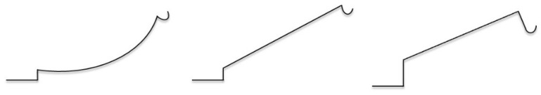
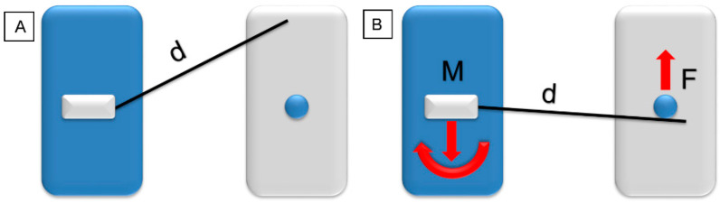
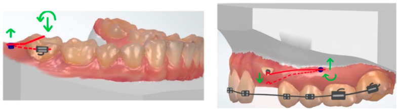
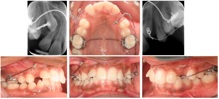
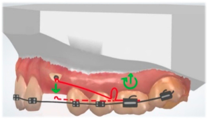
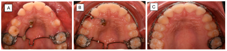
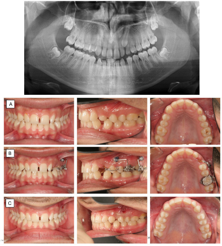
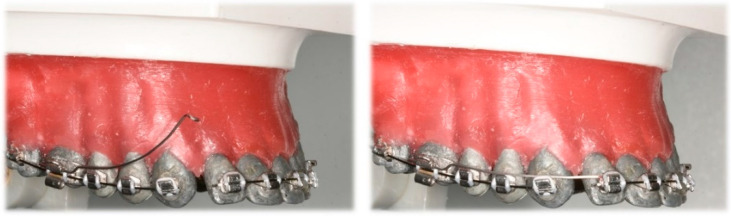
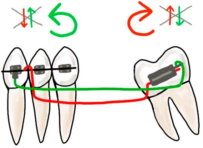
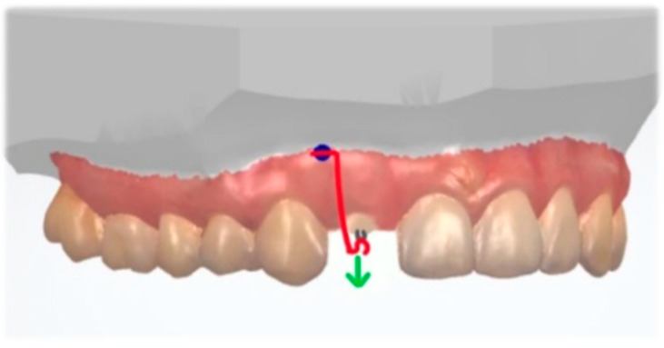
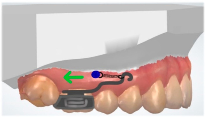
This review aims to discuss and illustrate various uses of cantilevers to solve multiple clinical issues and prove their versatility. Cantilevers are commonly used in the segmented arch technique, and they can be designed to solve various clinical problems with highly predictable results. Its design and shape can modify the various combinations of vertical and horizontal forces. The novel trend is to combine cantilevers with skeletal anchorage. Cantilevers offer a very simple and statically determined force system. The advantage is the control over side effects, which normally occur on the anchor teeth and the occlusion. The disadvantages include possible side effects on the anchorage unit, when the anchorage is poorly controlled. The review highlights the clear benefits of cantilever use in complex corrections of single teeth, segments, and entire arch with a diminished effect on the dentition, also with the use of skeletal anchorage. With their simple and easily tailored design, these springs can be called an orthodontic multi-tool.

Keywords: cantilevers; orthodontics; segmented arch technique; statically determine system.


Conflict of interest statement

The authors declare no conflict of interest.

Figures

Figure 1
Figure 1
Different cantilever designs (activated). From the left: deep curve, tip back, and utility arch.
Figure 2
Figure 2
Model of a statically determined force system (blue: anchorage unit with cantilever; grey: point of application). (A) Cantilever in a neutral position, length (d). (B) Activated cantilever a single force (F), and a force and a moment (M) on the other side (red arrows).
Figure 3
Figure 3
(Left): Placement of a TAD in line with or parallel to the dentition: no torquing moments on the TAD occurs. (Right) TAD is placed perpendicular to the long axis of the dentition; counterclockwise or clockwise torquing moments occurs (red: cantilever, green: force and moment).
Figure 4
Figure 4
Bilateral canine impaction: (right) buccal and (left) palatal. Right maxillary canine: traction with 0.017 × 0.025″ TMA cantilever activated for extrusion. Left maxillary canine: cantilever is inserted into the auxiliary tube of an upper molar, activated for canine extrusion and buccal displacement.
Figure 5
Figure 5
Cantilever with a loop activated for buccally impacted canine extrusion (red: cantilever, green: force and moment).
Figure 6
Figure 6
Traction of a palatally impacted right maxillary canine. (A) Canine extrusion with 0.017 × 0.025″ TMA cantilever inserted into the welded sheet on TPA, activated for extrusion. (B) Cantilever placed into the auxiliary tube of an upper molar, activated for canine extrusion and buccal displacement. (C) The canine is aligned into its final position.
Figure 7
Figure 7
Panoramic x-ray shows ectopic tooth 23 in a 16-year-old patient with only slight resorption of 63 (A) Palatally impacted left maxillary canine. The patient was satisfied with the smile-esthetic with the diastemas. Therefore, a sectional appliance was chosen for correction of position of 23 without changing neither the occlusion or position of the rest of the teeth. (B) Canine extrusion with 0.017 × 0.025″ TMA cantilever inserted into the tube of the molar, activated for extrusion and buccal displacement. TAD is applied to stabilize indirectly the molar (anchorage unit). (C) The canine is aligned into its final position.
Figure 8
Figure 8
Three-piece intrusion arch. Deep curve shape cantilevers activated for anterior segment intrusion cantilevers: (Left) activated, before ligation; (Right) ligated distally to lateral incisors.
Figure 9
Figure 9
Mouse trap appliance. Lever arms connected to TADs, activated for molar intrusion (red). Transpalatal arch (green).
Figure 10
Figure 10
Cantilever activated for canine intrusion, placed on the top of the mandibular canine bracket (red: cantilever, green: force and moment).
Figure 11
Figure 11
Statically determinate retraction system for space closure. Cantilever (red) is activated for anterior segment retraction (green: line of action).
Figure 12
Figure 12
Asymmetric cantilever activation: activation of utility arch (right side of typodont) and deep curve cantilever (left side of typodont), resulting intrusion and displacement of anterior segment (before: pink; after: blue).
Figure 13
Figure 13
Double cantilever mechanics. The red cantilever, from the molar to the anterior teeth, generates extrusion, clockwise rotation on the molar, and intrusion on the anterior teeth (red arrows). In order to control the vertical forces, a second cantilever, from a tube in the anterior, is required. This green cantilever is placed distally to the molar and it produces molar intrusion and a counter-clockwise movement with extrusion on the anterior teeth (green arrows). The resultant forces cancel each other, and two opposite moments occur.
Figure 14
Figure 14
Extrusive force (green) applied along the long tooth axis with a cantilever (red) connected to the TAD (blue).
Figure 15
Figure 15
Unilateral molar distalization with cantilever connected to the TAD with a closed-coil spring (green: force).

References

    1. Burstone C.J. Rationale of the segmented arch. Am. J. Orthod. 1962;48:805–822. doi: 10.1016/0002-9416(62)90001-5. - DOI - PubMed
    1. Laino A., Cacciafesta V., Martina R. Treatment of tooth impaction and transposition with a segmented-arch technique. J. Clin. Orthod. 2001;35:79–86. - PubMed
    1. Caballero G.M., Carvalho Filho O.A., Hargreaves B.O., Brito H.H., Magalhaes Junior P.A., Oliveira D.D. Mandibular canine intrusion with the segmented arch technique: A finite element method study. Am. J. Orthod. Dentofac. Orthop. 2015;147:691–697. doi: 10.1016/j.ajodo.2015.01.022. - DOI - PubMed
    1. Fischer T.J., Ziegler F., Lundberg C. Cantilever mechanics for treatment of impacted canines. J. Clin. Orthod. 2000;34:647–650. - PubMed
    1. Gurgel J.A., Pinzan-Vercelino C.R., Powers J.M. Mechanical properties of beta-titanium wires. Angle Orthod. 2011;81:478–483. doi: 10.2319/070510-379.1. - DOI - PMC - PubMed

LinkOut - more resources