. 2022 Jul 6;44(7):3089-3099.
doi: 10.3390/cimb44070213.

Sonicated Bordetella bronchiseptica Bacterin Can Protect Dendritic Cells from Differential Cytotoxicity Caused by Doxorubicin and Vincristine and Enhance Their Antigen-Presenting Capability

Affiliations

Sonicated Bordetella bronchiseptica Bacterin Can Protect Dendritic Cells from Differential Cytotoxicity Caused by Doxorubicin and Vincristine and Enhance Their Antigen-Presenting Capability

Ji Yun Sung et al. Curr Issues Mol Biol. .

Abstract

Doxorubicin (DOX) and vincristine (VC) are anti-cancer drugs commonly used for lymphoma in veterinary and human medicine. However, there are several side effects caused by these drugs. In this study, the protective effects of sonicated Bordetella bronchiseptica bacterin (sBb) on dendritic cells (DCs) damaged by two anti-cancer drugs were investigated. DCs play important roles in the innate and adaptive immunity of hosts, especially activating T cells that can suppress tumor growth. The metabolic activity of DCs significantly increased after the treatment with sBb compared to that of control DCs. In addition, there was a marked change in mitochondrial integrity between DOX-treated DC and DOX + sBb-treated DCs. Flow cytometric analysis also demonstrated that sBb upregulated the expression of the surface markers of DCs, particularly CD54. In mixed lymphocyte responses, sBb significantly increased the antigen-presenting capability of DCs. In particular, sBb increased the capability of control DCs by approximately 150% and that of VC-treated DCs by 221%. These results suggest that sBb can be used as a potential immunostimulatory agent to protect DCs from anti-cancer drug-induced damage and provide fundamental information about using a combination of DCs and vincristine in immunotherapy.

Keywords: dendritic cell; doxorubicin; protective effects; sonicated Bordetella bronchiseptica; vincristine.


Conflict of interest statement

The authors declare no conflict of interest.

Figures

Figure 1
Figure 1
Metabolic activity of DCs treated with DOX, VC, and sBb. The DCs were treated with DOX, VC, and sBb for 3 days, then the MTT assay was performed. The optical density was measured at 570 nm with a microplate reader. #, ##, and #### indicate p < 0.05, 0.01, and 0.0001 compared to the metabolic activity of DCs in the absence or presence of sBb, respectively.
Figure 2
Figure 2
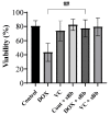
sBb increases the viability of DCs treated with DOX. The DCs were treated with 0.25 μg/mL DOX, 0.25 μg/mL VC and 0.1 μg/mL sBb for 2 days, then trypan blue exclusion assay was performed. The number of viable and dead cells were counted, and the viability rate was calculated. ## indicates p < 0.01 compared to the viability of DCs in the absence or presence of sBb.
Figure 3
Figure 3
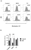
sBb increases the MMP of DOX-treated DCs. The DCs treated with DOX, VC, and/or sBb were stained with Rhodamine 123 solution and flow cytometric analysis was performed. (a) A representative histogram set was presented. The numbers in the upper part of the histogram indicate mean fluorescence intensity. (b) The graph was generated from 5 independent experiments. The mean fluorescence intensity of control DCs was set at 100%. ** indicates p  <  0.01 compared to control DCs, whereas #### indicates p  <  0.0001 compared to the treated DCs in the absence or presence of sBb.
Figure 4
Figure 4
sBb differentially up-regulates the expression of surface markers on DCs. The DCs treated with DOX, VC, and/or sBb were stained with specific antibodies in the presence of Fc blocker and then analyzed by flow cytometry, as described in the Materials and Methods. (a) A representative histogram set was presented. Numbers in the upper part of histograms indicate mean fluorescence intensity. (b) The graph was generated from 3 independent experiments. The mean fluorescence intensity of control DCs was set at 100%. *, **, and **** indicate p < 0.05, 0.01, and 0.0001 compared to control DCs, whereas #, #### indicate p < 0.05, p < 0.0001 compared to the treated DCs in the absence or presence of sBb.
Figure 4
Figure 4
sBb differentially up-regulates the expression of surface markers on DCs. The DCs treated with DOX, VC, and/or sBb were stained with specific antibodies in the presence of Fc blocker and then analyzed by flow cytometry, as described in the Materials and Methods. (a) A representative histogram set was presented. Numbers in the upper part of histograms indicate mean fluorescence intensity. (b) The graph was generated from 3 independent experiments. The mean fluorescence intensity of control DCs was set at 100%. *, **, and **** indicate p < 0.05, 0.01, and 0.0001 compared to control DCs, whereas #, #### indicate p < 0.05, p < 0.0001 compared to the treated DCs in the absence or presence of sBb.
Figure 5
Figure 5
sBb increases the antigen-presenting capability of DCs. The DCs treated with DOX, VC, and/or sBb prior to co-culture and allogeneic spleen cells were co-cultured as described in the Materials and Methods. Cell Counting Kit-8 solution was used. The optical density was measured at 450 nm with a microplate reader. *** indicates p < 0.001 compared to control DCs, whereas #### indicates p < 0.0001 compared to the treated-DCs in the absence or presence of sBb.
Figure 6
Figure 6
sBb increases the cytokine production of DCs. The supernatants of DOX, VC, and/or sBb treated DCs were harvested and used for ELISA. The amount of TNF-α, IL-1β, IL-6, and IL-12 produced by the treated DCs were measured by ELISA kits according to the manufacturer’s instructions.

Similar articles

References

    1. Steinman R.M. Decisions about dendritic cells: Past, present, and future. Annu. Rev. Immunol. 2012;30:1–22. doi: 10.1146/annurev-immunol-100311-102839. - DOI - PubMed
    1. Wculek S.K., Cueto F.J., Mujal A.M., Melero I., Krummel M.F., Sancho D. Dendritic cells in cancer immunology and immunotherapy. Nat. Rev. Immunol. 2020;20:7–24. doi: 10.1038/s41577-019-0210-z. - DOI - PubMed
    1. Kaneno R., Shurin G.V., Tourkova I.L., Shurin M.R. Chemomodulation of human dendritic cell function by antineoplastic agents in low noncytotoxic concentrations. J. Transl. Med. 2009;7:58. doi: 10.1186/1479-5876-7-58. - DOI - PMC - PubMed
    1. Shurin G.V., Tourkova I.L., Kaneno R., Shurin M.R. Chemotherapeutic agents in noncytotoxic concentrations increase antigen presentation by dendritic cells via an IL-12-dependent mechanism. J. Immunol. 2009;183:137–144. doi: 10.4049/jimmunol.0900734. - DOI - PMC - PubMed
    1. Palucka K., Banchereau J. Dendritic-cell-based therapeutic cancer vaccines. Immunity. 2013;39:38–48. doi: 10.1016/j.immuni.2013.07.004. - DOI - PMC - PubMed

LinkOut - more resources