

Clinically Inspired Skin Lesion Classification through the Detection of Dermoscopic Criteria for Basal Cell Carcinoma
- PMID: 35877641
- PMCID: PMC9319034
- DOI: 10.3390/jimaging8070197
Clinically Inspired Skin Lesion Classification through the Detection of Dermoscopic Criteria for Basal Cell Carcinoma
Abstract
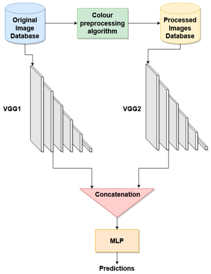
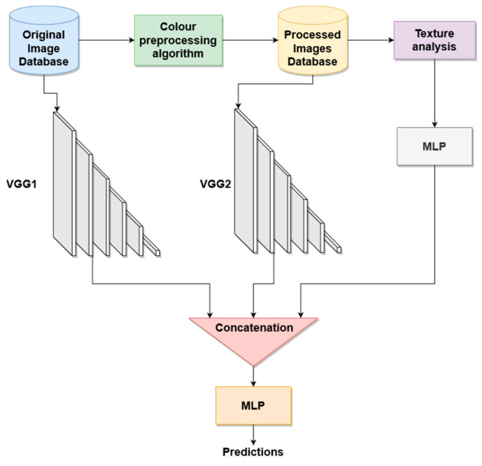
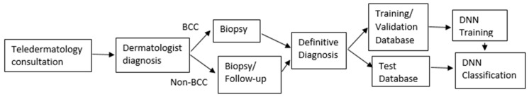
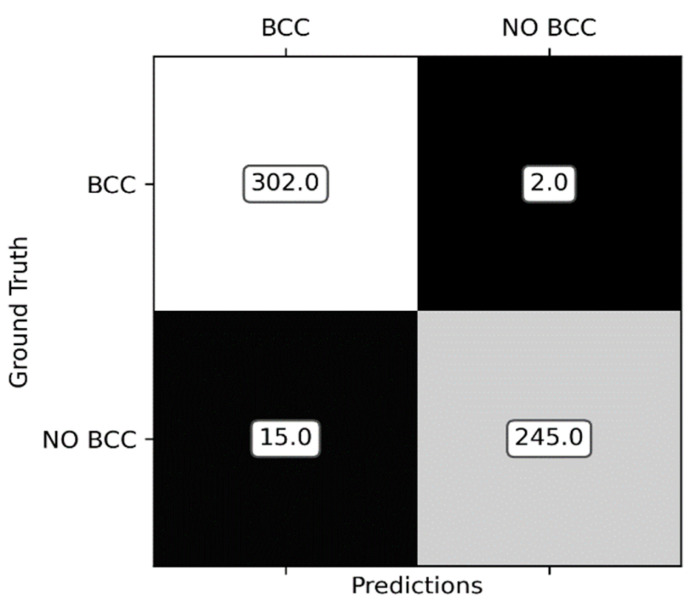
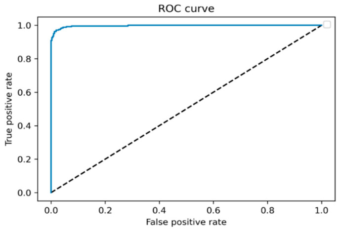
Background and Objective. Skin cancer is the most common cancer worldwide. One of the most common non-melanoma tumors is basal cell carcinoma (BCC), which accounts for 75% of all skin cancers. There are many benign lesions that can be confused with these types of cancers, leading to unnecessary biopsies. In this paper, a new method to identify the different BCC dermoscopic patterns present in a skin lesion is presented. In addition, this information is applied to classify skin lesions into BCC and non-BCC. Methods. The proposed method combines the information provided by the original dermoscopic image, introduced in a convolutional neural network (CNN), with deep and handcrafted features extracted from color and texture analysis of the image. This color analysis is performed by transforming the image into a uniform color space and into a color appearance model. To demonstrate the validity of the method, a comparison between the classification obtained employing exclusively a CNN with the original image as input and the classification with additional color and texture features is presented. Furthermore, an exhaustive comparison of classification employing different color and texture measures derived from different color spaces is presented. Results. Results show that the classifier with additional color and texture features outperforms a CNN whose input is only the original image. Another important achievement is that a new color cooccurrence matrix, proposed in this paper, improves the results obtained with other texture measures. Finally, sensitivity of 0.99, specificity of 0.94 and accuracy of 0.97 are achieved when lesions are classified into BCC or non-BCC. Conclusions. To the best of our knowledge, this is the first time that a methodology to detect all the possible patterns that can be present in a BCC lesion is proposed. This detection leads to a clinically explainable classification into BCC and non-BCC lesions. In this sense, the classification of the proposed tool is based on the detection of the dermoscopic features that dermatologists employ for their diagnosis.
Keywords: basal cell carcinoma; clinically inspired classification; color appearance models; color cooccurrence matrix; deep learning; dermatology.
Conflict of interest statement
The authors declare no conflict of interest.
Figures
References
-
- Skin Cancer Foundation, Skin Cancer Facts and Statistics. 2021. [(accessed on 30 March 2021)]. Available online: https://www.skincancer.org/skin-cancer-information/skin-cancer-facts/#:~....
-
- Peris K., Fargnoli M.C., Garbe C., Kaufmann R., Bastholt L., Seguin N.B., Bataille V., Marmol V.D., Dummer R., Harwood C.A., et al. Diagnosis and treatment of basal cell carcinoma: European consensus based interdisciplinary guidelines. Eur. J. Cancer. 2019;118:10–34. doi: 10.1016/j.ejca.2019.06.003. - DOI - PubMed
-
- Ferrante di Ruffano L., Takwoingi Y., Dinnes J., Chuchu N., Bayliss S.E., Davenport C., Matin R.N., Godfrey K., O'Sullivan C., Gulati A., et al. Cochrane Skin Cancer Diagnostic Test Accuracy Group. Computer-assisted diagnosis techniques (dermoscopy and spectroscopy-based) for diagnosing skin cancer in adults. Cochrane Database Syst. Rev. 2018;12:13186. doi: 10.1002/14651858.CD013186. - DOI - PMC - PubMed
