. 2022 Jul 25;18(7):e1010721.
doi: 10.1371/journal.ppat.1010721. eCollection 2022 Jul.

IFN-γ-independent control of M. tuberculosis requires CD4 T cell-derived GM-CSF and activation of HIF-1α

Affiliations

IFN-γ-independent control of M. tuberculosis requires CD4 T cell-derived GM-CSF and activation of HIF-1α

Erik Van Dis et al. PLoS Pathog. .

Abstract

The prevailing model of protective immunity to tuberculosis is that CD4 T cells produce the cytokine IFN-γ to activate bactericidal mechanisms in infected macrophages. Although IFN-γ-independent CD4 T cell based control of M. tuberculosis infection has been demonstrated in vivo it is unclear whether CD4 T cells are capable of directly activating macrophages to control infection in the absence of IFN-γ. We developed a co-culture model using CD4 T cells isolated from the lungs of infected mice and M. tuberculosis-infected murine bone marrow-derived macrophages (BMDMs) to investigate mechanisms of CD4 dependent control of infection. We found that even in the absence of IFN-γ signaling, CD4 T cells drive macrophage activation, M1 polarization, and control of infection. This IFN-γ-independent control of infection requires activation of the transcription factor HIF-1α and a shift to aerobic glycolysis in infected macrophages. While HIF-1α activation following IFN-γ stimulation requires nitric oxide, HIF-1α-mediated control in the absence of IFN-γ is nitric oxide-independent, indicating that distinct pathways can activate HIF-1α during infection. We show that CD4 T cell-derived GM-CSF is required for IFN-γ-independent control in BMDMs, but that recombinant GM-CSF is insufficient to control infection in BMDMs or alveolar macrophages and does not rescue the absence of control by GM-CSF-deficient T cells. In contrast, recombinant GM-CSF controls infection in peritoneal macrophages, induces lipid droplet biogenesis, and also requires HIF-1α for control. These results advance our understanding of CD4 T cell-mediated immunity to M. tuberculosis, reveal important differences in immune activation of distinct macrophage types, and outline a novel mechanism for the activation of HIF-1α. We establish a previously unknown functional link between GM-CSF and HIF-1α and provide evidence that CD4 T cell-derived GM-CSF is a potent bactericidal effector.


Conflict of interest statement

The authors have declared that no competing interests exist.

Figures

Fig 1
Fig 1. Lung-derived and in vitro-differentiated CD4 T cells control M. tuberculosis growth independent of IFN-γ.
(A) CFU/well at d 0, 2 and 4 postinfection for wild-type and Ifngr-/- BMDMs co-cultured with lung-derived CD4 T cells. (B) CFU fold-change at d 5 postinfection for wild-type and Ifngr-/- BMDMs co-cultured with lung-derived Ifng-/- CD4 T cells. (C) CFU fold-change at d 4 postinfection for wild-type and Ifngr-/- BMDMs cultured with lung-derived CD4 T cells in transwells. (D) CFU fold-change at d 5 postinfection for wild-type and Ifngr-/- BMDMs treated with conditioned media from lung-derived CD4 T cells (T cell conditioned media, TCCM). (E) CFU fold-change at d 5 postinfection for wild-type and Ifngr-/- BMDMs co-cultured with in vitro differentiated Th1 cells. (F) CFU fold-change at d 4 postinfection for wild-type and Ifngr-/- BMDMs treated with Th1 or Th17.1 supernatants (sups). (G) RLU fold-change at d 5 postinfection for Ifngr-/- BMDMs treated with Th1 or Th2 sups. (H) CFU fold-change at d 5 postinfection for Ifngr-/- BMDMs treated with Th1 sups +/- Proteinase K (PK). Figures are representative of two (H) or three or more (A)-(G) experiments. Error bars are SD from four (A)-(B), (D)-(H) or three (C) replicate samples, *p<0.05, **p<0.01, ***p<0.001, ****p<0.0001 by unpaired t-test.
Fig 2
Fig 2. IFN-γ is not required for T cell-mediated macrophage activation and polarization during M. tuberculosis infection.
(A)-(B) RNA-seq at 24 h postinfection for wild-type and Ifngr-/- BMDMs co-cultured with lung-derived CD4 T cells. (A) Principal component analysis plot, (B) Volcano plot of genes in Ifngr-/- BMDMs with >2-fold change in gene expression and adj. p-value >0.05 following cell co-culture. (C) Top ten enriched ontology clusters from Ingenuity Pathway Analysis of genes with >2-fold upregulation in both wild-type and Ifngr-/- BMDMs following co-culture. (D) Top ten enriched gene sets from the MSigDB H: Hallmark collection from Ifngr-/- BMDMs following co-culture. (E)-(F) RNA-seq reads (E) and heat map (F) of M1- or M2-associated transcripts. (G)-(H) Gene Set Enrichment Analysis plots of (G) Hypoxia or (H) Glycolysis gene sets in Ifngr-/- BMDMs following co-culture. Figures represent data from four replicate experiments, **p<0.01, ****p<0.0001 by Tukey post hoc test.
Fig 3
Fig 3. CD4 T cells induce IFN-γ- and NO-independent increases in glycolytic gene expression.
(A) RNA-seq reads of glycolytic genes. (B) Griess assay at 48 h postinfection for wild-type, Ifngr-/-, and Nos2-/- BMDMs co-cultured with a 10:1 ratio of wild-type or Nos2-/- lung-derived CD4 T cells to macrophages. (C) Griess assay at 48 h postinfection for wild-type and Ifngr-/- BMDMs co-cultured with Th1 cells. Figures represent data from four replicate experiments (A) or are representative of two (B) or three (C) independent experiments. Error bars are SD from four independent experiments (A) or four replicate samples (B)-(C), *p<0.05, **p<0.01, ****p<0.0001 by unpaired t-test.
Fig 4
Fig 4. IFN-γ-independent control requires the transcription factor HIF-1α.
(A)-(D) Microscopy for host lipid droplets (LDs) at d 3 postinfection for wild-type and Ifngr-/- BMDMs treated with Th1 supernatants (sups). (E)-(F) Quantification of (A)-(D) for (E) average number of LDs per BMDM and (F) total LD area. (G) RNA-seq reads of HIF-1α target genes. (H) Western blot for HIF-1α on cell lysates 12 h postinfection for wild-type and Ifngr-/- BMDMs treated with Th1 sups and 2-DG. (I) CFU fold-change at d 5 postinfection for wild-type and Hif1a-/- BMDMs co-cultured with wild-type or Ifng-/- lung-derived CD4 T cells. Figures represent data from four replicate experiments (G) or are representative of two (H) or three (A)-(F), (I) independent experiments. Error bars are SD from four independent experiments (G), four replicate samples (I), or 48 images from four replicate wells (E)-(F) *p<0.05, **p<0.01, ***p<0.001, ****p<0.0001 by unpaired t-test; p-values above bars are relative to UT for each genotype.
Fig 5
Fig 5. IFN-γ-independent control of M. tuberculosis requires CD4 T cell-derived GM-CSF.
(A) Concentration of select cytokines in in vitro differentiated Th1 supernatants (sups). (B) CFU fold-change at d 5 postinfection for wild-type, Ifngr-/-, and Tnfr1/2-/- BMDMs co-cultured with Ifng-/- lung-derived CD4 T cells. (C) CFU fold-change at d 5 postinfection for wild-type, Ifngr-/-, and Ifnar-/-/Ifngr-/- double-knockout BMDMs treated with Th1 sups. (D) CFU fold-change at d 5 postinfection for wild-type, Ifngr-/- and Tnfrsf8-/-/Ifngr-/- double-knockout BMDMs treated with Th1 sups. (E) ELISA for GM-CSF concentration at 24 h postinfection in wild-type and Ifngr-/- BMDMs treated with Th1 sups. (F) CFU fold-change at d 5 postinfection for wild-type and Csf2rb-/- BMDMs co-cultured with Ifng-/- lung-derived CD4 T cells. (G) CFU fold-change at d 5 postinfection for Ifngr-/- BMDMs co-cultured with wild-type or Csf2-/- lung-derived CD4 T cells. (H) CFU-fold change at d 4 postinfection for wild-type and Ifngr-/- BMDMs cultured with wild-type or Csf2-/- lung-derived CD4 T cells in transwells. Figures represent seven (A) or three (E) biological replicates or are representative of two (B), (D), (H) or at least three (C), (F)-(G) independent experiments. Error bars are SD from seven (A) or three (B) biological replicates or four (B)-(D), (F)-(G) or three (H) replicate samples, *p<0.05, **p<0.01, ***p<0.001, ****p<0.0001 by unpaired t-test.
Fig 6
Fig 6. GM-CSF activates HIF-1α to restrict M. tuberculosis in peritoneal macrophages.
(A) CFU fold-change at d 4 postinfection for BMDMs and peritoneal macrophages treated with IFN-γ or 10 or 100 ng/mL GM-CSF. (B) CFU fold-change at d 5 postinfection for Ifngr-/- BMDMs co-cultured with wild-type or Csf2-/- lung CD4 T cells and GM-CSF. (C) CFU fold-change at d 4 postinfection for alveolar macrophages treated with GM-CSF. (D) Glucose consumption at 24 h postinfection for peritoneal macrophages treated with GM-CSF. (E)-(H) Microscopy for host lipid droplets (LDs) at 24 h postinfection for wild-type or Hif1a-/- peritoneal macrophages treated with GM-CSF. (I) Quantification of (E)-(H) for LD signal per macrophage. (J) Western blot for HIF-1α on cell lysates 20 h postinfection for peritoneal macrophages treated with GM-CSF. HIF-1α/β-actin ratio is indicated and was quantified using ImageJ. (K) CFU fold-change at d 4 postinfection for wild-type and Hif1a-/- peritoneal macrophages treated with GM-CSF. Figures are representative of two (E)-(J) or three or more (A)-(D), (K) experiments. Error bars are SD from four replicate samples (A)-(D), (K) or four replicate wells (I), *p<0.05, **p<0.01, ***p<0.001, ****p<0.0001 by unpaired t-test.
Fig 7
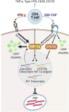
Fig 7. Model for IFN-γ-independent CD4 T cell-mediated control of M. tuberculosis by GM-CSF and HIF-1α.
CD4 T cells secrete immune activating cytokines including TNF-α and Type I IFN and express the cell surface molecules CD40 and CD153, none of which mediate control of M. tuberculosis in macrophages. IFN-γ activates macrophages to control M. tuberculosis in part by stabilizing HIF-1α expression in a nitric oxide (NO)-dependent manner to drive a metabolic switch to glycolysis and the production of macrophage lipid droplets, and HIF-1α is required for IFN-γ-mediated control. In the absence of IFN-γ, CD4 T cell-derived GM-CSF is necessary for M. tuberculosis control in BMDMs and GM-CSF requires HIF-1α expression for control. HIF-1α activation during GM-CSF-mediated control, however, is NO-independent, indicating distinct mechanisms of activation following IFN-γ and GM-CSF signaling. Similarly, both IFN-γ and GM-CSF mediate lipid droplet biogenesis, but this is HIF-1α-dependent following IFN-γ activation and HIF-1α-independent in the context of GM-CSF. Importantly, lipid droplets do not mediate bacterial control following IFN-γ or GM-CSF activation. With or without IFN-γ, CD4 T cells producing GM-CSF upregulate macrophage expression of M1 and glycolytic transcripts leading to an activated, polarized, and antibacterial state which controls bacterial growth through an undefined mechanism. Model created using BioRender.com.

Similar articles

Cited by

References

    1. Green AM, Difazio R, Flynn JL. IFN-γ from CD4 T cells is essential for host survival and enhances CD8 T cell function during Mycobacterium tuberculosis infection. Journal of Immunology. 2013;190(1):270–7. doi: 10.4049/jimmunol.1200061 - DOI - PMC - PubMed
    1. Amelio P, Portevin D, Hella J, Reither K, Kamwela L, Lweno O, et al.. HIV Infection Functionally Impairs Mycobacterium tuberculosis-Specific CD4 and CD8 T-Cell Responses. Journal of virology. 2019;93(5). doi: 10.1128/JVI.01728-18 - DOI - PMC - PubMed
    1. Flesch IE, Kaufmann SH. Mycobacterial growth inhibition by interferon-gamma-activated bone marrow macrophages and differential susceptibility among strains of Mycobacterium tuberculosis. Journal of immunology (Baltimore, Md: 1950). 1987;138(12). - PubMed
    1. Flesch IE, Kaufmann SH. Mechanisms involved in mycobacterial growth inhibition by gamma interferon-activated bone marrow macrophages: role of reactive nitrogen intermediates. Infection and Immunity. 1991;59(9):3213–8. doi: 10.1128/iai.59.9.3213-3218.1991 - DOI - PMC - PubMed
    1. Flynn JL, Chan J, Triebold KJ, Dalton DK, Stewart TA, Bloom BR. An essential role for interferon gamma in resistance to Mycobacterium tuberculosis infection. The Journal of Experimental Medicine. 1993;178(6):2249–54. doi: 10.1084/jem.178.6.2249 - DOI - PMC - PubMed

Publication types

Substances