Review
. 2022 Jun 29;12(7):673.
doi: 10.3390/membranes12070673.

Amphiphilic Gold Nanoparticles: A Biomimetic Tool to Gain Mechanistic Insights into Peptide-Lipid Interactions

Affiliations
Review

Amphiphilic Gold Nanoparticles: A Biomimetic Tool to Gain Mechanistic Insights into Peptide-Lipid Interactions

Ester Canepa et al. Membranes (Basel). .

Abstract

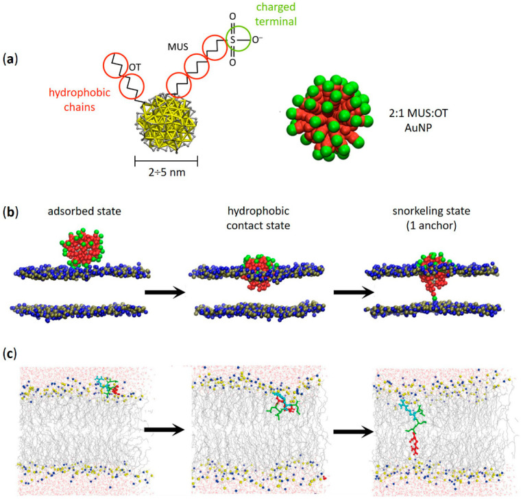
Functional peptides are now widely used in a myriad of biomedical and clinical contexts, from cancer therapy and tumor targeting to the treatment of bacterial and viral infections. Underlying this diverse range of applications are the non-specific interactions that can occur between peptides and cell membranes, which, in many contexts, result in spontaneous internalization of the peptide within cells by avoiding energy-driven endocytosis. For this to occur, the amphipathicity and surface structural flexibility of the peptides play a crucial role and can be regulated by the presence of specific molecular residues that give rise to precise molecular events. Nevertheless, most of the mechanistic details regulating the encounter between peptides and the membranes of bacterial or animal cells are still poorly understood, thus greatly limiting the biomimetic potential of these therapeutic molecules. In this arena, finely engineered nanomaterials-such as small amphiphilic gold nanoparticles (AuNPs) protected by a mixed thiol monolayer-can provide a powerful tool for mimicking and investigating the physicochemical processes underlying peptide-lipid interactions. Within this perspective, we present here a critical review of membrane effects induced by both amphiphilic AuNPs and well-known amphiphilic peptide families, such as cell-penetrating peptides and antimicrobial peptides. Our discussion is focused particularly on the effects provoked on widely studied model cell membranes, such as supported lipid bilayers and lipid vesicles. Remarkable similarities in the peptide or nanoparticle membrane behavior are critically analyzed. Overall, our work provides an overview of the use of amphiphilic AuNPs as a highly promising tailor-made model to decipher the molecular events behind non-specific peptide-lipid interactions and highlights the main affinities observed both theoretically and experimentally. The knowledge resulting from this biomimetic approach could pave the way for the design of synthetic peptides with tailored functionalities for next-generation biomedical applications, such as highly efficient intracellular delivery systems.

Keywords: antimicrobial peptides; cell membranes; cell-penetrating peptides; lipid bilayers; molecular dynamics; non-specific interactions; spontaneous membrane translocation; thiol-protected gold nanoparticles.


Conflict of interest statement

The authors declare no conflict of interest.

Figures

Figure 1
Figure 1
Amphiphilic AuNPs and amphiphilic peptides: similarities between their spontaneous penetration mechanism into lipid membranes. (a) Structure of an amphiphilic MUS:OT AuNP with its coarse-grained (CG) representation (2:1 MUS:OT ligand ratio). Red beads represent hydrophobic carbon groups, while green beads represent the charged MUS terminals. (b) Different stages of penetration of a MUS:OT AuNP into a 1-palmitoyl-2-oleoyl-glycero-3-phosphocholine (POPC) lipid bilayer obtained from CG molecular dynamics simulations. From left to right: adsorbed state, hydrophobic contact state, and snorkeling of the first MUS ligand which binds to the opposite leaflet. Eventually—through a sequential anchoring process—more and more MUS ligands are dropped leading to the fully snorkeled configuration of the NP-membrane complex. Lipid heads are blue (choline) and tan (phosphate), lipid tails and water are not shown. (c) Translocation process of an amphiphilic ‘Spontaneous Membrane-Translocating Peptide’ (SMTP) into a POPC lipid bilayer obtained from united atom bias-exchange metadynamics simulations. Specifically, the SMTP contains a LRLLR sequence composed of two Arg (R) and three leucines (L) residues. From left to right: SMTP located in the lipid head region, SMTP on its way towards the opposite leaflet, and final snorkeled configuration. The first Arg is shown in cyan, the second Arg is shown in red, and leucine hydrophobic residues are shown in green. Nitrogen and phosphorus atoms in the lipid head region are shown in blue and yellow, respectively. The lipid tails are shown as thin gray lines, while water is shown as red (oxygen) and gray (hydrogen) cylinders. (a,b) adapted with permission from Simonelli et al. [38]—Copyright © 2015 American Chemical Society. (c) reprinted with permission from Cao et al. [51]—Copyright © 2020 Elsevier B.V. All rights reserved.
Figure 2
Figure 2
Amphiphilic AuNPs and amphiphilic peptides: a common affinity for the disordered domains of phase−separated lipid membranes. (a) Phase−separated SLBs containing 1,2-dioleoyl-sn-glycero-3-phosphocholine (DOPC), sphingomyelin (SM), cholesterol and ganglioside GM1 (63:31:1:5 molar ratio) imaged in liquid by atomic force microscopy (AFM) after addition of ~3 nm MUS:OT AuNPs (40 min and 15 h). After hours, large clusters of amphiphilic AuNPs (white arrows) slowly formed on the darker disordered phase and at the edges of the lighter (i.e., higher) ordered lipid domains. (b) Potential of mean force (PMF) profiles calculated for the adsorption of a single MUS:OT AuNPs on the surface of the Ld and Lo phase. The Ld phase, with a binding free energy of ~18 kJ/mol (~9 kBT), is favoured over the Lo phase (~11 kJ/mol, ~5 kBT). (c) Giant plasma membrane vesicles (GPMVs) derived from rat basophilic leukemia cells incubated at low temperature with three examples of fluorescein-labeled CPPs—i.e., MAP (model amphipathic peptide), penetratin (pAntp) and transportan 10 (TP10) (green). All these CPPs are amphipatic and contain Lys or Arg residues. Lo and Ld phases are labeled with AF594-labeled cholera toxin B subunit (CtxB, red) and AF647-labeled annexin V (AnV, pseudocolored as white), respectively. (a,b) contain images by Canepa et al. [44] reprinted with minor modifications under a CC BY-NC 3.0 license with permission from the Royal Society of Chemistry. (c) is reproduced and adapted with permission from Säälik et al. [81]—Copyright © 2011 Elsevier B.V. All rights reserved.
Figure 3
Figure 3
Perturbation of membrane ordered–disordered phase separation upon interaction with amphiphilic AuNPs and amphiphilic peptides. (a) Topographic AFM images showing fragmentation of ordered domains induced by ~3 nm MUS:OT AuNPs on phase-separated lipid bilayers containing DOPC:SM:chol:GM1 (63:31:1:5 molar ratio). Two height profiles of the phase-separated membrane without NPs are also reported. On the right: comparison of height difference distributions (Δz) between ordered and disordered domains before and after NP/membrane interaction. (b) Fluorescence images of the same region of a POPC:1,2-dimyristoyl-sn-glycero-3-PG (DMPG) 1:1 phase-separated SLB with PG-enriched ordered domains recorded before and after exposure to the lipopetide DAP (1 μM). Two solid ordered domains (dark regions) in a liquid disordered background (bright region) containing the fluorescence lipid probe DHPE-Texas Red (TR, 1%) are shown. The channel of kynurenine (KYN)—an intrinsically fluorescent DAP residue—is used to visualize the morphology of ordered domains since DAP strongly interacts with PG lipids. Overall, the SLB/DAP interaction induces an extensive size reduction of the solid domains equal to 59%. (c) Force spectroscopy analysis and topographic AFM images of the same POPC:DMPG 1:1 SLB before and after exposure to increasing DAP concentrations (0–8 μM). Jump-through (J–T) force maps (resolution 32 × 32 pixels) are reported next to each AFM image, together with the comparison of solid and fluid domains jump-through force upon increasing concentrations of DAP. (a) contains images by Canepa et al. [44] reprinted with minor modifications under a CC BY-NC 3.0 license with permission from the Royal Society of Chemistry. (b,c) are reproduced and adapted with permission from Mescola et al. [99]—Copyright © 2020 American Chemical Society. All rights reserved.
Figure 4
Figure 4
Translocation of amphiphilic AuNPs and amphiphilic peptides is favoured into lipid membranes with lower cholesterol content. (a) Left: coarse-grained structure of hydrophilic (MUS) and hydrophobic (OT) ligands and an amphiphilic MUS:OT AuNP in water (2 nm core size; water not shown). Right: simulation snapshots showing the ligand anchoring typical of these AuNPs (see Figure 1b). The NP goes from the hydrophobic contact state (top) to the anchored state (bottom) in which one MUS charged terminal is in contact with the lipid heads (transparent gray) of the distal leaflet. Cholesterol molecules—intercalated between the apolar tails of membrane phospholipids (DOPC)—are shown in tan in the membrane detail on the bottom left. (b) Anchoring free energy barriers calculated with well-tempered metadynamics simulations at different membrane cholesterol concentrations. (c) Average anchoring time (Δtanchor) and average number of anchored ligands after 1 μs (Nanchors) obtained from unbiased MD simulations as a function of membrane cholesterol content. (d) Translocation of the Arg-rich CPP nona-arginine (Arg9)—labeled with fluorescein (green)—into GPMVs derived from MDA-MB-231 (MDA GPMV) and RBL-2H3 (RBL GPMV) cells. Left images: GPMVs labeled with filipin (pseudo-colored as white) to bind membrane cholesterol and enable its visualization. Right images: GPMVs labeled with Alexa Fluor 555-conjugated cholera toxin B subunit (CtxB, red) and Alexa Fluor 647-conjugated annexin V (AnV, pseudocolored as white) to visualize, respectively, the Lo membrane domains and phosphatidylserine contained in the outer leaflet of the limiting membrane of both vesicle types. Quantification of the filipin signal shows that MDA GPMVs contain approximately 30% less membrane cholesterol than RBL GPMVs; in addition, RBL GPMVs show several large cholesterol-enriched subdomains (white arrows) that are rarer in MDA GPMVs. Overall, Arg9 translocation is significantly reduced in vesicles characterized by higher membrane cholesterol content and more cholesterol-rich membrane microdomains. *** p-value < 0.0001 and 0.0005 for filipin signal and fluo-Arg9 translocation, respectively. (ac) contain images by Canepa et al. [45] reprinted with minor modifications—Copyright © 2021 The Authors, published by American Chemical Society. (d) is reproduced and adapted with permission from Lorents et al. [111]—Copyright © 2018 American Chemical Society.
Figure 5
Figure 5
The tendency for membrane aggregation is shared by amphiphilic AuNPs and amphiphilic peptides. (a) Top, sketch of two curvature inducing membrane inclusions; due to the elastic energy of the membrane, an effective interacting potential can arise and, depending on the system, the interaction can be attractive and thus lead to aggregation. Bottom, system in which the inclusions suppress the natural fluctuations of the membrane; aggregation can minimize this region. (b) Aggregation induced by lipid depletion. (c) Aggregation induced by capillary forces. In configuration (1), there are two regions of modified lipid density around the inclusion; dimerization allows to minimize their total area, as shown in configuration (2). (d) Ordered aggregate of MUS:OT NPs adsorbed on a DOPC membrane. The snapshot is taken from unbiased MD simulations (Martini CG). Nanoparticles are represented with yellow beads (Au), pink beads (S), cyan (MUS ligands) and blue (OT ligands); membrane lipids are represented with red beads. (e) Dimer of adsorbed NPs on DOPC membrane from unbiased MD simulations (Martini CG). The extended ligand configuration can be observed. Representation as in (d). (f) Cryo-EM image of MUS:OT aggregation on the surface of a DOPC liposome. The NP-NP is compatible with the extended ligand configuration. (g) Ordered aggregate of MUS:OT NPs embedded in a model neuronal plasma membrane. The snapshot is taken from unbiased MD simulations (Martini CG). The NP are represented with a yellow core, blue OT ligands and cyan MUS ligands. The membrane is represented with red DliPC lipids, light pink sphingomyelin, yellow ganglioside and grey cholesterol. (h) Dimer of MUS:OT NPs embedded in a model neuronal plasma membrane. The deformation of the ligand shell and the presence of the stabilizing layer of ions (red beads) can be observed. The membrane headgroups are shown as semi-transparent surface, lipid tails are not shown for clarity. (i) Supramolecular lattice formed by M1 bilayer-embedded MUS:OT NPS, imaged by AFM. The digital zoom of the area with blue contour shows the lattice order at higher magnification. (ac) adapted with permission from Johannes et al. [152] Copyright © 2022 Elsevier B.V. All rights reserved. (df) adapted from Lavagna et al. [173] with permission from the Royal Society of Chemistry. (gi) adapted from Canepa et al. [44] under a CC BY-NC 3.0 license with permission from the Royal Society of Chemistry.

Similar articles

References

    1. Apostolopoulos V., Bojarska J., Chai T.-T., Elnagdy S., Kaczmarek K., Matsoukas J., New R., Parang K., Lopez O.P., Parhiz H., et al. A Global Review on Short Peptides: Frontiers and Perspectives. Molecules. 2021;26:430. doi: 10.3390/molecules26020430. - DOI - PMC - PubMed
    1. Ko J., Auyeung K. Identification of Functional Peptides from Natural and Synthetic Products on Their Anticancer Activities by Tumor Targeting. CMC. 2014;21:2346–2356. doi: 10.2174/0929867321666140205132124. - DOI - PubMed
    1. Wang X.-Y., Wang Y.-H., Song Z., Hu X.-Y., Wei J.-P., Zhang J., Wang H.-S. Recent Progress in Functional Peptides Designed for Tumor-Targeted Imaging and Therapy. J. Mater. Chem. C. 2021;9:3749–3772. doi: 10.1039/D0TC05405D. - DOI
    1. Rong L., Qin S.-Y., Zhang C., Cheng Y.-J., Feng J., Wang S.-B., Zhang X.-Z. Biomedical Applications of Functional Peptides in Nano-Systems. Mater. Today Chem. 2018;9:91–102. doi: 10.1016/j.mtchem.2018.06.001. - DOI
    1. Oyston P.C.F., Fox M.A., Richards S.J., Clark G.C. Novel Peptide Therapeutics for Treatment of Infections. J. Med. Microbiol. 2009;58:977–987. doi: 10.1099/jmm.0.011122-0. - DOI - PubMed

LinkOut - more resources