

Towards Biohybrid Lung Development: Establishment of a Porcine In Vitro Model
- PMID: 35877890
- PMCID: PMC9325277
- DOI: 10.3390/membranes12070687
Towards Biohybrid Lung Development: Establishment of a Porcine In Vitro Model
Abstract
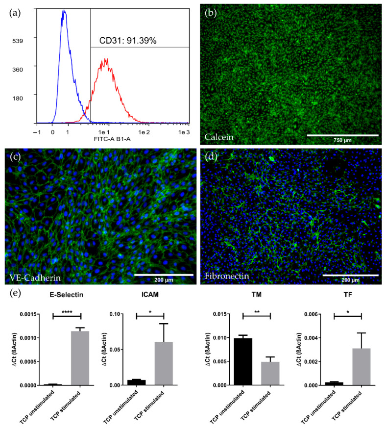
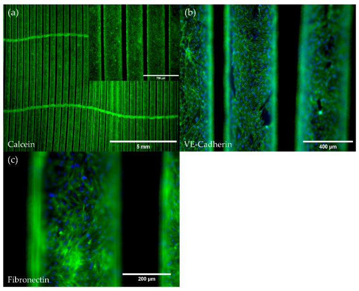
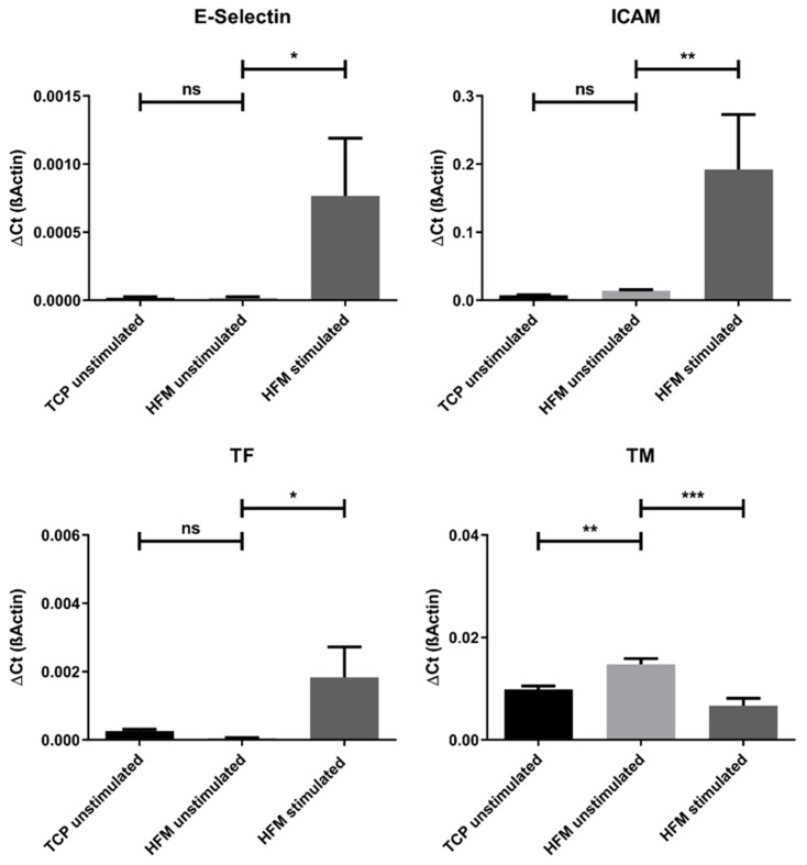
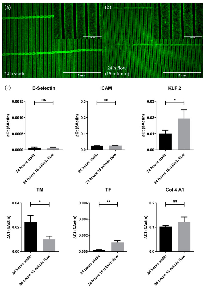
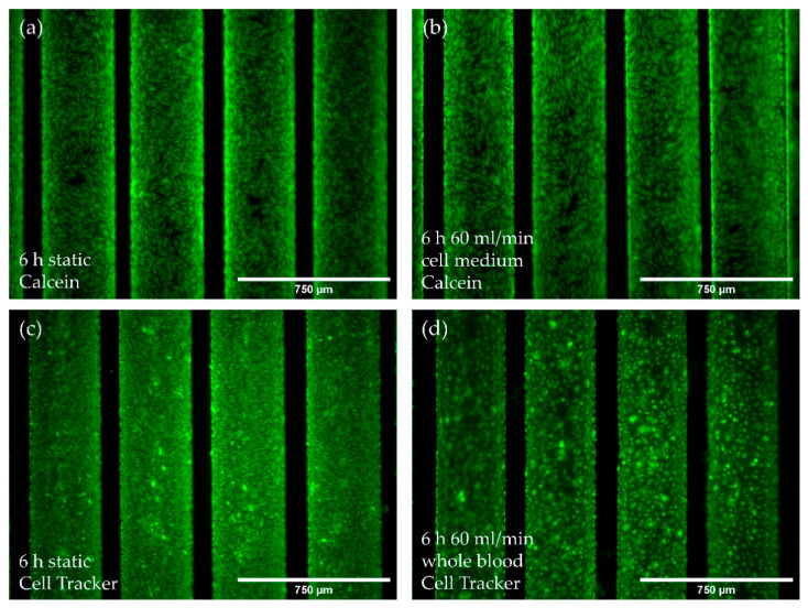
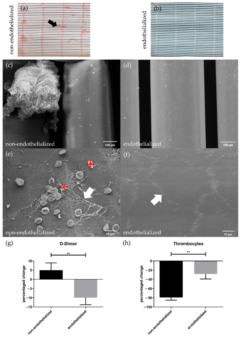
Lung transplantation (LTx) is the only curative therapy option for patients with end-stage lung diseases, though only available for chosen patients. To provide an alternative treatment option to LTx, we aim for the development of an implantable biohybrid lung (BHL) based on hollow fiber membrane (HFM) technology used in extracorporeal membrane oxygenators. Crucial for long-lasting BHL durability is complete hemocompatibility of all blood contacting surfaces, which can be achieved by their endothelialization. In continuation to successful in vitro investigations using human endothelial cells (ECs), indicating general feasibility, the appropriate porcine in vivo model needs to be prepared and established to fill the translational data gap prior to patient's application. Therefore, isolation of porcine ECs from carotid arteries (pCECs) was established. Following, pCECs were used for HFM endothelialization and examined under static and dynamic conditions using cell medium or heparinized blood, to assess their proliferation capacity, flow resistance and activation state, especially under clinically relevant conditions. Additionally, comparative hemocompatibility tests between native and endothelialized HFMs were performed. Overall, pure pCECs formed a viable and confluent monolayer, which resisted applied flow conditions, in particular due to physiological extracellular matrix synthesis. Additionally, pCECs remained the non-inflammatory and anti-thrombogenic status, significantly improving the hemocompatibility of endothelialized HFMs. Finally, as relevant for reliable porcine to human translation, pCECs behaved in the same way as human ECs. Concluding, generated in vitro data justify further steps towards pre-clinical BHL examination, in particular BHL application to porcine lung injury models, reflecting the clinical scenario with end-stage lung-diseased patients.
Keywords: biohybrid lung; endothelialization; hemocompatibility; hollow fiber membrane; porcine endothelial cells.
Conflict of interest statement
The authors declare no conflict of interest. The funders had no role in the design of the study; in the collection, analyses or interpretation of data; in the writing of the manuscript, or in the decision to publish the results.
Figures
Similar articles
-
Biohybrid lung Development: Towards Complete Endothelialization of an Assembled Extracorporeal Membrane Oxygenator.Bioengineering (Basel). 2023 Jan 5;10(1):72. doi: 10.3390/bioengineering10010072. Bioengineering (Basel). 2023. PMID: 36671644 Free PMC article.
-
Towards Biohybrid Lung: Induced Pluripotent Stem Cell Derived Endothelial Cells as Clinically Relevant Cell Source for Biologization.Micromachines (Basel). 2021 Aug 19;12(8):981. doi: 10.3390/mi12080981. Micromachines (Basel). 2021. PMID: 34442603 Free PMC article.
-
Towards Biohybrid Lung Development-Fibronectin-Coating Bestows Hemocompatibility of Gas Exchange Hollow Fiber Membranes by Improving Flow-Resistant Endothelialization.Membranes (Basel). 2021 Dec 27;12(1):35. doi: 10.3390/membranes12010035. Membranes (Basel). 2021. PMID: 35054561 Free PMC article.
-
Advances in Enhancing Hemocompatibility of Hemodialysis Hollow-Fiber Membranes.Adv Fiber Mater. 2023 Apr 3:1-43. doi: 10.1007/s42765-023-00277-5. Online ahead of print. Adv Fiber Mater. 2023. PMID: 37361105 Free PMC article. Review.
-
Anti-thrombogenic Surface Coatings for Extracorporeal Membrane Oxygenation: A Narrative Review.ACS Biomater Sci Eng. 2021 Sep 13;7(9):4402-4419. doi: 10.1021/acsbiomaterials.1c00758. Epub 2021 Aug 26. ACS Biomater Sci Eng. 2021. PMID: 34436868 Review.
Cited by
-
Citrate-Coated Iron Oxide Nanoparticles Facilitate Endothelialization of Left Ventricular Assist Device Impeller for Improved Antithrombogenicity.Adv Sci (Weinh). 2025 Feb;12(6):e2408976. doi: 10.1002/advs.202408976. Epub 2024 Dec 20. Adv Sci (Weinh). 2025. PMID: 39707689 Free PMC article.
-
Biohybrid lung Development: Towards Complete Endothelialization of an Assembled Extracorporeal Membrane Oxygenator.Bioengineering (Basel). 2023 Jan 5;10(1):72. doi: 10.3390/bioengineering10010072. Bioengineering (Basel). 2023. PMID: 36671644 Free PMC article.
References
-
- World Health Organization . World Health Statistics 2021: Monitoring Health for the SDGs, Sustainable Development Goals. WHO; Geneva, Switzerland: 2021.
-
- Weill D., Benden C., Corris P.A., Dark J.H., Davis R.D., Keshavjee S., Lederer D.J., Mulligan M.J., Patterson G.A., Singer L.G., et al. A consensus document for the selection of lung transplant candidates: 2014—An update from the Pulmonary Transplantation Council of the International Society for Heart and Lung Transplantation. J. Heart Lung Transplant. 2015;34:1–15. doi: 10.1016/j.healun.2014.06.014. - DOI - PubMed
Grants and funding
LinkOut - more resources
Full Text Sources
Miscellaneous
