. 2022 Jul 14;12(7):707.
doi: 10.3390/membranes12070707.

Roles of Sodium Hydrogen Exchanger (NHE1) and Anion Exchanger (AE2) across Chondrocytes Plasma Membrane during Longitudinal Bone Growth

Affiliations

Roles of Sodium Hydrogen Exchanger (NHE1) and Anion Exchanger (AE2) across Chondrocytes Plasma Membrane during Longitudinal Bone Growth

Adamu Abdul Abubakar et al. Membranes (Basel). .

Abstract

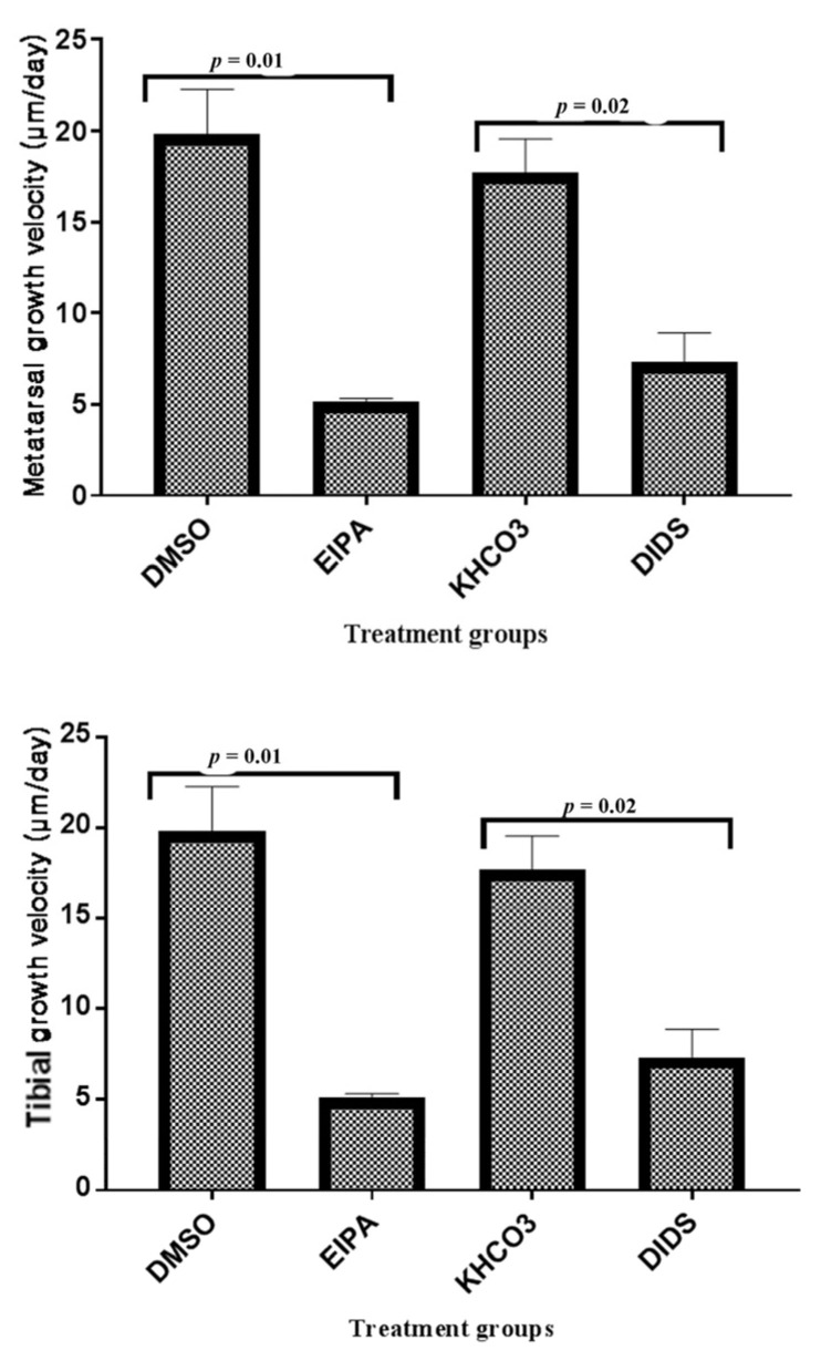
Mammalian long bone growth occurs through endochondral ossification, majorly regulated by the controlled enlargement of chondrocytes at the growth plate (GP). This study aimed to investigate the roles of Na+/H+ (sodium hydrogen exchanger (NHE1)) and HCO3− (anion exchanger [AE2]) during longitudinal bone growth in mammals. Bones from P10 SpragueDawley rat pups were cultured exvivo in the presence or absence of NHE1 and AE2 inhibitors to determine their effect on long bone growth. Gross morphometry, histomorphometry, and immunohistochemistry were used to assess the bone growth. The results revealed that the culture of the bones in the presence of NHE1 and AE2 inhibitors reduces bone growth significantly (p < 0.05) by approximately 11%. The inhibitor significantly (p < 0.05) reduces bone growth velocity and the length of the hypertrophic chondrocyte zone without any effect on the total GP length. The total GP chondrocyte density was significantly (p < 0.05) reduced, but hypertrophic chondrocyte densities remained constant. NHE1 fluorescence signaling across the GP length was higher than AE2, and their localization was significantly (p < 0.05) inhibited at the hypertrophic chondrocytes zone. The GP lengthening was majorly driven by an increase in the overall GP chondrocyte and hypertrophic chondrocyte densities apart from the regulatory volume phenomenon. This may suggest that NHE1 and AE2 could have a regulatory role in long bone growth.

Keywords: chondrocytes; growth plate; long bone growth; plasma membrane inhibitors.


Conflict of interest statement

The authors declare no conflict of interest.

Figures

Figure 1
Figure 1
Representative of Serial stereomicroscopic images of metatarsal bone before and after treatments with EIPA and DIDS at respective concentrations of 444 µM and 250 µM. The lengths of the bones were measured in centimeter (cm) at X6.5 objective magnification.
Figure 2
Figure 2
The bar chart shows metatarsal and tibial length (cm) growth changes at 0 and 48 h incubation with different treatments. Data were pooled from the left and right three middle metatarsals and their corresponding tibia from 20 rats. Each group represent fifteen metatarsal (n = 15) and ten tibial bones (n = 10). * indicates significant differences (p < 0.05; paired Student’s t-test) between baseline and the corresponding 48 h bone growth rates, data were expressed as means ± SEM.
Figure 3
Figure 3
The bar chart shows the mean metatarsal and tibial growth velocity (μm/day) trend of changes between treated groups and their corresponding control. The p-value above the bars indicatesa significant difference between the treated and control groups (unpaired Student t-test).
Figure 4
Figure 4
Histomicrograph showing the height of the tibial GP and HCZ of GP in different treatment groups after 48 h treatments with EIPA or DIDS. The straight vertical line at the -mid-GP section was used to determine the total GP length. Panel (A,B) are a comparison of HCZ length in EIPA-treated and its control group. Panels (C,D) are a comparison of HCZ length in DIDS-treated and its control group. Scale bar = 100 μm in all panels, X10 objective. PCZ, HCZ, and MB stand for proliferative chondrocytes zone, hypertrophic chondrocytes zone, and mineralized bone, respectively. Slides were stained with toluidine blue O.
Figure 5
Figure 5
(a) Immunostaining micrographs showing the appearance of immunoperoxidase (IP) staining intensity of different treatment groups across the GP. Panels (AD) are a comparison of immunoperoxidase localisations of N+/H+ or HCO3 in the GP chondrocytes treated with EIPA or DIDS and their corresponding controls. Scale bar = 100 μm in all panels, X10 objective. (b) Immunofluorescence micrographs showing the appearance of fluorescence labeling intensity of different treatment groups across the GP. Panels (AD) are the comparison of fluorescence labeling intensity in EIPA and DIDS with their respective control groups. Scale bar = 100 μm in all panels, X10 objective.
Figure 5
Figure 5
(a) Immunostaining micrographs showing the appearance of immunoperoxidase (IP) staining intensity of different treatment groups across the GP. Panels (AD) are a comparison of immunoperoxidase localisations of N+/H+ or HCO3 in the GP chondrocytes treated with EIPA or DIDS and their corresponding controls. Scale bar = 100 μm in all panels, X10 objective. (b) Immunofluorescence micrographs showing the appearance of fluorescence labeling intensity of different treatment groups across the GP. Panels (AD) are the comparison of fluorescence labeling intensity in EIPA and DIDS with their respective control groups. Scale bar = 100 μm in all panels, X10 objective.

Similar articles

References

    1. Mackie E.J., Ahmed Y.A., Tatarczuch L., Chen K.S., Mirams M. Endochondral ossification: How cartilage is converted into bone in the developing skeleton. Int. J. Biochem. Cell Biol. 2008;40:46–62. doi: 10.1016/j.biocel.2007.06.009. - DOI - PubMed
    1. Cooper K.L., Oh S., Sung Y., Dasari R.R., Marc W., Tabin C.J. Multiple phases of chondrocytes enlargement underlie differences in Skeletal Proportions. Nature. 2013;495:375–378. doi: 10.1038/nature11940. - DOI - PMC - PubMed
    1. Staines K.A., Pollard A.S., McGonnell I.M., Farquharson C., Pitsillides A.A. Cartilage to bone transitions in health and disease. J. Endocrinol. 2013;219:R1–R12. doi: 10.1530/JOE-13-0276. - DOI - PMC - PubMed
    1. Long F., Chung U., Ohba S., McMahon J., Kronenberg H.M., McMahon A.P. Ihh signaling is directly required for the osteoblast lineage in the endochondral skeleton. Development. 2004;131:1309–1318. doi: 10.1242/dev.01006. - DOI - PubMed
    1. Emons J., Chagin A.S., Hultenby K., Zhivotovsky B., Wit J.M., Karperien M.S., Vendahl L. Epiphyseal fusion in the human growth plate does not involve classical apoptosis. Pediatr. Res. 2009;66:654–659. doi: 10.1203/PDR.0b013e3181beaa8c. - DOI - PubMed

LinkOut - more resources