. 2022 Jul 20;12(7):719.
doi: 10.3390/membranes12070719.

Small Intestinal Submucosa Biomimetic Periosteum Promotes Bone Regeneration

Affiliations

Small Intestinal Submucosa Biomimetic Periosteum Promotes Bone Regeneration

Yanlin Su et al. Membranes (Basel). .

Abstract

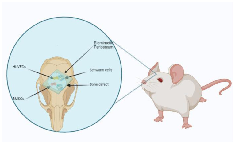
Background: Critical bone defects are a significant problem in clinics. The periosteum plays a vital role in bone regeneration. A tissue-engineered periosteum (TEP) has received increasing attention as a novel strategy for bone defect repairs.

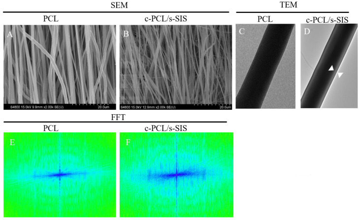
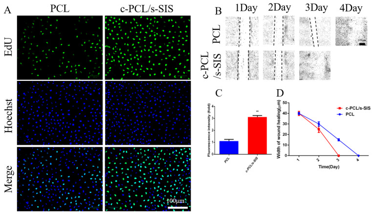
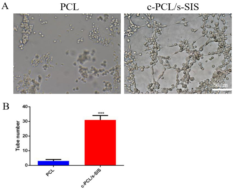
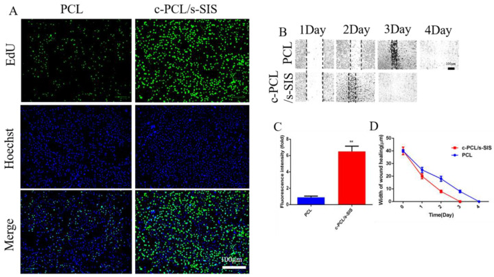
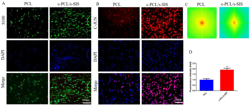
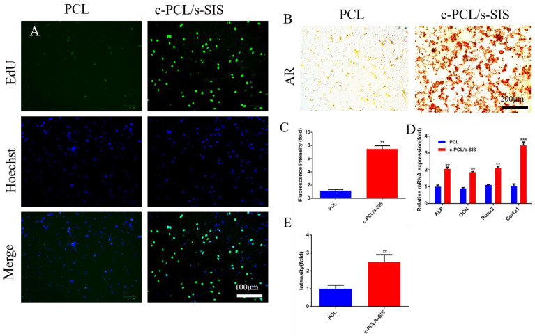
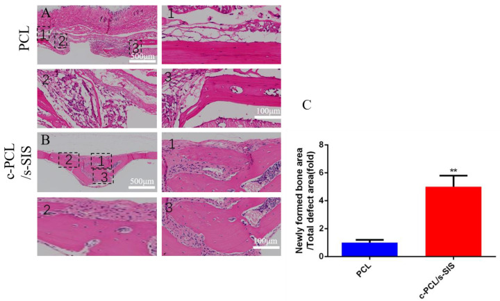
Methods: In this experiment, a biomimetic periosteum was fabricated by using coaxial electrospinning technology with decellularized porcine small intestinal submucosa (SIS) as the shell and polycaprolactone (PCL) as the core. In vitro, the effects of the biomimetic periosteum on Schwann cells, vascular endothelial cells, and bone marrow mesenchymal stem cells were detected by a scratch test, an EdU, a tube-forming test, and an osteogenesis test. In vivo, we used HE staining to evaluate the effect of the biomimetic periosteum on bone regeneration.

Results: In vitro experiments showed that the biomimetic periosteum could significantly promote the formation of angiogenesis, osteogenesis, and repaired Schwann cells (SCs). In vivo experiments showed that the biomimetic periosteum could promote the repair of bone defects.

Conclusions: The biomimetic periosteum could simulate the structural function of the periosteum and promote bone repair. This strategy may provide a promising method for the clinical treatment of skull bone defects.

Keywords: Schwann cells; angiogenesis; biomimetic periosteum; bone regeneration; ordered coaxial electrospinning.


Conflict of interest statement

The authors declare no conflict of interest.

Figures

Scheme 1
Scheme 1
Schematic diagram of biomimetic periosteum in the treatment of a bone defect repair.
Figure 1
Figure 1
Characteristics of the biomimetic periosteum. SEM images of (A) the PCL and (B) the c-PCL/s-SIS membrane. Scale bar: 200 μm. (C,D) TEM images of the c-PCL/s-SIS membrane. Scale bar: 2 μm. FFT (Fast Fourier transform) analyses of (E) the PCL and (F) the c-PCL/s-SIS membrane.
Figure 2
Figure 2
In vitro biomimetic periosteum with HUVECs. (A) Cell proliferation for 3 days of culture. Scale bar: 100 μm. (B) Wound scratch test was used to assess the migration of HUVECs. Scale bar: 100 μm. (C) Quantification of the EdU test. (D) Quantification of (B). n = 3; ** p < 0.01.
Figure 3
Figure 3
In vitro biomimetic periosteum with HUVECs. (A) Tube-forming test. Scale bar: 50 μm. (B) Quantification of (A). n = 3; *** p < 0.001.
Figure 4
Figure 4
In vitro biomimetic periosteum with Schwann cells. (A) Cell proliferation for 3 days of culture. Scale bar: 100 μm. (B) A wound scratch test was used to assess the migration of SCs. (C) Quantification of the EdU test. (D) Quantification of (B). n = 3; ** p < 0.01.
Figure 5
Figure 5
In vitro biomimetic periosteum with Schwann cells. (A) Detection of the guidance of the bionic periosteum to Schwann cells. Scale bar: 100 μm. (B) Schwann cell phenotypic transformation. (C) FFT analyses of (A). (D) Quantification of (B). n = 3; ** p < 0.01.
Figure 6
Figure 6
In vitro osteogenesis evaluations of BMSCs cultured on the biomimetic membranes. (A) Cell proliferation for 3 days of culture. Scale bar: 100 μm. (B) Alizarin Red staining. (C) Quantification of the EdU test. (D) Relative osteogenesis-related gene expressions (ALP, OCN, RUNX2, and COL1a1) of BMSCs cultured on the membranes for 14 days. (E) Quantification of (B). n = 3; ** p < 0.01; *** p < 0.001.
Figure 7
Figure 7
Histological analysis of H&E staining at 8 weeks of (A) the PCL group or (B) the c-PCL/s-SIS group (the other images represent higher magnification images of the corresponding black square boxes in the first image). (C) Quantification of (A,B). n = 5; ** p < 0.01.scale bar: 500 μm and 100 μm.

Similar articles

Cited by

References

    1. Koons G.L., Diba M., Mikos A.G. Materials design for bone-tissue engineering. Nat. Rev. Mater. 2020;5:584–603. doi: 10.1038/s41578-020-0204-2. - DOI
    1. Zhang M., Matinlinna J.P., Tsoi J.K.H., Liu W., Cui X., Lu W.W., Pan H. Recent developments in biomaterials for long-bone segmental defect reconstruction: A narrative overview. J. Orthop. Transl. 2022;22:26–33. doi: 10.1016/j.jot.2019.09.005. - DOI - PMC - PubMed
    1. Maia F.R., Bastos A.R., Oliveira J.M., Correlo V.M., Reis R.L. Recent approaches towards bone tissue engineering. Bone. 2022;154:116256. doi: 10.1016/j.bone.2021.116256. - DOI - PubMed
    1. Diomede F., Marconi G.D., Fonticoli L., Pizzicanella J., Merciaro I., Bramanti P., Mazzon E., Trubiani O. Functional Relationship between Osteogenesis and Angiogenesis in Tissue Regeneration. Int. J. Mol. Sci. 2020;21:3242. doi: 10.3390/ijms21093242. - DOI - PMC - PubMed
    1. Schott N.G., Friend N.E., Stegemann J.P. Coupling Osteogenesis and Vasculogenesis in Engineered Orthopedic Tissues. Tissue Eng. Rev. 2021;27:199–214. doi: 10.1089/ten.teb.2020.0132. - DOI - PMC - PubMed

LinkOut - more resources