Review
. 2022 Jul 6;14(7):462.
doi: 10.3390/toxins14070462.

Novel Anatomical Proposal for Botulinum Neurotoxin Injection Targeting Lateral Canthal Rhytids

Affiliations
Review

Novel Anatomical Proposal for Botulinum Neurotoxin Injection Targeting Lateral Canthal Rhytids

Kyu-Ho Yi et al. Toxins (Basel). .

Abstract

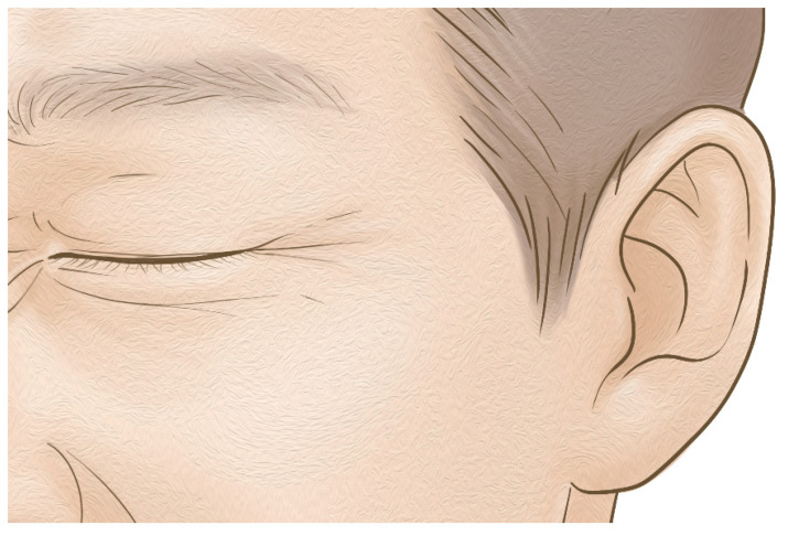
Botulinum neurotoxin injections near the lateral canthal rhytids are commonly used in cosmetic settings; however, there is a lack of thorough anatomical knowledge, and an effective way to treat them with accumulating knowledge is needed. The anatomical characteristics concerning the injection of botulinum neurotoxin into the orbicularis oculi muscle were evaluated in this review. Current knowledge on the identification of botulinum neurotoxin injection points from recent anatomical research was assessed. The lateral canthal lines are involved with the orbicularis oculi muscle and nearby anatomical structures, and the injection points can be more precisely defined. The best possible injection sites were provided, and the injection procedure was described. This review proposes evidence for injection sites associated with the surface anatomy of the orbicularis oculi muscles to enhance the effectiveness of easing lateral canthal rhytids.

Keywords: crow’s feet; facial wrinkle; infraorbital wrinkles; injection point; orbicularis oculi muscle.


Conflict of interest statement

The authors declare no conflict of interest.

Figures

Figure 1
Figure 1
The wrinkles in the eye region are lateral canthal rhytids in many individuals.
Figure 2
Figure 2
The external anatomical landmark of the eye regions. LOR, lateral orbital rim at the level of lateral canthus; S, sellion; Z, zygion; Zp, zygomatic point located on the outer orbital region; L, lateral canthus; IC, interciliary point located on the frontal notch.
Figure 3
Figure 3
Schematic image of the orbicularis oculi muscle. The orbicularis oculi muscle is divided into the orbital portion and the palpebral portion according to their position. The palpebral portion is subdivided into preseptal and pretarsal portions.
Figure 4
Figure 4
Dissected image of orbicularis orculi muscle. The muscle is composed of lateral and medial muscular band, preseptal and pretarsal part, and orbital portion.
Figure 5
Figure 5
The probe of the ultrasonography has been placed longitudinally at lateral orbital rim at the level of lateral canthus (A). The thin hypoechoic orbicularis oculi muscle observed in ultrasonography during resting state (B). The thick hypoechoic orbicularis oculi muscle is observed in contracted state when asked to smile (C).
Figure 6
Figure 6
The injection points for lateral canthal rhytids (Crow’s feet). The light blue dot represents dose of 2 U, while green dot represents dose of 1 U. The degree of the superior and inferior injection is by 30 degrees. The total is 5 U, which is a satisfactory dose for each side. LOR, lateral orbital rim at the level of lateral canthus.
Figure 7
Figure 7
The veins of periorbital regions should be delicately avoided with superficial injection sentinel vein (medial zygomaticotemporal vein) and inferior palpebral vein.
Figure 8
Figure 8
Four types of lateral canthal rhytids classified by kane. (A) full type, full dispersal of rhytid; (B) lower type, rhytid predominantly on the lower side of the lateral canthus; (C) lateral type, rhytids that are limited to the lateral canthus region and (D) upper type, rhytid predominantly on the upper side of the lateral canthus.

References

    1. Dessy L.A., Mazzocchi M., Rubino C., Mazzarello V., Spissu N., Scuderi N. An objective assessment of botulinum toxin A effect on superficial skin texture. Ann. Plast. Surg. 2007;58:469–473. doi: 10.1097/01.sap.0000244968.16977.1f. - DOI - PubMed
    1. Childers M.K. Targeting the neuromuscular junction in skeletal muscles. Am. J. Phys. Med. Rehabil. 2004;83:S38–S44. doi: 10.1097/01.PHM.0000141129.23219.42. - DOI - PubMed
    1. Wutthiphan S., Kowal L., O’Day J., Jones S., Price J. Diplopia following subcutaneous injections of botulinum A toxin for facial spasms. J. Pediatr. Ophthalmol. Strabismus. 1997;34:229–234. doi: 10.3928/0191-3913-19970701-10. - DOI - PubMed
    1. Lee J.J., Jeon H.S., Choi H.Y. Three Cases of Esotropia after Cosmetic Botulinum Toxin A Use in the Eyelid. J. Korean Ophthalmol. Soc. 2015;56:799–802. doi: 10.3341/jkos.2015.56.5.799. - DOI
    1. Chen C.S., Miller N.R. Botulinum toxin injection causing lateral rectus palsy. Br. J. Ophthalmol. 2007;91:843. doi: 10.1136/bjo.2006.109926. - DOI - PMC - PubMed

Publication types

Substances