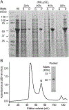
Preparation of carotenoid cleavage dioxygenases for X-ray crystallography
- PMID: 35878980
- PMCID: PMC10809780
- DOI: 10.1016/bs.mie.2021.10.020
Preparation of carotenoid cleavage dioxygenases for X-ray crystallography
Abstract
Carotenoid cleavage dioxygenases (CCDs) constitute a superfamily of enzymes that are found in all domains of life where they play key roles in the metabolism of carotenoids and apocarotenoids as well as certain phenylpropanoids such as resveratrol. Interest in these enzymes stems not only from their biological importance but also from their remarkable catalytic properties including their regioselectivity, their ability to accommodate diverse substrates, and the additional activities (e.g., isomerase) that some of these enzyme possess. X-ray crystallography is a key experimental approach that has allowed detailed investigation into the structural basis behind the interesting biochemical features of these enzymes. Here, we describe approaches used by our lab that have proven successful in generating single crystals of these enzymes in resting or ligand-bound states for high-resolution X-ray diffraction analysis.
Keywords: Apocarotenoid; Carotenoid; Cleavage dioxygenase; Cobalt; Crystallization; Enzyme; Metalloenzyme; Non-heme iron; X-ray crystallography.
Copyright © 2022 Elsevier Inc. All rights reserved.
Figures


Similar articles
-
Structural basis for carotenoid cleavage by an archaeal carotenoid dioxygenase.Proc Natl Acad Sci U S A. 2020 Aug 18;117(33):19914-19925. doi: 10.1073/pnas.2004116117. Epub 2020 Aug 3. Proc Natl Acad Sci U S A. 2020. PMID: 32747548 Free PMC article.
-
Enzymology of the carotenoid cleavage dioxygenases: reaction mechanisms, inhibition and biochemical roles.Arch Biochem Biophys. 2014 Feb 15;544:105-11. doi: 10.1016/j.abb.2013.10.005. Epub 2013 Oct 19. Arch Biochem Biophys. 2014. PMID: 24144525 Review.
-
Structural and mechanistic aspects of carotenoid cleavage dioxygenases (CCDs).Biochim Biophys Acta Mol Cell Biol Lipids. 2020 Nov;1865(11):158590. doi: 10.1016/j.bbalip.2019.158590. Epub 2019 Dec 23. Biochim Biophys Acta Mol Cell Biol Lipids. 2020. PMID: 31874225 Free PMC article. Review.
-
Key Residues for Catalytic Function and Metal Coordination in a Carotenoid Cleavage Dioxygenase.J Biol Chem. 2016 Sep 9;291(37):19401-12. doi: 10.1074/jbc.M116.744912. Epub 2016 Jul 24. J Biol Chem. 2016. PMID: 27453555 Free PMC article.
-
Analysis of carotenoid isomerase activity in a prototypical carotenoid cleavage enzyme, apocarotenoid oxygenase (ACO).J Biol Chem. 2014 May 2;289(18):12286-99. doi: 10.1074/jbc.M114.552836. Epub 2014 Mar 19. J Biol Chem. 2014. PMID: 24648526 Free PMC article.
Cited by
-
Spectroscopy and crystallography define carotenoid oxygenases as a new subclass of mononuclear non-heme FeII enzymes.J Biol Chem. 2025 May;301(5):108444. doi: 10.1016/j.jbc.2025.108444. Epub 2025 Mar 25. J Biol Chem. 2025. PMID: 40147775 Free PMC article.
-
Carotenoid cleavage enzymes evolved convergently to generate the visual chromophore.Nat Chem Biol. 2024 Jun;20(6):779-788. doi: 10.1038/s41589-024-01554-z. Epub 2024 Feb 14. Nat Chem Biol. 2024. PMID: 38355721 Free PMC article.
References
-
- Auldridge ME, Block A, Vogel JT, Dabney-Smith C, Mila I, Bouzayen M, et al. (2006). Characterization of three members of the Arabidopsis carotenoid cleavage dioxygenase family demonstrates the divergent roles of this multifunctional enzyme family. Plant Journal, 45(6), 982–993. - PubMed
-
- Brefort T, Scherzinger D, Limon MC, Estrada AF, Trautmann D, Mengel C, et al. (2011). Cleavage of resveratrol in fungi: characterization of the enzyme Rco1 from Ustilago maydis. Fungal Genetics and Biology, 48(2), 132–143. - PubMed
-
- Bruno M, Vermathen M, Alder A, Wust F, Schaub P, van der Steen R, et al. (2017). Insights into the formation of carlactone from in-depth analysis of the CCD8-catalyzed reactions. FEBS Letters, 591(5), 792–800. - PubMed
-
- Copeland RA (2000). Enzymes : a practical introduction to structure, mechanism, and data analysis (2nd ed.). New York: Wiley.
Publication types
MeSH terms
Substances
Grants and funding
LinkOut - more resources
Full Text Sources
Miscellaneous

