Review
. 2022 Jul 25;71(11):2337-2349.
doi: 10.1136/gutjnl-2021-326257. Online ahead of print.

Immunopathogenesis and environmental triggers in coeliac disease

Affiliations
Review

Immunopathogenesis and environmental triggers in coeliac disease

Anais Levescot et al. Gut. .

Abstract

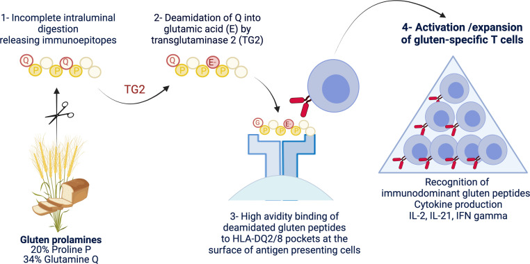
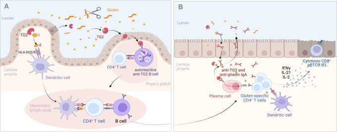
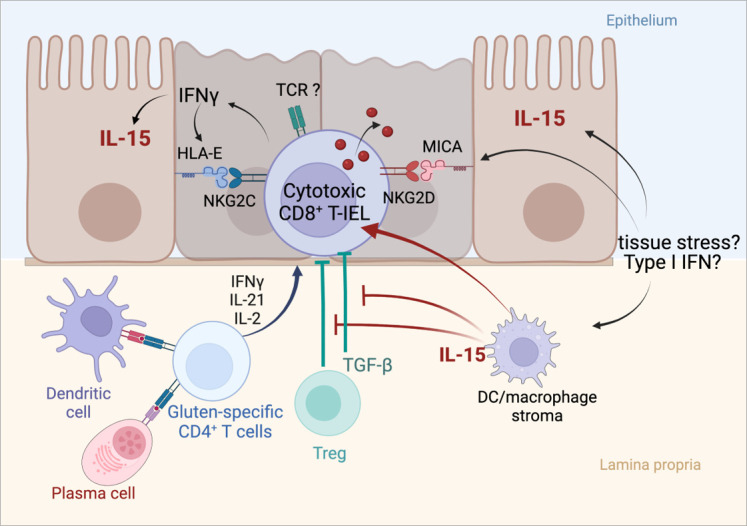
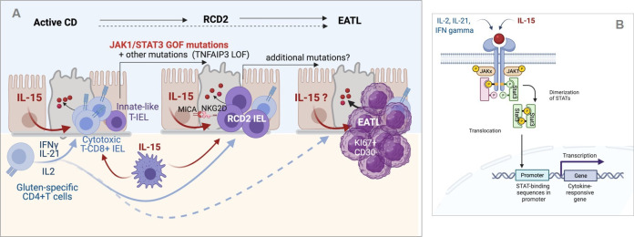
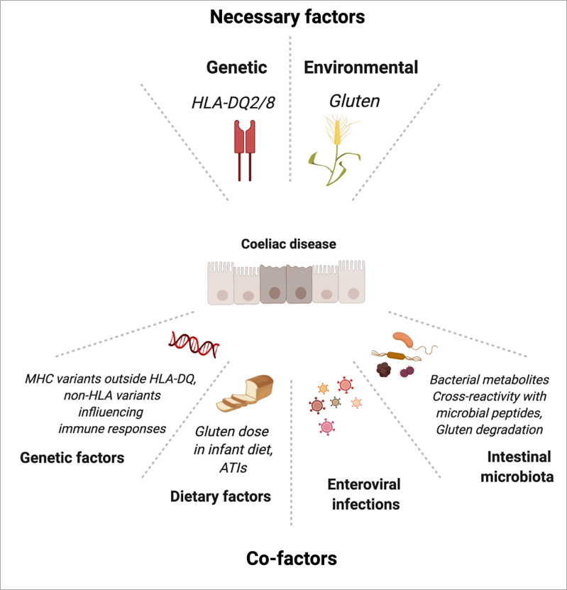
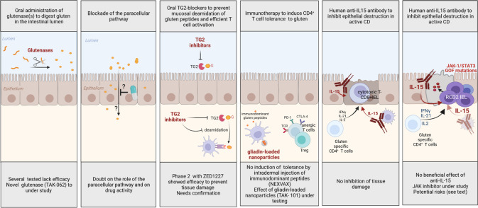
Coeliac disease (CD) is a frequent immune enteropathy induced by gluten in genetically predisposed individuals. Its pathogenesis has been extensively studied and CD has emerged as a model disease to decipher how the interplay between environmental and genetic factors can predispose to autoimmunity and promote lymphomagenesis. The keystone event is the activation of a gluten-specific immune response that is driven by molecular interactions between gluten, the indispensable environmental factor, HLA-DQ2/8, the main predisposing genetic factor and transglutaminase 2, the CD-specific autoantigen. The antigluten response is however not sufficient to induce epithelial damage which requires the activation of cytotoxic CD8+ intraepithelial lymphocytes (IEL). In a plausible scenario, cooperation between cytokines released by gluten-specific CD4+ T cells and interleukin-15 produced in excess in the coeliac gut, licenses the autoimmune-like attack of the gut epithelium, likely via sustained activation of the Janus kinase-signal transducer and activator of transcription (JAK/STAT) pathway in IEL. Demonstration that lymphomas complicating CD arise from IEL that have acquired gain-of-function JAK1 or STAT3 mutations stresses the key role of this pathway and explains how gluten-driven chronic inflammation may promote this rare but most severe complication. If our understanding of CD pathogenesis has considerably progressed, several questions and challenges remain. One unsolved question concerns the considerable variability in disease penetrance, severity and presentation, pointing to the role of additional genetic and environmental factors that remain however uneasy to untangle and hierarchize. A current challenge is to transfer the considerable mechanistic insight gained into CD pathogenesis into benefits for the patients, notably to alleviate the gluten-free diet, a burden for many patients.

Keywords: T lymphocytes; autoimmunity; coeliac disease; gluten sensitive enteropathy; lymphoma.


Conflict of interest statement

Competing interests: None declared.

Figures

Figure 1
Figure 1
Schematic representation of the driver antigluten response. The richness of gluten proteins in proline and glutamine underlies their ‘toxicity’ for patients with coeliac disease. Due to the lack of luminal endo-prolylpeptidases, proline-rich gluten proteins are incompletely digested and release large immunogenic peptides which can be modified by transglutaminase 2 (TG2). This enzyme selectively targets a subset of proline-rich and glutamine-rich sequences in gluten peptides and deamidate neutral glutamine residues (Q) into negatively charged glutamic acid (E). Gluten peptides harbouring negative charges on specific positions can bind with high avidity the peptide pockets of HLA-DQ2 or HLA-DQ8 molecules at the surface of antigen-presenting cells. The latter cells can then efficiently stimulate activation and expansion of gluten-specific CD4+ T cells that produce proinflammatory cytokines such as interleukin (IL)-2, interferon gamma (IFNγ) and IL-21. Created with Biorender.com.
Figure 2
Figure 2
Proposed mechanisms of the antigluten and antitransglutaminase 2 (TG2) responses in the coeliac intestine. (A) Initiation of the adaptive immune response against gluten and TG2. Gluten peptides left undigested are deamidated by TG2 either in the intestinal lumen where TG2 can be released by extruded dead enterocytes or after crossing the epithelium by TG2 liberated in the extracellular matrix. Deamidated peptides bind HLA-DQ2/8 molecules at the surface of dendritic cells in Peyer’s patches (not shown) or in lamina propria. Dendritic cells can then initiate the activation of gluten-specific T cells in the Peyer’s patches (not shown) or after migration in the mesenteric lymph nodes. Gluten-specific CD4+ T cells cooperate with naïve antigluten B cells to induce the antigluten humoral response. Alternatively, TG2-gluten complexes may cross the Peyer’s patch epithelium and bind to the B cell receptor of autoreactive anti-TG2 B cells present in the lymphoid follicle. TG2-gluten complexes bound to the B cell receptor are endocytosed, processed in B cell endolysosomal compartments, where released gluten peptides can be loaded on HLA-DQ molecules. HLA-DQ-gluten complexes are then translocated at the surface of autoreactive B cells, allowing the activation of gluten-specific CD4+ T cells and simultaneously cooperative interactions that license the IgA response against TG2. (B) Activation of the effector antigliadin CD4+ T cell response. Following their activation in gut lymphoid tissues and mesenteric lymph nodes, gluten-specific effector CD4+ T cells, antigluten and anti-TG2 IgA plasma blasts migrate into the lamina propria. On re-encounter with HLA-DQ-gluten complexes displayed by dendritic cells and by anti-TG2 plasma cells, effector CD4+ T cells secrete cytokines and notably interferon gamma (IFNγ), interleukin (IL)-21 and IL-2, which participate in the activation of CD8+ cytotoxic T intraepithelial lymphocytes (IEL). Plasma cells produce IgA antibodies against gluten and TG2 antibodies that are transported into the intestinal lumen. IgA-gluten and IgA-TG2-gluten complexes may be retrotransported across the epithelium and further foster the antigluten response. Created with Biorender.com.
Figure 3
Figure 3
Activation of cytotoxic intraepithelial lymphocytes and induction of tissue damage in active coeliac disease (CD). Cooperation between the cytokines released by activated gluten-specific CD4+ T cells and interleukin (IL)-15 produced by epithelial and myeloid cells drive the activation of cytotoxic CD8+ T intraepithelial lymphocytes (IEL) and license epithelial cell killing. Cytotoxic CD8+ T IEL synthesise interferon gamma (IFNγ) and cytotoxic molecules such as granzyme B and perforin, upregulate the activating natural killer receptor NKG2D and CD94/NKG2C, thereby facilitating the interactions of cytotoxic CD8+ T IEL with enterocytes which, in active CD, display MICA (MHC class I polypeptide-related sequence A), the ligand of NKG2D (induced by tissue stress) and HLA-E, the ligand of NKG2C (induced by IFNγ). DC, dendritic cell; TCR, T cell receptor; TGF-β, transforming growth factor beta; Treg, regulatory T cell. Created with Biorender.com.
Figure 4
Figure 4
Inflammation-driven lymphomagenesis in coeliac disease (CD). (A) Left panel: in active CD, cytokines produced by gluten-specific CD4+ T cells cooperate with interleukin (IL)-15 to stimulate the expansion and cytotoxic activation of polyclonal CD8+ T intraepithelial lymphocytes (IEL) that drive a cytolytic attack of epithelium. Middle panel: in type 2 refractory CD (RCD2), innate-like T IEL that have acquired somatic mutations and notably gain-of-function (GOF) mutations in Janus kinase 1 (JAK1) or signal transducer and activator of transcription 3 (STAT3) can clonally expand and outcompete CD8+ T IEL in the cytokine-rich CD intestine. Transformation is fostered by the acquisition of additional mutations, notably in TNFAIP/A20, which enhances the nuclear factor kappa B pathway, and in epigenetic regulators. Due to their natural killer (NK)-like functions, RCD2 IEL can induce severe epithelial lesions. Right panel: further accumulation of mutations in the clonally expanded RCD2 IEL ultimately lead to enteropathy-associated T cell lymphoma (EATL). While EATL generally develops from RCD2, it can also develop without a prior step of RCD2. (B) Schematic representation of the JAK1-STAT3 pathway. Its activation both by cytokines produced by CD4+ T cells and IL-15 explains its importance in the activation of normal CD8+ T IEL and RCD2 IEL. Created with Biorender.com.
Figure 5
Figure 5
Genetic and environmental factors in coeliac disease. ATI, amylase trypsin inhibitor; MHC, major histocompatibility complex. Created with Biorender.com.
Figure 6
Figure 6
Proposed rationale-based therapies for coeliac disease (CD). GOF, gain-of-function; IFN, interferon; IL, interleukin; JAK, Janus kinase; TG2, tissue transglutaminase 2. Created with BioRender.com.

References

    1. Al-Toma A, Volta U, Auricchio R, et al. . European Society for the study of coeliac disease (ESsCD) guideline for coeliac disease and other gluten-related disorders. United European Gastroenterol J 2019;7:583–613. 10.1177/2050640619844125 - DOI - PMC - PubMed
    1. Singh P, Arora A, Strand TA, et al. . Global Prevalence of Celiac Disease: Systematic Review and Meta-analysis. Clin Gastroenterol Hepatol 2018;16:823–36. 10.1016/j.cgh.2017.06.037 - DOI - PubMed
    1. Malamut G, Cording S, Cerf-Bensussan N. Recent advances in celiac disease and refractory celiac disease. F1000Research 2019;8:969. 10.12688/f1000research.18701.1 - DOI - PMC - PubMed
    1. Jabri B, Sollid LM. T cells in celiac disease. J.i. 2017;198:3005–14. 10.4049/jimmunol.1601693 - DOI - PMC - PubMed
    1. Abadie Valérie, Kim SM, Lejeune T, et al. . Il-15, gluten and HLA-DQ8 drive tissue destruction in coeliac disease. Nature 2020;578:600–4. 10.1038/s41586-020-2003-8 - DOI - PMC - PubMed