. 2022 Jul;54(7):1024-1037.
doi: 10.1038/s12276-022-00806-z. Epub 2022 Jul 25.

Circular RNA ROCK1, a novel circRNA, suppresses osteosarcoma proliferation and migration via altering the miR-532-5p/PTEN axis

Affiliations

Circular RNA ROCK1, a novel circRNA, suppresses osteosarcoma proliferation and migration via altering the miR-532-5p/PTEN axis

Yize Liu et al. Exp Mol Med. 2022 Jul.

Abstract

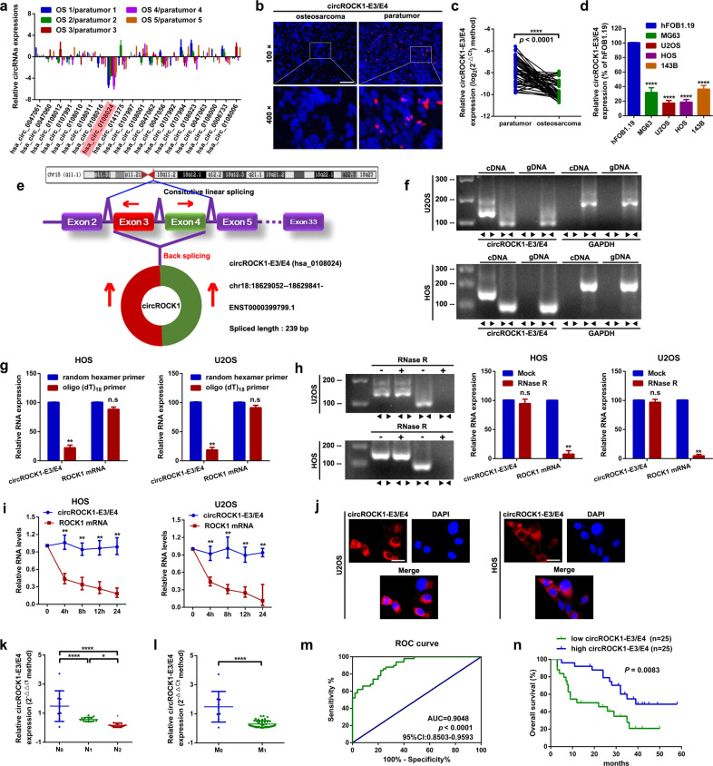
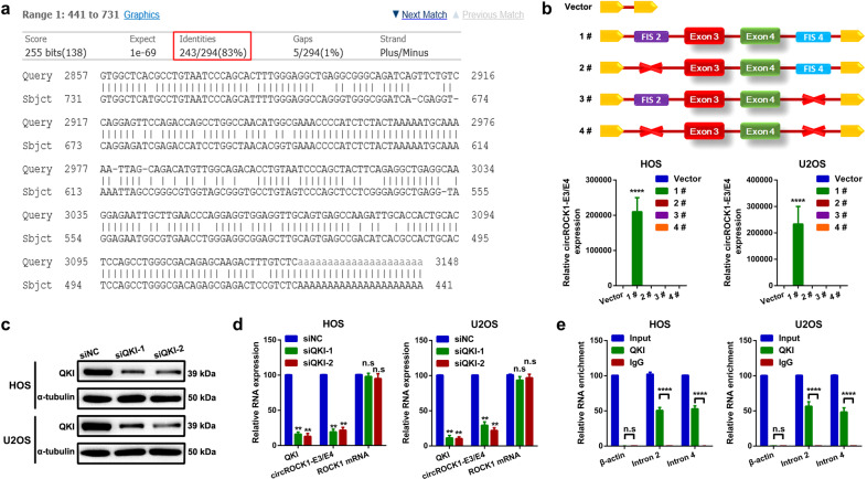
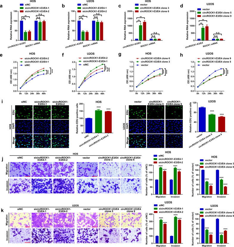
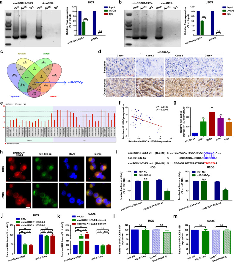
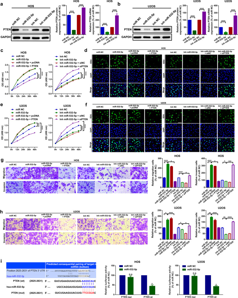
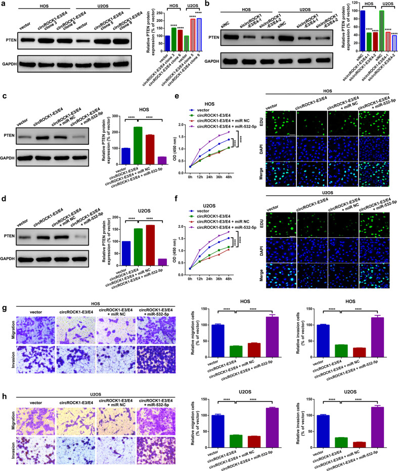
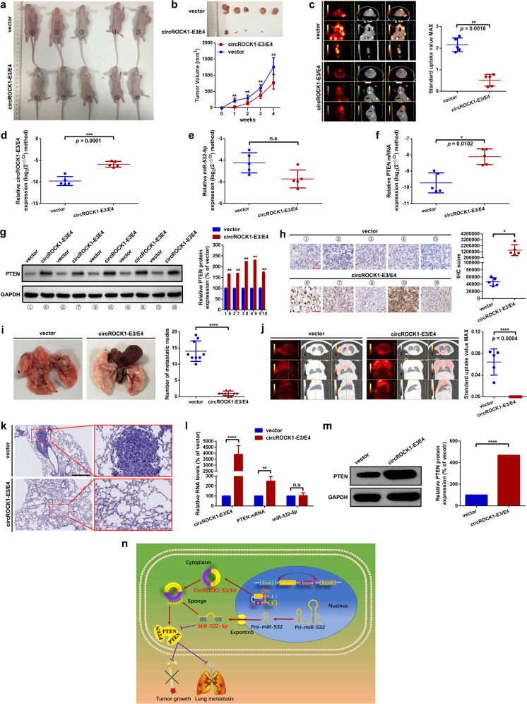
As the most prevalent bone tumor in children and adolescents, the pathogenesis and metastasis of osteosarcoma (OS) remain largely unclear. Here, we investigated the expression and function of a novel circular RNA (circRNA), circROCK1-E3/E4, which is back-spliced from exons 3 and 4 of Rho-associated coiled-coil containing protein kinase 1 (ROCK1) in OS. We found that circROCK1-E3/E4, regulated by the well-known RNA-binding protein quaking (QKI), was downregulated in OS and correlated with unfavorable clinical features of patients with OS. Functional proliferation and cell motility assays indicated that circROCK1-E3/E4 serves as a tumor suppressor in OS cells. Mechanistically, circROCK1-E3/E4 suppressed proliferation and migration by upregulating phosphatase and tensin homolog (PTEN) through microRNA-532-5p (miR-532-5p) sponging. In the constructed nude mouse model, circROCK1-E3/E4 inhibited tumor growth and lung metastasis in vivo. This study demonstrates the functions and molecular mechanisms of circROCK1-E3/E4 in the progression of OS. These findings may identify novel targets for the molecular therapy of OS.


Conflict of interest statement

The authors declare no competing interests.

Figures

Fig. 1
Fig. 1. CircROCK1-E3/E4 is downregulated and associated with poor prognosis in patients with osteosarcoma.
a The expression of certain ROCK1-derived circRNAs in osteosarcoma tissue specimens and paired paratumor tissue specimens was determined by an RT–qPCR assay. b Representative images of circROCK1-E3/E4 expression in osteosarcoma and paratumor tissues as displayed by a FISH assay. c The expression of circROCK1 in 50 osteosarcoma and paired paratumor tissue specimens was detected by RT–qPCR assay. ****p < 0.0001. d CircROCK1-E3/E4 was downregulated in osteosarcoma cell lines, as confirmed by an RT–qPCR assay. ****p < 0.0001. e CircROCK1-E3/E4 is circularized from exons 3 and 4 in ROCK1, as demonstrated by a schematic illustration. f CircROCK1-E3/E4 was amplified by divergent primers in cDNA but not gDNA by using GAPDH as a negative control, as validated by an RT–qPCR assay. g Compared with oligo (dT)18 primers, the expression of circROCK1-E3/E4 was significantly decreased when random hexamer primers were applied. n.sp > 0.05 and **p < 0.01. h Compared with linear ROCK1 mRNA, the expression changes of circROCK1-E3/E4 were unremarkable in the presence of RNase R. n.sp > 0.05 and **p < 0.01. i The RNA levels of circROCK1-E3/E4 and ROCK1 mRNA after 2 μg/ml actinomycin D treatment were quantified at different time points by qRT–PCR assays. **p < 0.01. j CircROCK1-E3/E4 was mainly localized in the cytoplasm of U2OS and HOS cells. k CircROCK1-E3/E4 is frequently downregulated in patients with lymph node metastasis (N1 and N2). *p < 0.05, ****p < 0.0001. l CircROCK1-E3/E4 is downregulated in patients with distant metastasis (M1 stage). ****p < 0.0001. m ROC curve analysis of the area under the curve (AUC) and 95% confidence interval (CI) of circROCK1-E3/E4 in patients with osteosarcoma. n A low expression level of circROCK1-E3/E4 was correlated with shorter survival rates in patients with osteosarcoma as determined by Kaplan–Meier analysis. All data are presented as the mean ± SD from three independent experiments.
Fig. 2
Fig. 2. CircROCK1-E3/E4 expression is partially regulated by QKI.
a The complementary sequences of intron 2 of ROCK1 were blasted with reverse sequences of intron 4 of ROCK1. Very highly similar sequences (83% identity over 294 nt) were found, as illustrated by using BLAST (https://blast.ncbi.nlm.nih.gov/Blast.cgi). b Four vectors (vector #1 containing FIS2 and FIS4; vector #2 with FIS2 deletion; vector #3 with FIS4 deletion; vector #4 with FIS2 and FIS4 deletion) were transfected into U2OS and HOS cells, and the expression level of circROCK1-E3/E4 was checked by an RT–qPCR assay. ****p < 0.0001. c QKI was knocked down as determined by a western blot assay. d Expression of QKI mRNA, circROCK1-E3/E4 and ROCK1 mRNA after transfection of QKI siRNAs was analyzed by an RT–qPCR assay. n.sp > 0.05 and **p < 0.01. e By using IgG as a control, partial segments of intron 2 and intron 4 of ROCK1 rather than β-actin were abundantly enriched in QKI, as measured by an RIP assay. n.sp > 0.05 and ****p < 0.0001. All data are presented as the mean ± SD from three independent experiments.
Fig. 3
Fig. 3. CircROCK1-E3/E4 suppresses osteosarcoma cell proliferation and migration in vitro.
a, b CircROCK1-E3/E4 expression levels after transfection of specific siRNAs were measured by an RT–qPCR assay. n.sp > 0.05 and **p < 0.01. c, d Expression levels of circROCK1-E3/E4 and ROCK1 mRNA were analyzed by an RT–qPCR assay. n.sp > 0.05 and **p < 0.01. eh Changes in cell proliferation ability after various circROCK1-E3/E4 interventions were determined by a CCK8 assay. ****p < 0.0001. i An EdU assay was performed to evaluate changes in cell proliferation ability after up- and downregulation of circROCK1-E3/E4. ****p < 0.0001. j, k Cell motility ability changes after different circROCK1-E3/E4 interventions were determined by a transwell assay. ****p < 0.0001. All data are presented as the mean ± SD from three independent experiments.
Fig. 4
Fig. 4. CircROCK1-E3/E4 sponges miR-532-5p in U2OS and HOS cells.
a, b Compared with circANRIL, circROCK1 was abundantly enriched in AGO2 by using IgG as an internal control. n.sp > 0.05 and ***p < 0.001 c A Venn diagram exhibiting overlapping miRNAs that might interact with circROCK1-E3/E4 and presented in GSE65071. d Expression of miR-532-5p in osteosarcoma tissues was illustrated by an in situ hybridization assay. e Expression of miR-532-5p in osteosarcoma tissues and paired healthy tissues in the osteosarcoma-related genome-wide study GSE65071. Probe ID 49 represents miR-532-5p. f Spearman correlation analysis showed that circROCK1-E3/E4 was inversely correlated with miR-532-5p. r = –0.5406 and p < 0.0001. g Expression levels of miR-532-5p in osteosarcoma cell lines were confirmed by an RT–qPCR assay. h The subcellular localization of circROCK1-E3/E4 and miR-532-5p was confirmed by a FISH assay. i The targeted binding effect and binding sites between circROCK1-E3/E4 and miR-532-5p were tested by a luciferase assay. n.sp > 0.05 and *p < 0.05. j, k Expression of miR-532-5p after up- and downregulation of circROCK1-E3/E4 was analyzed by RT–qPCR. n.sp > 0.05 and **p < 0.01. l, m Relative expression of circROCK1-E3/E4 after different miR-532-5p interventions was measured by RT–qPCR. n.sp > 0.05. All data are presented as the mean ± SD from three independent experiments.
Fig. 5
Fig. 5. MiR-532-5p promotes proliferation and migration by targeting PTEN in U2OS and HOS cells.
a, b Expression of PTEN after treatment with different miR-532-5p concentrations was determined by a western blot assay and an RT–qPCR assay. ****p < 0.0001. cf Cell proliferation ability changes in HOS and U2OS cells were determined by CCK8 and EdU assays, respectively. ****p < 0.0001. g, h Cell migration and invasion ability changes in HOS and U2OS cells were measured by a transwell assay. *p < 0.05, **p < 0.01, ***p < 0.001, and ****p < 0.0001. i The targeted binding effect between miR-532-5p and PTEN was confirmed by a luciferase assay. The left panel shows the online prediction result by using TargetScan (http://www.targetscan.org/vert_72/), and the middle and right panels display the results of relative luminance changes. n.sp > 0.05 and *p < 0.05. All data are presented as the mean ± SD from three independent experiments.
Fig. 6
Fig. 6. CircROCK1-E3/E4 suppresses proliferation and migration via regulation of the miR-532-5p/PTEN axis in U2OS and HOS cells.
a, b Expression of PTEN protein was analyzed by western blot assay. ****p < 0.0001. c, d PTEN protein expression after different circROCK1-E3/E4 and miR-532-5p interventions was measured by western blot. ****p < 0.0001. e, f Cell proliferation ability changes were checked by a CCK8 assay and an EdU assay. ****p < 0.0001. g, h Cell motility changes were evaluated by a transwell assay. ****p < 0.0001. All data are presented as the mean ± SD from three independent experiments.
Fig. 7
Fig. 7. CircROCK1-E3/E4 suppresses proliferation and lung metastasis in vivo.
a Macroscopic appearance of nude mice injected with osteosarcoma cells with stable overexpression of circROCK1-E3/E4 or with control cells (vector). b Overexpression of circROCK1-E3/E4 obviously restricted subcutaneous nodule formation. **p < 0.01. c Representative 18F-FDG micro positron emission tomography/computed tomography (PET/CT) images of formatted subcutaneous nodes in mice with diverse circROCK1-E3/E4 expression. p = 0.0016. df Expression of circROCK1-E3/E4, miR-532-5p and PTEN mRNA in the formatted subcutaneous nodes was analyzed by RT–qPCR. n.sp > 0.05, and p-values are presented in the figures accordingly. g, h PTEN protein expression in formatted subcutaneous nodes was determined by Western blot and IHC. *p < 0.05 and **p < 0.01. i Macroscopic appearance of formatted lung metastatic nodes in mice. ****p < 0.0001. j Representative 18F-FDG micro positron emission tomography/computed tomography (PET/CT) images of formatted lung metastatic nodes in mice with diverse circROCK1-E3/E4 expression. p = 0.0004. k H&E staining of formatted lung metastatic nodes; scale bars, 200 and 50 µm for magnifications of ×100 and ×400, respectively. l Expression levels of circROCK1-E3/E4, miR-532-5p, and PTEN mRNA in the formatted lung metastatic nodes were analyzed by RT–qPCR. n.sp > 0.05, **p < 0.01, and ****p < 0.0001. m PTEN protein expression in formatted lung metastatic nodes was determined by western blot. ****p < 0.0001. n Diagram indicating that circROCK1-E3/E4, back-spliced from exons 3 and exon 4 of the ROCK1 gene, which is regulated by QKI, suppressed cell proliferation and migration by altering the miR-532-5p/PTEN axis in osteosarcoma. All data are presented as the mean ± SD from three independent experiments.

Similar articles

Cited by

References

    1. Philip T, et al. Osteosarcoma. Br. J. Cancer. 2001;84:78–80. doi: 10.1054/bjoc.2000.1770. - DOI - PMC - PubMed
    1. Ottaviani G, Jaffe N. The epidemiology of osteosarcoma. Cancer Treat. Res. 2009;152:3–13. doi: 10.1007/978-1-4419-0284-9_1. - DOI - PubMed
    1. Li X, Yang L, Chen LL. The Biogenesis, Functions, and Challenges of Circular RNAs. Mol. Cell. 2018;71:428–442. doi: 10.1016/j.molcel.2018.06.034. - DOI - PubMed
    1. Memczak S, et al. Circular RNAs are a large class of animal RNAs with regulatory potency. Nature. 2013;495:333–338. doi: 10.1038/nature11928. - DOI - PubMed
    1. Hansen TB, et al. Natural RNA circles function as efficient microRNA sponges. Nature. 2013;495:384–388. doi: 10.1038/nature11993. - DOI - PubMed

Publication types