Review
. 2023 Mar;23(3):159-173.
doi: 10.1038/s41577-022-00760-x. Epub 2022 Jul 25.

Mitochondrial control of inflammation

Affiliations
Review

Mitochondrial control of inflammation

Saverio Marchi et al. Nat Rev Immunol. 2023 Mar.

Abstract

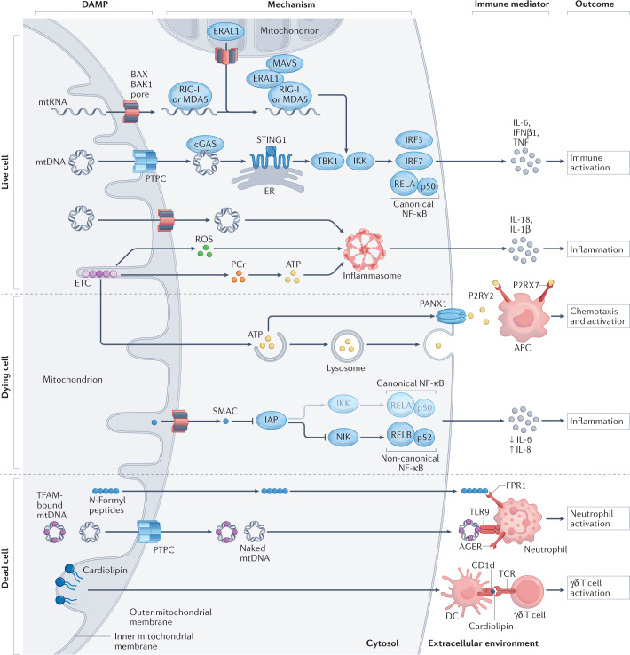
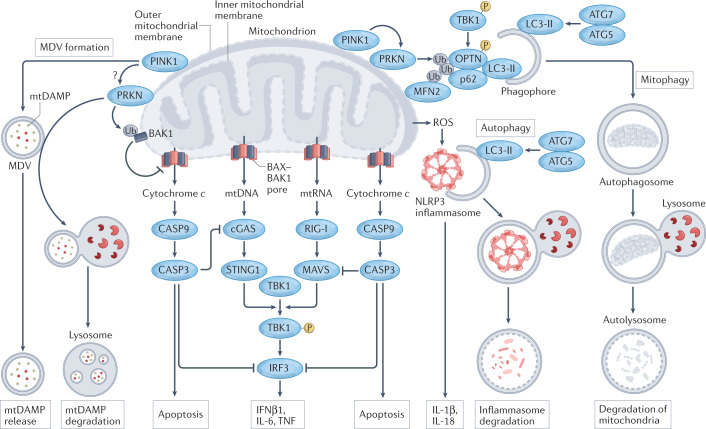
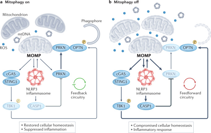
Numerous mitochondrial constituents and metabolic products can function as damage-associated molecular patterns (DAMPs) and promote inflammation when released into the cytosol or extracellular milieu. Several safeguards are normally in place to prevent mitochondria from eliciting detrimental inflammatory reactions, including the autophagic disposal of permeabilized mitochondria. However, when the homeostatic capacity of such systems is exceeded or when such systems are defective, inflammatory reactions elicited by mitochondria can become pathogenic and contribute to the aetiology of human disorders linked to autoreactivity. In addition, inefficient inflammatory pathways induced by mitochondrial DAMPs can be pathogenic as they enable the establishment or progression of infectious and neoplastic disorders. Here we discuss the molecular mechanisms through which mitochondria control inflammatory responses, the cellular pathways that are in place to control mitochondria-driven inflammation and the pathological consequences of dysregulated inflammatory reactions elicited by mitochondrial DAMPs.


Conflict of interest statement

L.G. has held research contracts with Lytix Biopharma and Promontory, has received consulting/advisory honoraria from Boehringer Ingelheim, AstraZeneca, OmniSEQ, Onxeo, The Longevity Labs, Inzen, Sotio, Promontory, Noxopharm, EduCom and the Luke Heller TECPR2 Foundation, and holds Promontory stock options. All other authors declare no conflicts of interest.

Figures

Fig. 1
Fig. 1. Main mechanisms of mitochondrial DAMP signalling.
Various mitochondrial components and products that are released as a consequence of mitochondrial dysfunction (and potentially cell death) drive inflammatory responses upon accumulation in the cytosol or the extracellular environment. Mitochondrial DNA (mtDNA), which can exit mitochondria via BCL-2-associated X, apoptosis regulator (BAX) and BCL-2 antagonist/killer 1 (BAK1) pores or via the permeability transition pore complex (PTPC), is a potent activator of cyclic GMP–AMP synthase (cGAS), resulting in stimulator of interferon response cGAMP interactor 1 (STING1) signalling and consequent synthesis of cytokines such as interferon-β1 (IFNβ1), IL-6 and tumour necrosis factor (TNF). Cytosolic mitochondrial RNA (mtRNA) has similar effects, although they depend on retinoic acid-inducible gene I protein (RIG-I), melanoma differentiation-associated protein 5 (MDA5) and mitochondrial antiviral signalling protein (MAVS). This pathway is promoted by the BAX–BAK1-dependent release of Era-like 12S mitochondrial rRNA chaperone 1 (ERAL1), which favours MAVS stabilization at the mitochondrial surface. Moreover, upon release from dysfunctional mitochondria, both mtDNA and reactive oxygen species (ROS) can drive IL-1β and IL-18 secretion as a consequence of inflammasome signalling. Electron transport chain (ETC) functions also seem to affect inflammasome activation independently of ROS as they preserve intracellular ATP availability through phosphocreatine (PCr). ATP can be released by dying cells through lysosomal secretion and pannexin 1 (PANX1) channels, mediating both chemotactic and immunostimulatory effects on antigen-presenting cells (APCs) by binding to the purinergic receptors P2RX7 and P2RY2. Upon BAX–BAK1 oligomerization during apoptosis, diablo IAP-binding mitochondrial protein (DIABLO; best known as SMAC) release not only favours caspase activation (not shown) but also rewires NF-κB signalling from canonical to non-canonical programmes. Along similar lines, mtDNA (be it naked or complexed with the mitochondrial transcription factor TFAM) and N-formyl peptides (other mitochondrial components), which accumulate in the extracellular milieu upon regulated cell death, cause neutrophil activation via Toll-like receptor 9 (TLR9) or advanced glycosylation end product-specific receptor (AGER) and formyl peptide receptor 1 (FPR1), respectively. Finally, extracellular cardiolipin (which is normally restricted to the inner mitochondrial membrane) can be presented by dendritic cells (DCs) on the MHC class I-like molecule CD1d, resulting in the activation of γδ T cells. DAMP, damage-associated molecular pattern; ER, endoplasmic reticulum; IKK, IκB kinase; IRF, interferon regulatory factor; NIK (official name MAP3K14), mitogen-activated protein kinase kinase kinase 14; TBK1, TANK-binding kinase 1; TCR, T cell receptor.
Fig. 2
Fig. 2. Roles of apoptosis and autophagy in the inhibition of inflammatory responses elicited by mitochondria.
Intrinsic apoptosis proceeds with the formation of BCL-2-associated X, apoptosis regulator (BAX) and BCL-2 antagonist/killer 1 (BAK1) pores at the outer mitochondrial membrane, resulting in the cytosolic accumulation of cytochrome c and consequent activation of apoptotic caspases. Caspase 9 (CASP9)-driven CASP3 activation inhibits inflammatory responses elicited by dysfunctional mitochondria by catalysing the cleavage of cyclic GMP–AMP synthase (cGAS), mitochondrial antiviral signalling protein (MAVS) and interferon regulatory factor 3 (IRF3). A similar inhibitory effect results from the disposal of dysfunctional mitochondria via PTEN-induced putative kinase 1 (PINK1)- and parkin (PRKN)-dependent mitophagy. This is promoted (at least initially) by the capacity of TANK-binding kinase 1 (TBK1) — which is activated by phosphorylation (P) following cGAS-driven stimulator of interferon response cGAMP interactor 1 (STING1) signalling and retinoic acid-inducible gene I (RIG-I)-driven MAVS signalling downstream of the cytosolic accumulation of mitochondrial DNA (mtDNA) or mitochondrial RNA (mtRNA) — to phosphorylate optineurin (OPTN) and hence improve the ability of OPTN to recruit sequestosome 1 (SQSTM1; best known as p62) to ubiquitylated proteins at the outer mitochondrial membrane such as mitofusin 2 (MFN2). Engagement of p62 culminates in the recruitment of forming autophagosomes via lipidated microtubule-associated protein 1 light chain 3 beta (MAP1LC3B; best known as LC3-II in its lipidated form). Of note, PRKN also mediates the ubiquitylation (Ub)-dependent inactivation of BAK1. Moreover, PRKN-dependent mitophagy seems to prevent mitochondrial damage-associated molecular patterns (mtDAMPs) from being incorporated in the mitochondria-derived vesicles (MDVs) that are normally released as part of mitochondrial quality control in a PINK1-dependent manner. The underlying mechanisms, however, remain to be fully elucidated. General autophagy can likewise suppress inflammatory responses driven by mitochondrial dysfunction, at least in part reflecting its ability to degrade NLR family pyrin domain-containing 3 (NLRP3) inflammasomes. ATG, autophagy-related protein; IFNβ1, interferon-β1; ROS, reactive oxygen species; TNF, tumour necrosis factor.
Fig. 3
Fig. 3. The mitophagy rheostat in the control of mitochondria-driven inflammation.
a | When only a small number of mitochondria are permeabilized, limited signalling via cyclic GMP–AMP synthase (cGAS) and the NLR family pyrin domain-containing 3 (NLRP3) inflammasome promotes mitophagy associated with the recruitment of parkin (PRKN) to dysfunctional mitochondria, TANK-binding kinase 1 (TBK1)-dependent optineurin (OPTN) phosphorylation (P) and consequent engulfment of mitochondria by forming autophagosomes. This engages a feedback mechanism that enables the restoration of cellular homeostasis in the absence of robust inflammatory responses. b | Conversely, when the functions and integrity of a large number of mitochondria are compromised, robust cGAS and NLRP3 inflammasome signalling is accompanied by mitophagy inhibition — as a consequence of caspase 1 (CASP1)-dependent cleavage of PRKN, despite OPTN phosphorylation — and increased NLRP3-dependent mitochondrial dysfunction, resulting in a feedforward loop that maximizes inflammation in the context of lost cellular homeostasis. Taken together, these mechanisms identify a rheostat that determines a threshold for recovered cellular homeostasis in the context of suppressed inflammation versus compromised cellular homeostasis in the context of acute inflammatory responses. MOMP, mitochondrial outer membrane permeabilization; mtDNA, mitochondrial DNA; ROS, reactive oxygen species; STING1, stimulator of interferon response cGAMP interactor 1.

Similar articles

Cited by

References

    1. Roda G, et al. Crohn’s disease. Nat. Rev. Dis. Prim. 2020;6:22. doi: 10.1038/s41572-020-0156-2. - DOI - PubMed
    1. Tansey MG, et al. Inflammation and immune dysfunction in Parkinson disease. Nat. Rev. Immunol. 2022 doi: 10.1038/s41577-022-00684-6. - DOI - PMC - PubMed
    1. Stark K, Massberg S. Interplay between inflammation and thrombosis in cardiovascular pathology. Nat. Rev. Cardiol. 2021;18:666–682. doi: 10.1038/s41569-021-00552-1. - DOI - PMC - PubMed
    1. Basso PJ, Andrade-Oliveira V, Camara NOS. Targeting immune cell metabolism in kidney diseases. Nat. Rev. Nephrol. 2021;17:465–480. doi: 10.1038/s41581-021-00413-7. - DOI - PubMed
    1. Marchi S, Morroni G, Pinton P, Galluzzi L. Control of host mitochondria by bacterial pathogens. Trends Microbiol. 2022;30:452–465. doi: 10.1016/j.tim.2021.09.010. - DOI - PubMed

Publication types