Review
. 2022 Jul;4(7):813-825.
doi: 10.1038/s42255-022-00610-z. Epub 2022 Jul 25.

Glial cells as integrators of peripheral and central signals in the regulation of energy homeostasis

Affiliations
Review

Glial cells as integrators of peripheral and central signals in the regulation of energy homeostasis

Sreekala Nampoothiri et al. Nat Metab. 2022 Jul.

Abstract

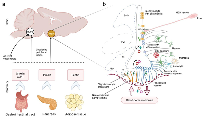
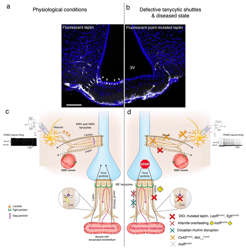
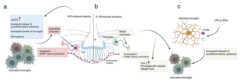
Communication between the periphery and the brain is key for maintaining energy homeostasis. To do so, peripheral signals from the circulation reach the brain via the circumventricular organs (CVOs), which are characterized by fenestrated vessels lacking the protective blood-brain barrier (BBB). Glial cells, by virtue of their plasticity and their ideal location at the interface of blood vessels and neurons, participate in the integration and transmission of peripheral information to neuronal networks in the brain for the neuroendocrine control of whole-body metabolism. Metabolic diseases, such as obesity and type 2 diabetes, can disrupt the brain-to-periphery communication mediated by glial cells, highlighting the relevance of these cell types in the pathophysiology of such complications. An improved understanding of how glial cells integrate and respond to metabolic and humoral signals has become a priority for the discovery of promising therapeutic strategies to treat metabolic disorders. This Review highlights the role of glial cells in the exchange of metabolic signals between the periphery and the brain that are relevant for the regulation of whole-body energy homeostasis.


Conflict of interest statement

Competing interest

The authors have no competing interests to declare.

Figures

Figure 1
Figure 1. The cross-talk between the brain and the peripheral organs via metabolic signals for energy homeostasis.
(a) The peripheral signals from the visceral organs (such as ghrelin and GLP1 from the gastrointestinal tract, insulin from the pancreas and leptin from the adipose tissue shown here or other metabolites and regulators discussed in section 3.2.2) reach two key regions of the brain: the MBH via pituitary-portal circulation, and/or the DVC constituting the AP and NTS via the circulation or through the sensory vagus nerve. (b) Schematic representation of the transport of blood-borne molecules to the MBH via the fenestrated capillaries of the ME. Tanycytes at the floor of the third ventricle (ME tanycytes) adapt their plasticity to permit the passage of molecules. Tanycytes at the ARH and VMH sense the uptake of peripheral signals and transmit them to the neighbouring neurons. Astrocytes at the interface of BBB capillaries also sense and transmit the systemic metabolic information to neurons. MCH neuronal projections from the LHA release MCH at the base or the pole of ependymal cells controlling ciliary beat frequency. AP, area postrema; ARH, arcuate nucleus of the hypothalamus; BBB, blood-brain barrier; CVO, circumventricular organ; DVC, dorsal vagus complex; LHA, lateral hypothalamic area; MBH, mediobasal hypothalamus; MCH, melanin-concentrating hormone; ME, median eminence; NTS, nucleus of the solitary tract; VMH, ventromedial nucleus of the hypothalamus.
Figure 2
Figure 2. Leptin and other circulating signals are transported from the periphery to hypothalamic and extra-hypothalamic areas around the 3V by tanycytes.
(a) Representative photomicrographs with fluorescent leptin (25 nmol/mouse) labelled tanycytic processes (arrows) and cell bodies (arrowheads; white labeling), where fluorescent bioactive leptin was administered intravenously in wild-type mice. (b) Similar experiment using fluorescent point-mutated leptin and showing that the mutated leptin is taken up by tanycytes, but remains blocked at the tanycytic end-feet (asterisk) due to its inability to stimulate LepR-mediated EGFR activation, as detailed in the schematics (c,d). The cell nuclei are counterstained with Hoechst solution. Adapted with permission from. (c) Schematic showing tanycytes as the gateway for the passage of leptin and other blood-borne metabolic signals from fenestrated blood vessels to the CSF of the 3V. Tanycytes of the ARH sense CSF-borne molecules transported by tanycytes from the periphery, translate and transmit the information to neurons under the influence of the tanycytic network. (d) Diseased state or the experimental conditions in which mechanisms related to leptin transport,, insulin, ghrelin transport, or action are altered in tanycytes. Cx43 gap junction–mediated tanycytic metabolic networks are required for the transport of lactate, produced and released by tanycytes, to glucose-insensitive POMC neurons. Tanycytes also play a role in the control of daily circadian changes. 3V, third ventricle; ARH, arcuate nucleus of the hypothalamus; Cx43, connexin 43; DIO, diet-induced obesity; Egfr, epidermal growth factor receptor; Insr, insulin receptor; LepR, leptin receptor; Mct, monocarboxylate transporters; POMC, pro-opiomelanocortin; SCN, suprachiasmatic nucleus; TanKO, gene selectively knocked out in tanycytes; TanKD, gene selectively knocked down in tanycytes.
Figure 3
Figure 3. Microglia in HFD-induced obesity and IL-1β/LPS-induced activation.
(a) In HFD-induced obesity, ARH microglia are activated, leading to hypothalamic inflammation with increased release of proinflammatory cytokines, increased expression of microglial UCP2, increased number of microglia and altered microglial morphology (microgliosis),,. Of note, invalidating IKKB/ NF-KB signalling selectively in AgRP neurons results in an anti-obesity and anti-diabetic phenotype in mice fed a HFD. (b) Anorexia induced by systemic administration of IL-1β prominently activates NF-KB in tanycytes and microglia. This increases Cox2 expression and prostaglandin release mediating weight loss. NF-KB activation also directly stimulates POMC transcription in LPS-induced illness. (c) Microglia of the ARH are sensitive to LPS (used to model sickness-induced anorexia) and saturated fatty acids (SFAs, which stimulate dietary inflammation), activating TLR4 to stimulate the release of proinflammatory cytokines and microglial activation. ARH, arcuate nucleus of hypothalamus; HFD, high fat diet; LPS, lipopolysaccharide; POMC, pro-opiomelanocortin; SFA, saturated fatty acids; TLR4, toll-like receptor; UCP2, uncoupled protein 2.

References

    1. Garcia-Caceres C, et al. Role of astrocytes, microglia, and tanycytes in brain control of systemic metabolism. Nat Neurosci. 2019;22:7–14. - PubMed
    1. Chowen JA, Frago LM, Fernandez-Alfonso MS. Physiological and pathophysiological roles of hypothalamic astrocytes in metabolism. J Neuroendocrinol. 2019;31:e12671. - PubMed
    1. Clasadonte J, Prevot V. The special relationship: glia-neuron interactions in the neuroendocrine hypothalamus. Nat Rev Endocrinol. 2018;14:25–44. - PubMed
    1. Al Massadi O, Lopez M, Tschop M, Dieguez C, Nogueiras R. Current Understanding of the Hypothalamic Ghrelin Pathways Inducing Appetite and Adiposity. Trends Neurosci. 2017;40:167–180. - PubMed
    1. Friedman JM. Leptin and the endocrine control of energy balance. Nat Metab. 2019;1:754–764. - PubMed

Publication types