Review
. 2022 Dec;20(12):750-766.
doi: 10.1038/s41579-022-00763-4. Epub 2022 Jul 25.

Immune evasion and provocation by Mycobacterium tuberculosis

Affiliations
Review

Immune evasion and provocation by Mycobacterium tuberculosis

Pallavi Chandra et al. Nat Rev Microbiol. 2022 Dec.

Abstract

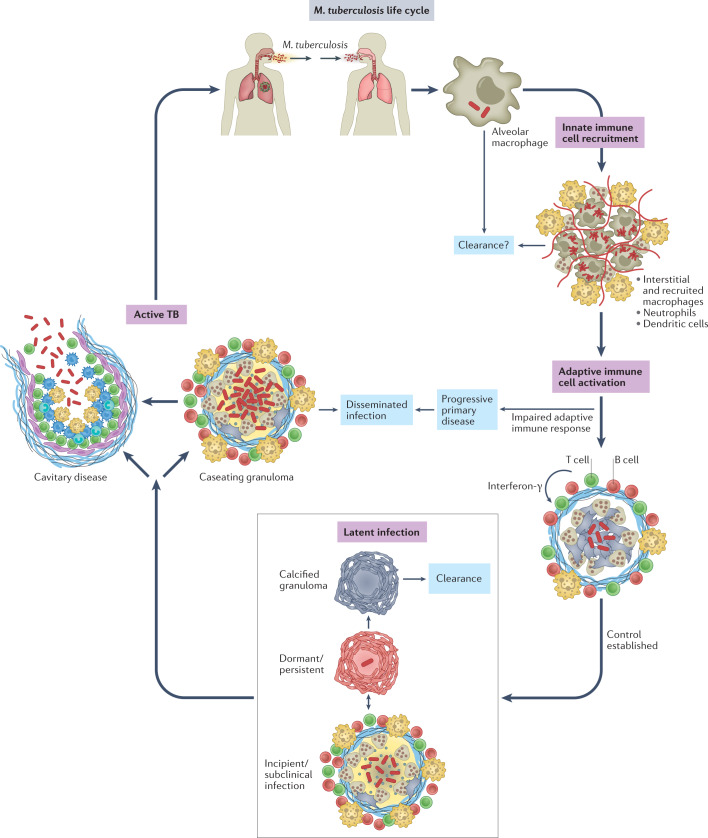
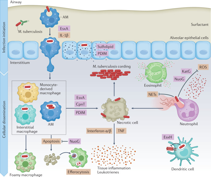
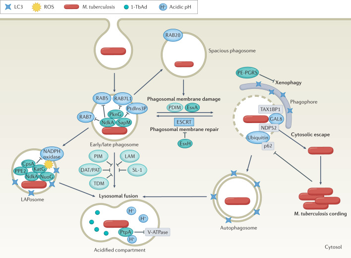
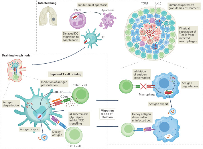
Mycobacterium tuberculosis, the causative agent of tuberculosis, has infected humans for millennia. M. tuberculosis is well adapted to establish infection, persist in the face of the host immune response and be transmitted to uninfected individuals. Its ability to complete this infection cycle depends on it both evading and taking advantage of host immune responses. The outcome of M. tuberculosis infection is often a state of equilibrium characterized by immunological control and bacterial persistence. Recent data have highlighted the diverse cell populations that respond to M. tuberculosis infection and the dynamic changes in the cellular and intracellular niches of M. tuberculosis during the course of infection. M. tuberculosis possesses an arsenal of protein and lipid effectors that influence macrophage functions and inflammatory responses; however, our understanding of the role that specific bacterial virulence factors play in the context of diverse cellular reservoirs and distinct infection stages is limited. In this Review, we discuss immune evasion and provocation by M. tuberculosis during its infection cycle and describe how a more detailed molecular understanding is crucial to enable the development of novel host-directed therapies, disease biomarkers and effective vaccines.


Conflict of interest statement

The authors declare no competing interests.

Figures

Fig. 1
Fig. 1. Life cycle of Mycobacterium tuberculosis.
Mycobacterium tuberculosis is transmitted by aerosol from an individual with active pulmonary infection. The first cells infected are alveolar macrophages. After infected alveolar macrophages migrate into the lung interstitium, the bacilli infect a variety of monocyte-derived and tissue-resident macrophages, dendritic cells and neutrophils. Whether the innate immune response clears infection in some individuals is not clear. Dendritic cells travel to the draining lymph node, where antigen-specific T cells are primed. T cells return to the site of infection and are crucial to establish control and prevent dissemination. With an effective adaptive immune response, most infected individuals develop latent infection, a spectrum of outcomes ranging from sterilized infection to subclinical disease. For reasons that are not well understood, ~5–10% of infected individuals will develop active tuberculosis (TB), most often cavitary disease in the lungs. Most transmission occurs from individuals with cavitary pulmonary disease.
Fig. 2
Fig. 2. Infection establishment and innate immune evasion by Mycobacterium tuberculosis.
In the airways, Mycobacterium tuberculosis first encounters alveolar macrophages (AMs), which present a permissive niche for infection establishment. In addition, M. tuberculosis infects pulmonary epithelial cells and sheds virulence lipids such as phthiocerol dimycoserosate (PDIM) and sulfolipids into epithelial host cell membranes. Infected AMs migrate into the lung interstitium, in a manner that depends on the ESX-1 secretion system of M. tuberculosis and production of host IL-1β. When M. tuberculosis enters the lung interstitium, it infects additional macrophage populations. Neutrophils respond to M. tuberculosis infection by inducing reactive oxygen species (ROS) and neutrophil extracellular traps (NETs), which do little to control bacterial replication and exacerbate inflammation instead. Some macrophages appear to be better at controlling infection than others, using antimicrobial mechanisms such as phagolysosomal fusion, autophagy and oxidative stress to kill M. tuberculosis, and exhibit a pro-inflammatory metabolic shift. M. tuberculosis detoxifies reactive oxygen with the catalase–peroxidase KatG, and also inhibits ROS production in macrophages and neutrophils using NuoG. When infected macrophages undergo an apoptotic mode of cell death, they can be cleared by efferocytosis, which limits pathogen spread. M. tuberculosis uses virulence factors such as EsxA, CpnT and PDIM to induce necrosis and promote M. tuberculosis dissemination, extracellular replication and immunopathology. M. tuberculosis also induces a foamy macrophage phenotype by enhancing accumulation of host lipids, which support bacterial nutrition and persistence. Host cytokines, such as type I interferons and TNF, and leukotrienes contribute to tissue inflammation, which in turn recruits more cells. Further, M. tuberculosis EsxH suppresses antigen presentation by dendritic cells to delay the onset of adaptive immunity.
Fig. 3
Fig. 3. Mycobacterium tuberculosis resides in diverse intracellular compartments.
Mycobacterium tuberculosis is taken up in a single membrane-bound phagosome, which is targeted by the host to promote bacterial clearance. The interactions between host proteins (blue) and M. tuberculosis virulence factors (proteins in dark green; lipids in light green) shape infection outcomes. Sequential recruitment of molecular markers such as RAB5, RAB7 and phosphatidylinositol 3-phosphate (PtdIns3P) normally promote maturation of early to late phagosomes, but M. tuberculosis actively evades phagosomal maturation. In a process called ‘LC3-associated phagocytosis’ (LAP), NADPH oxidase assembles on the M. tuberculosis phagosome (LAPosome) and induces oxidative stress, but M. tuberculosis effectors impair the recruitment of the NADPH oxidase. M. tuberculosis can also be targeted to spacious phagosomes in a RAB20-dependent manner. In addition, the M. tuberculosis ESX-1 substrate EsxA and phthiocerol dimycoserosate (PDIM) promote phagosomal damage. M. tuberculosis EsxH inhibits the host endosomal sorting complex required for transport (ESCRT) machinery to prevent membrane repair. M. tuberculosis escapes into the cytosol and can replicate to form cords. The host attempts to recapture cytosol-exposed M. tuberculosis in double-membrane autophagosomes, which is inhibited by effectors, including PE-PGRS family members. M. tuberculosis contained within single-membrane or double-membrane compartments is targeted for lysosomal killing. M. tuberculosis has mechanisms to resist acidification, such as blocking vacuolar-type ATPase (V-ATPase) and producing the antacid 1-tuberculosinyladenosine (1-TbAd) to neutralize the pH. DAT, diacyltrehalose; LAM, lipoarabinomannan; PAT, polyacyltrehalose; PIM, phosphatidylinositol mannoside; ROS, reactive oxygen species; SL-1, sulfoglycolipid 1; TDM, trehalose dimycolate.
Fig. 4
Fig. 4. Mycobacterium tuberculosis delays and impairs the adaptive immune response.
During Mycobacterium tuberculosis infection, there is a delay in migration of dendritic cells (DCs) to the lungs. NuoG contributes to this delay by inhibiting apoptosis of infected polymorphonuclear neutrophils (PMNs) and antigen uptake by DCs. Once infected DCs arrive in the lymph node, M. tuberculosis impairs their ability to prime CD4+ T cells by degrading antigens (for example, Hip1 degrades GroEL2), exporting antigens out of the cell, and inhibiting antigen processing (mediated by EsxH). Exported M. tuberculosis lipoglycans such as lipoarabinomannan also interfere directly with T cell responses. Phthiocerol dimycoserosate inhibits CD4+ T cell priming and differentiation by inhibiting expression of CD86 and IL-12. M. tuberculosis also preferentially elicits CD4+ and CD8+ responses to decoy antigens (for example, Ag85B and TB10.4), whose expression is downregulated after T cell priming or whose targeting by T cells is not protective. T cells in the infected lung are physically separated from infected macrophages. High levels of IL-10 and transforming growth factor-β (TGFβ) in granulomas also inhibit T cell effector functions. Finally, by inhibiting antigen presentation in infected macrophages, M. tuberculosis also prevents recognition of infected cells. TCR, T cell receptor.
Fig. 5
Fig. 5. Potential vaccine strategies to promote protection from Mycobacterium tuberculosis infection.
a | Possible strategies for generating a protective vaccine. A vaccine could generate antibodies that neutralize a panel of essential cell surface or secreted virulence factors that are required for Mycobacterium tuberculosis to establish infection. Opsonizing antibodies could promote bacterial uptake specifically into protective macrophages or direct the bacilli to a bactericidal path intracellularly. By generating epigenetic changes, macrophages could be trained to a more protective phenotype. It may be possible to identify antigens that generate a protective T cell response, as opposed to the decoy antigens that are dominant during natural infection. Finally, generating lymphocytes that are poised in the lung, ready to respond to infection, would overcome the delay in T cell priming. b | The approaches proposed in part a could protect against M. tuberculosis infection by restoring macrophages’ effector functions, such as lysosomal trafficking, reactive oxygen species (ROS) production and signalling, optimal metabolic responses and protective cell death pathways. Antibodies or training could also skew cellular recruitment towards protective myeloid cells and away from permissive macrophages and neutrophils. Finally, restoring functional interactions with the adaptive immune system could enhance the antimicrobial capacity of myeloid cells and their protective inflammatory responses. LAM, lipoarabinomannan; LAP, LC3-associated phagocytosis; Me1, 1-methylation.

References

    1. Comas I, et al. Out-of-Africa migration and Neolithic coexpansion of Mycobacterium tuberculosis with modern humans. Nat. Genet. 2013;45:1176–1182. doi: 10.1038/ng.2744. - DOI - PMC - PubMed
    1. Huang L, Nazarova EV, Tan S, Liu Y, Russell DG. Growth of Mycobacterium tuberculosis in vivo segregates with host macrophage metabolism and ontogeny. J. Exp. Med. 2018;215:1135–1152. doi: 10.1084/jem.20172020. - DOI - PMC - PubMed
    1. Pisu D, et al. Single cell analysis of M. tuberculosis phenotype and macrophage lineages in the infected lung. J. Exp. Med. 2021 doi: 10.1084/jem.20210615. - DOI - PMC - PubMed
    1. Pisu D, Huang L, Grenier JK, Russell DG. Dual RNA-Seq of Mtb-infected macrophages in vivo reveals ontologically distinct host-pathogen interactions. Cell Rep. 2020;30:335–350.e4. doi: 10.1016/j.celrep.2019.12.033. - DOI - PMC - PubMed
    1. Lee J, et al. CD11cHi monocyte-derived macrophages are a major cellular compartment infected by Mycobacterium tuberculosis. PLoS Pathog. 2020;16:e1008621. doi: 10.1371/journal.ppat.1008621. - DOI - PMC - PubMed

Publication types

Substances