

Decreased visual acuity is related to thinner cortex in cognitively normal adults: cross-sectional, single-center cohort study
- PMID: 35879770
- PMCID: PMC9310451
- DOI: 10.1186/s13195-022-01045-0
Decreased visual acuity is related to thinner cortex in cognitively normal adults: cross-sectional, single-center cohort study
Abstract
Background: Decreased visual acuity (VA) is reported to be a risk factor for dementia. However, the association between VA and cortical thickness has not been established. We investigated the association between VA and cortical thickness in cognitively normal adults.
Method: We conducted a cross-sectional, single-center cohort study with cognitively normal adults (aged ≥ 45) who received medical screening examinations at the Health Promotion Center at Samsung Medical Center. Subjects were categorized as bad (VA ≤ 20/40), fair (20/40 < VA ≤ 20/25), and good (VA > 20/25) VA group by using corrected VA in the Snellen system. Using 3D volumetric brain MRI, cortical thickness was calculated using the Euclidean distance between the linked vertices of the inner and outer surfaces. We analyzed the association between VA and cortical thickness after controlling for age, sex, hypertension, diabetes, dyslipidemia, intracranial volume, and education level.
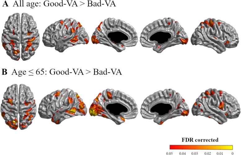
Results: A total of 2756 subjects were analyzed in this study. Compared to the good VA group, the bad VA group showed overall thinner cortex (p = 0.015), especially in the parietal (p = 0.018) and occipital (p = 0.011) lobes. Topographical color maps of vertex-wise analysis also showed that the bad VA group showed a thinner cortex in the parieto-temporo-occipital area. These results were more robust in younger adults (aged 45 to 65) as decreased VA was associated with thinner cortex in more widespread regions in the parieto-temporo-occipital area.
Conclusion: Our results suggest that a thinner cortex in the visual processing area of the brain is related to decreased visual stimuli.
Keywords: Cortical thickness; Dementia; Visual acuity.
© 2022. The Author(s).
Conflict of interest statement
The authors declare that they have no competing interests.
Figures
Similar articles
-
Different regional patterns of cortical thinning in Alzheimer's disease and frontotemporal dementia.Brain. 2007 Apr;130(Pt 4):1159-66. doi: 10.1093/brain/awm016. Epub 2007 Mar 12. Brain. 2007. PMID: 17353226 Free PMC article.
-
Analysis of the volumetric relationship among human ocular, orbital and fronto-occipital cortical morphology.J Anat. 2015 Oct;227(4):460-73. doi: 10.1111/joa.12364. Epub 2015 Aug 7. J Anat. 2015. PMID: 26250048 Free PMC article.
-
Altered cortical morphology of visual cortex in adults with monocular amblyopia.J Magn Reson Imaging. 2019 Nov;50(5):1405-1412. doi: 10.1002/jmri.26708. Epub 2019 Mar 10. J Magn Reson Imaging. 2019. PMID: 30854758
-
Helicobacter Pylori Infection Is Associated with Neurodegeneration in Cognitively Normal Men.J Alzheimers Dis. 2021;82(4):1591-1599. doi: 10.3233/JAD-210119. J Alzheimers Dis. 2021. PMID: 34180413
-
Thinner cortex in the frontal lobes in mentally disordered offenders.Psychiatry Res. 2012 Aug-Sep;203(2-3):126-31. doi: 10.1016/j.pscychresns.2011.12.011. Epub 2012 Sep 1. Psychiatry Res. 2012. PMID: 22947310
Cited by
-
Comparing retinotopic maps of children and adults reveals a late-stage change in how V1 samples the visual field.Nat Commun. 2023 Mar 21;14(1):1561. doi: 10.1038/s41467-023-37280-8. Nat Commun. 2023. PMID: 36944643 Free PMC article.
-
Distinct brain age gradients across the adult lifespan reflect diverse neurobiological hierarchies.Commun Biol. 2025 May 25;8(1):802. doi: 10.1038/s42003-025-08228-z. Commun Biol. 2025. PMID: 40415122 Free PMC article.
References
Publication types
MeSH terms
LinkOut - more resources
Full Text Sources
Medical
Research Materials
