. 2022 Jul 25;8(1):50.
doi: 10.1186/s40942-022-00400-5.

Alternative management of central serous chorioretinopathy using intravitreal metoprolol

Affiliations

Alternative management of central serous chorioretinopathy using intravitreal metoprolol

Annelise Nicotti Gonçalves et al. Int J Retina Vitreous. .

Abstract

Background: Beta-blockers may counteract the effect of catecholamines on central serous chorioretinopathy (CSC) pathology and accelerate the improvement of neurosensory retinal detachment. Oral propranolol has been associated with decreased duration of CSC in some studies. We describe two patients with visually symptomatic chronic CSC (cCSC) treated successfully with intravitreal metoprolol.

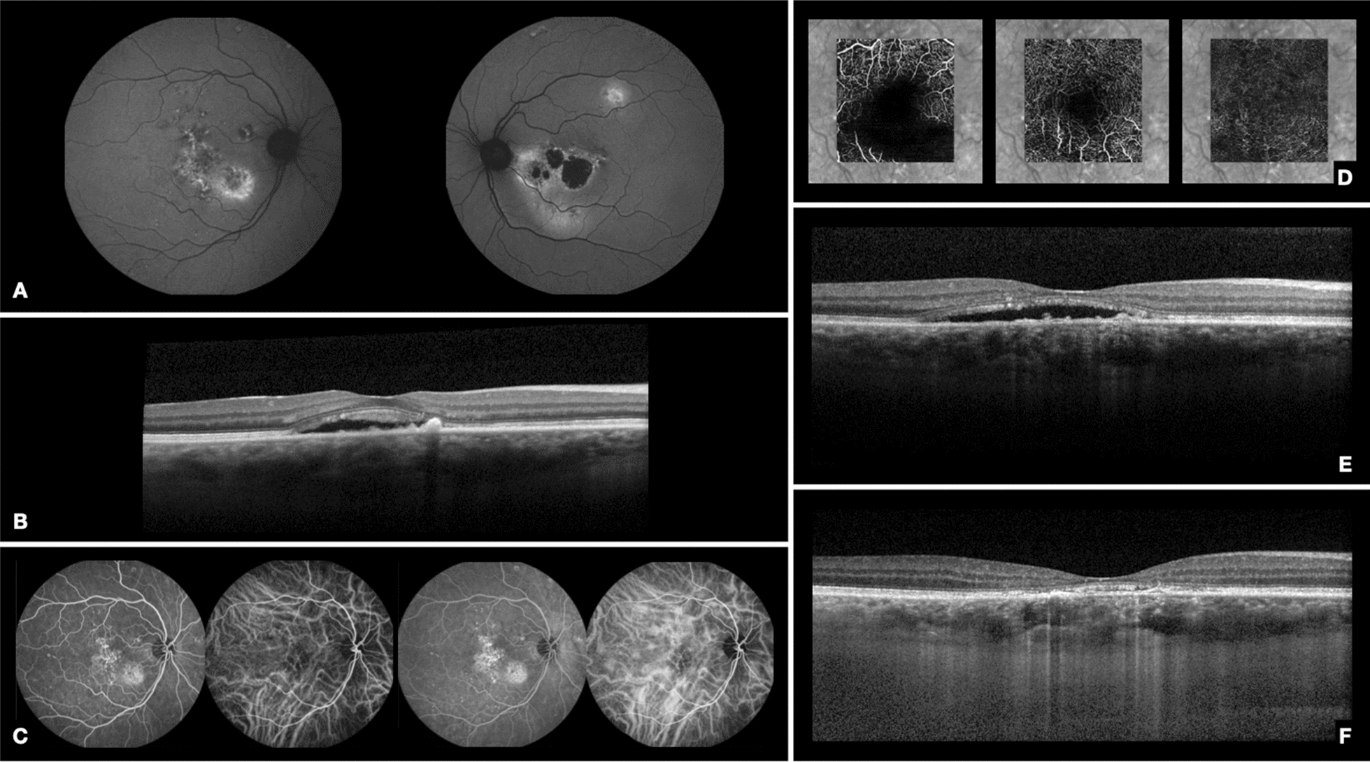
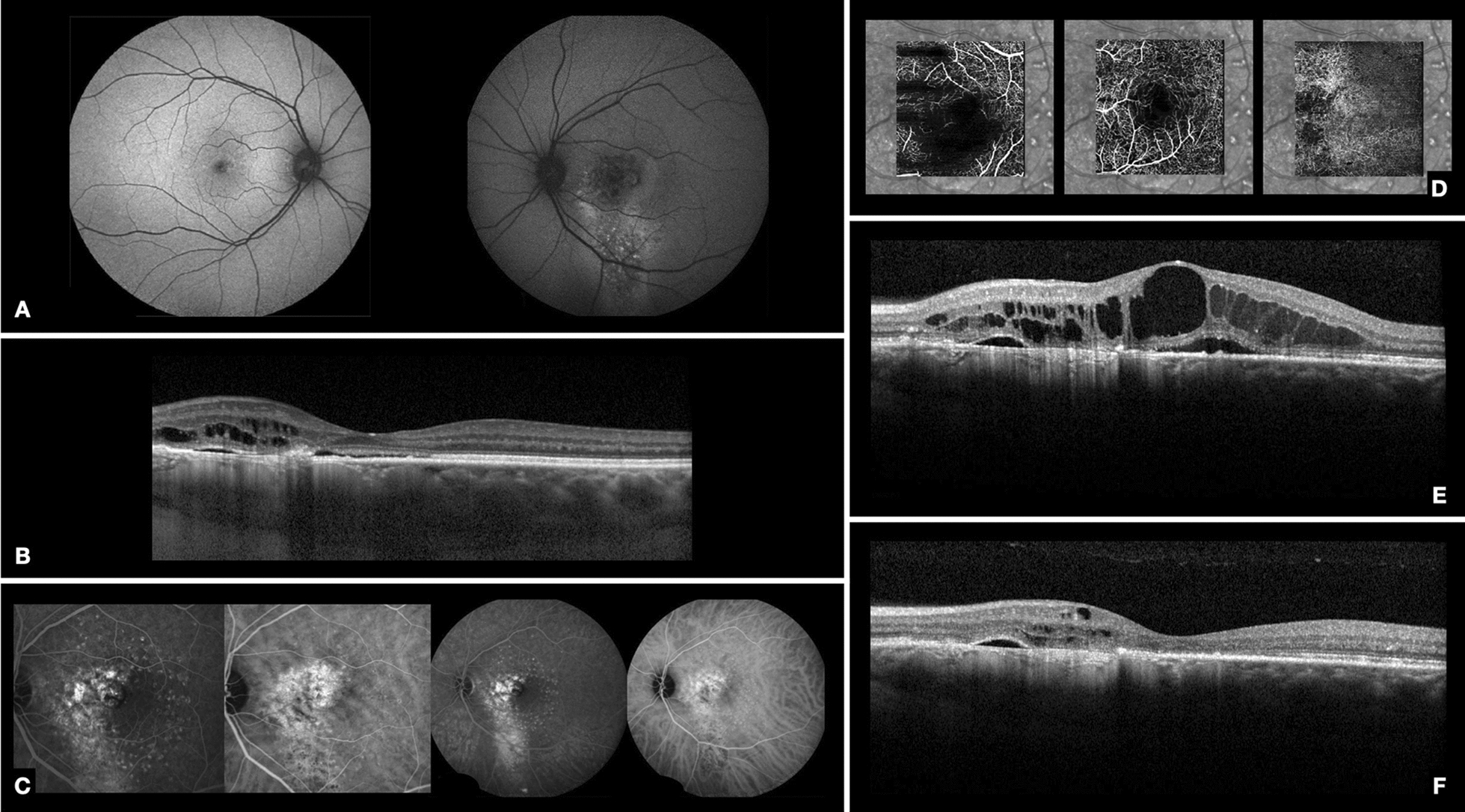
Case presentations: After obtaining the patients' informed consent, two eyes of two 43-year-old men diagnosed with cCSC treated unsuccessfully with oral spirolactone, micropulse laser and intravitreal anti-vascular endothelial growth factor (anti-VEGF) agents were treated with one off-label intravitreal injection of metoprolol (50 µg/0.05 ml). Baseline (pre-injection) and follow-up examinations (at 1 month post-injection) included best-corrected visual acuity (BCVA), anterior and posterior segment biomicroscopy, fundus autofluorescence, spectral domain optical coherence tomography (Spectralis, Heidelberg), and electroretinogaphy (ERG) according to International Society for Clinical Electrophysiology of Vision (ISCEV) full-field scotopic and photopic standard protocols. ERG results at baseline (pre-injection) and at 1 month post-injection were compared using paired t-tests.

Results: There was no significant difference in any of the ISCEV recommended ERG parameters with respect to a- and b-wave amplitude and implicit time, and oscillatory potentials maximal amplitude. BCVA improved in both patients. Neither patient developed clinical evidence of intraocular inflammation. Subretinal and/or intraretinal fluid had improved in both patients at 1 month after the metoprolol injection.

Conclusion: These preliminary findings suggest that intravitreal metoprolol may be a safe alternative therapy for patients with cCSC.

Keywords: B-blockers; Central serous chorioretinopathy; Intravitreal; Retina; Subretinal fluid.


Conflict of interest statement

None related to the discussed topic.

Figures

Fig. 1
Fig. 1
Case 1. A Baseline autofluorescence of both eyes. In the OD, there is a mixture of stippled hyper- and hypo-autofluorescence in the posterior pole, including the macula, with extension inferonasally. In the OS, there is a hypo-autofluorescent area in the fovea, with pronounced hyper-autofluorescent regions inferonasal to the fovea. B Baseline spectral domain optical coherence tomography (SD-OCT) of the RE shows shaggy photoreceptors and subretinal fluid in the foveal area. CE 39 months after baseline, and 1 week before metoprolol injection. C Fluorescein and indocyanine green (ICG) angiography of the OD reveal multifocal areas of granular window defect with hyperfluorescent patches. On ICG angiography, there are focal areas of hyperfluorescnce in the initial and middle phases corresponding to areas of RPE changes and choriocapillaris hypermeability. A foveal hyperfluorescent fine-vessel choroidal network was suspected. D Optical coherence tomography angiography of the OD shows no signs of choroidal neovascularization. E SD-OCT of the OD before intravitreal metoprolol injection demonstrates shaggy photoreceptors, disruption of the ellipsoid zone, and subretinal fluid in the foveal area with RPE irregularity. F SD-OCT of the OD 12 weeks after metoprolol injection demonstrates resolution of the subretinal fluid
Fig. 2
Fig. 2
Case 2. A Baseline autofluorescence of both eyes demonstrates no significant RPE changes in the OD and, in the left eye, shows focal hypo-autofluorescent dots in the posterior pole, including the macula, and forming a descending tract. B Baseline spectral domain optical coherence tomography (SD-OCT) of the OS reveals foveal subretinal fluid. In the nasal macular region, there is outer retina and RPE atrophy, associated with intraretinal and subretinal fluid. CE 31 months after baseline, and 1 week before metoprolol injection. C Fluorescein angiography reveals transmission hyperfluorescence in the areas corresponding to the RPE changes on autofluorescence. There are also small hyperfluorescent dots surrounding the foveal area, which corresponded to short-pulse laser burns. OS Indocyanine green angiography demonstrates areas of hyperfluorescence secondary to RPE changes and choriocapillaris hyperpermeability in the early and late phases. D Optical coherence tomography angiography of the OS shows no signs of choroidal neovascularization. E Spectral domain optical coherence tomography (SD-OCT) of the OS before intravitreal metoprolol demonstrates outer retinal atrophy and RPE defects in the foveal area associated with chronic cystoid macular edema. F SD-OCT of the OS five weeks after metoprolol injection shows a significant reduction of the intraretinal fluid

Similar articles

Cited by

References

    1. Nicholson B, Noble J, Forooghian F, Meyerle C. Central serous chorioretinopathy: update on pathophysiology and treatment. Surv Ophthalmol. 2013;58(2):103–126. doi: 10.1016/j.survophthal.2012.07.004. - DOI - PMC - PubMed
    1. Baran NV, Gürlü VP, Esgin H. Long-term macular function in eyes with central serous chorioretinopathy. Clin Exp Ophthalmol. 2005;33(4):369–372. doi: 10.1111/j.1442-9071.2005.01027.x. - DOI - PubMed
    1. Stefánsson E, Geirsdóttir A, Sigurdsson H. Metabolic physiology in age related macular degeneration. Prog Retin Eye Res. 2011;30(1):72–80. doi: 10.1016/j.preteyeres.2010.09.003. - DOI - PubMed
    1. Lee WJ, Lee JH, Lee BR. Fundus autofluorescence imaging patterns in central serous chorioretinopathy according to chronicity. Eye. 2016;30(10):1336–1342. doi: 10.1038/eye.2016.113. - DOI - PMC - PubMed
    1. Mrejen S, Balaratnasingam C, Kaden TR, et al. Long-term visual outcomes and causes of vision loss in chronic central serous chorioretinopathy. Ophthalmology. 2019;126(4):576–588. doi: 10.1016/j.ophtha.2018.12.048. - DOI - PubMed

LinkOut - more resources