

Advanced MRI to differentiate schwannomas and metastases in the cerebellopontine angle/internal auditory canal
- PMID: 35879866
- PMCID: PMC9796724
- DOI: 10.1111/jon.13028
Advanced MRI to differentiate schwannomas and metastases in the cerebellopontine angle/internal auditory canal
Abstract
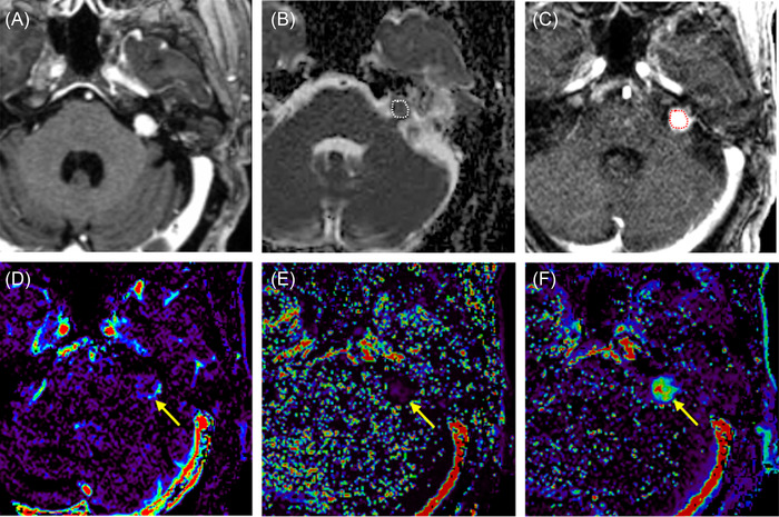
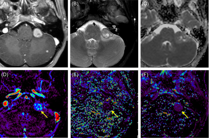
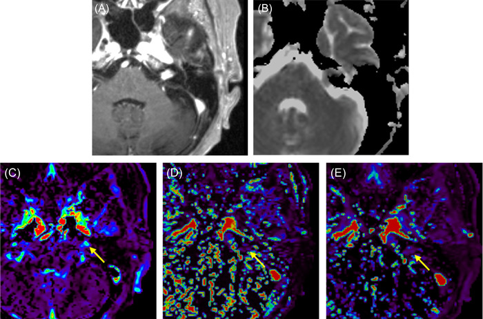
Background and purpose: Differentiating schwannomas and metastases in the cerebellopontine angles (CPA)/internal auditory canals (IAC) can be challenging. This study aimed to assess the role of diffusion-weighted imaging (DWI) and dynamic contrast-enhanced MRI (DCE-MRI) to differentiate schwannomas and metastases in the CPA/IAC.
Methods: We retrospectively reviewed 368 patients who were diagnosed with schwannomas or metastases in the CPA/IAC between April 2017 and February 2022 in a single academic center. Forty-three patients had pretreatment DWI and DCE-MRI along with conventional MRI. Normalized mean apparent diffusion coefficient ratio (nADCmean) and DCE-MRI parameters of fractional plasma volume (Vp), flux rate constant (Kep), and forward volume transfer constant were compared along with patients' demographics and conventional imaging features between schwannomas and metastases as appropriate. The diagnostic performances and multivariate logistic regression analysis were performed using the significantly different values.
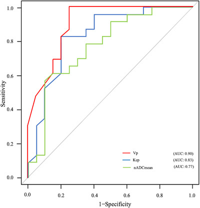
Results: Between 23 schwannomas (15 males; median 48 years) and 20 metastases (9 males; median 61 years), nADCmean (median: 1.69 vs. 1.43; p = .002), Vp (median: 0.05 vs. 0.20; p < .001), and Kep (median: 0.41 vs. 0.81 minute-1 ; p < .001) were significantly different. The diagnostic performances of nADCmean, Vp, and Kep were 0.77, 0.90, and 0.83 area under the curves, with cutoff values of 1.68, 0.12, and 0.53, respectively. Vp was identified as the most significant parameter for the tumor differentiation in the multivariate logistic regression analysis (p < .001).
Conclusions: DWI and DCE-MRI can help differentiate CPA/IAC schwannomas and metastases, and Vp is the most significant parameter.
Keywords: CPA; DCE-MRI; DWI; metastasis; schwannoma.
© 2022 The Authors. Journal of Neuroimaging published by Wiley Periodicals LLC on behalf of American Society of Neuroimaging.
Figures
Similar articles
-
Precise differentiation between jugular foramen paragangliomas and metastases: utility of diffusion-weighted and dynamic contrast-enhanced magnetic resonance imaging.Neuroradiology. 2023 Apr;65(4):805-813. doi: 10.1007/s00234-023-03113-0. Epub 2023 Jan 13. Neuroradiology. 2023. PMID: 36635515
-
Neurofibromatosis type 2 versus sporadic vestibular schwannoma: The utility of MR diffusion and dynamic contrast-enhanced imaging.J Neuroimaging. 2022 May;32(3):554-560. doi: 10.1111/jon.12966. Epub 2022 Jan 17. J Neuroimaging. 2022. PMID: 35037337
-
MR diffusion and dynamic-contrast enhanced imaging to distinguish meningioma, paraganglioma, and schwannoma in the cerebellopontine angle and jugular foramen.J Neuroimaging. 2022 May;32(3):502-510. doi: 10.1111/jon.12959. Epub 2021 Dec 22. J Neuroimaging. 2022. PMID: 34936708
-
Cerebellopontine Angle and Internal Auditory Canal Lipomas: Case Series and Systematic Review.Laryngoscope. 2021 Sep;131(9):2081-2087. doi: 10.1002/lary.29434. Epub 2021 Feb 10. Laryngoscope. 2021. PMID: 33567134
-
Radiological Biomarkers for Brain Metastases Prognosis: Quantitative Magnetic Resonance Imaging (MRI) Modalities As Non-invasive Biomarkers for the Effect of Radiotherapy.Cureus. 2023 Apr 30;15(4):e38353. doi: 10.7759/cureus.38353. eCollection 2023 Apr. Cureus. 2023. PMID: 37266043 Free PMC article. Review.
Cited by
-
Precise differentiation between jugular foramen paragangliomas and metastases: utility of diffusion-weighted and dynamic contrast-enhanced magnetic resonance imaging.Neuroradiology. 2023 Apr;65(4):805-813. doi: 10.1007/s00234-023-03113-0. Epub 2023 Jan 13. Neuroradiology. 2023. PMID: 36635515
-
Sacral intraosseous schwannoma in an adolescent patient: A case report.Radiol Case Rep. 2025 May 12;20(8):3655-3661. doi: 10.1016/j.radcr.2025.04.066. eCollection 2025 Aug. Radiol Case Rep. 2025. PMID: 40475044 Free PMC article.
-
MRI findings for the pretreatment diagnosis of small Meckel's cave tumors: comparison of meningiomas and schwannomas.BMC Med Imaging. 2025 Feb 22;25(1):57. doi: 10.1186/s12880-025-01597-1. BMC Med Imaging. 2025. PMID: 39987052 Free PMC article.
-
Solitary metastasis in the internal auditory canal from non-small cell lung carcinoma: A case report.Respir Med Case Rep. 2025 Jan 28;53:102175. doi: 10.1016/j.rmcr.2025.102175. eCollection 2025. Respir Med Case Rep. 2025. PMID: 39980611 Free PMC article.
References
-
- Bonneville F, Savatovsky J, Chiras J. Imaging of cerebellopontine angle lesions: an update. Part 1: enhancing extra‐axial lesions. Eur Radiol. 2007;17:2472‐82. - PubMed
-
- Bonneville F, Sarrazin JL, Marsot‐Dupuch K, et al. Unusual lesions of the cerebellopontine angle: a segmental approach. Radiographics. 2001;21:419‐38. - PubMed
-
- Skolnik AD, Loevner LA, Sampathu DM, et al. Cranial nerve schwannomas: diagnostic imaging approach. Radiographics. 2016;36:1463‐77. - PubMed
MeSH terms
Substances
LinkOut - more resources
Full Text Sources
Miscellaneous
