

Triphasic developmentally guided protocol to generate retinal pigment epithelium from induced pluripotent stem cells
- PMID: 35880133
- PMCID: PMC9307589
- DOI: 10.1016/j.xpro.2022.101582
Triphasic developmentally guided protocol to generate retinal pigment epithelium from induced pluripotent stem cells
Abstract
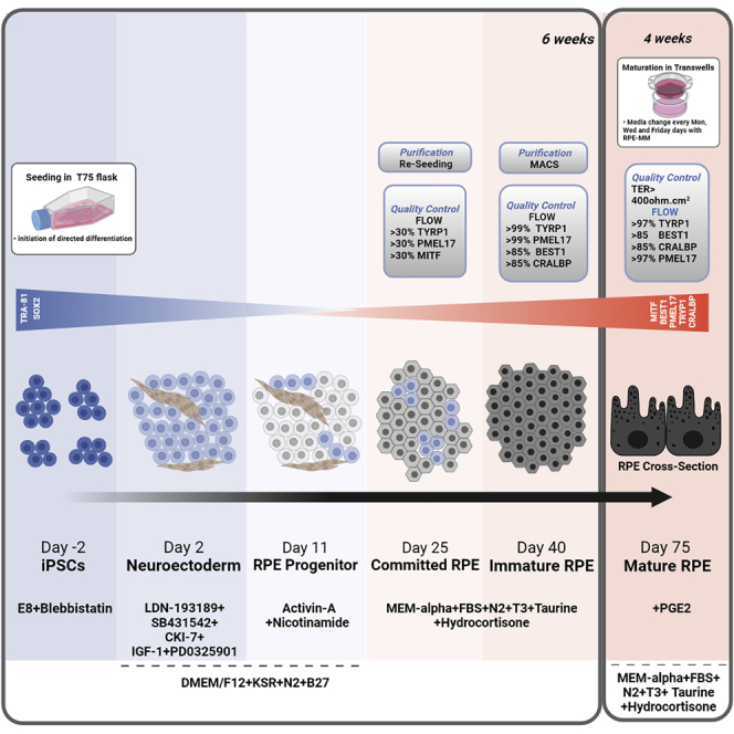
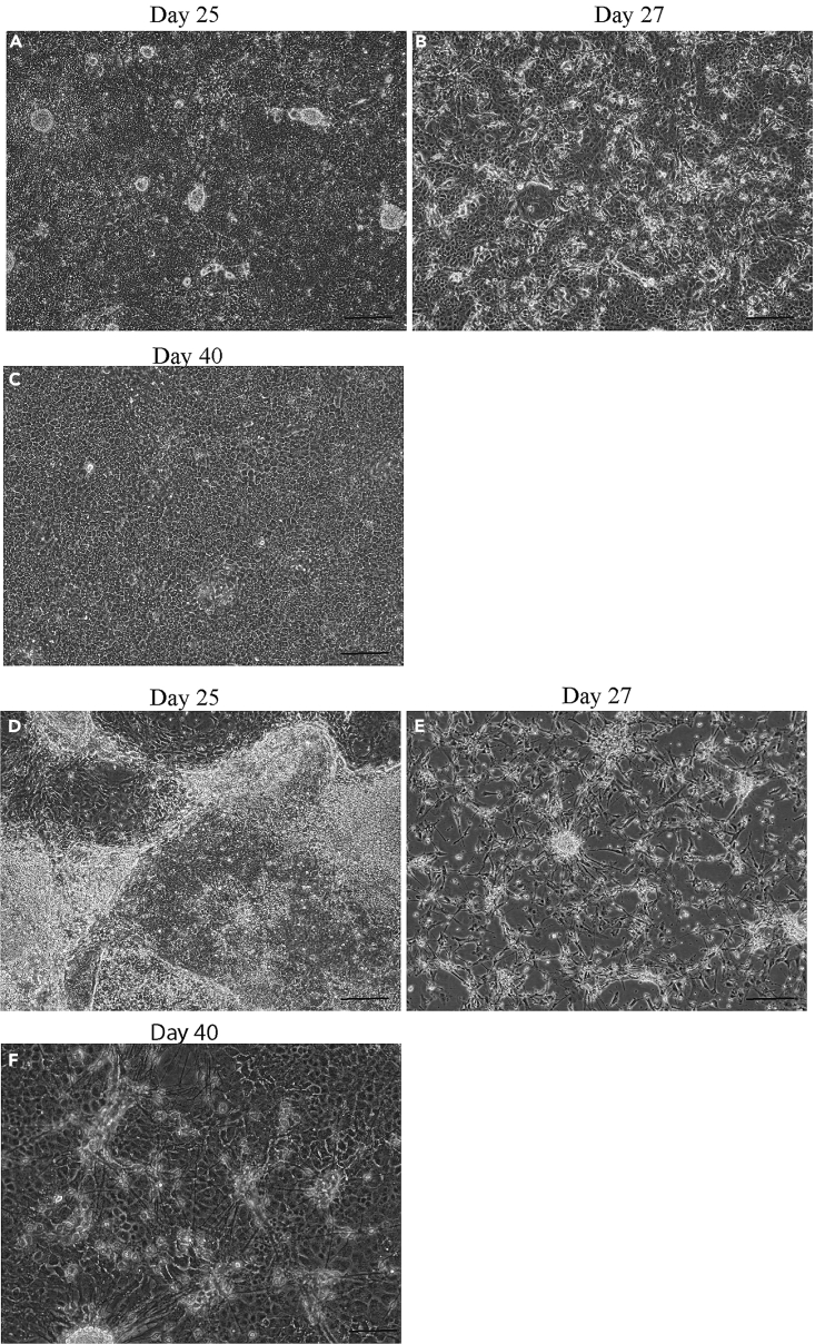
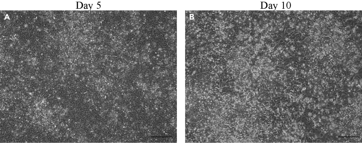
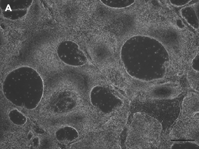
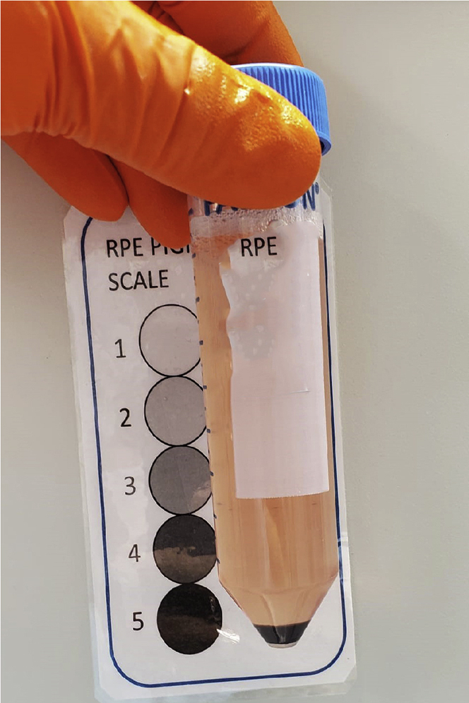
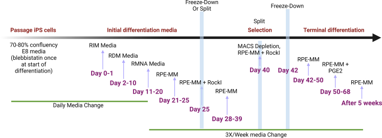
RPE tissues are derived from induced pluripotent stem cells (iPSCs) to model retinal diseases and as a replacement therapy for macular degeneration. Here, we developed a robust and efficient directed differentiation protocol to generate pure RPE cells that form a polarized monolayer. This protocol describes how to set up RPE differentiation and to obtain a pure population that expresses mature RPE markers and forms strong tight junctions. For complete details on the use and execution of this protocol, please refer to Sharma et al., 2019, Sharma et al., 2021 and Miyagishima et al. (2021).
Keywords: Cell Biology; Cell Differentiation; Cell culture; Cell isolation; Flow Cytometry/Mass Cytometry; Health Sciences; Stem Cells.
Conflict of interest statement
We, the authors, have a patent related to this work.
Figures
References
-
- Miyagishima K.J., Sharma R., Nimmagadda M., Clore-Gronenborn K., Qureshy Z., Ortolan D., Bose D., Farnoodian M., Zhang C., Fausey A., et al. AMPK modulation ameliorates dominant disease phenotypes of CTRP5 variant in retinal degeneration. Commun. Biol. 2021;4:1360. doi: 10.1038/S42003-021-02872-X. - DOI - PMC - PubMed
-
- Sharma R., Khristov V., Rising A., Jha B.S., Dejene R., Hotaling N., Li Y., Stoddard J., Stankewicz C., Wan Q., et al. Clinical-grade stem cell-derived retinal pigment epithelium patch rescues retinal degeneration in rodents and pigs. Sci. Transl. Med. 2019;11:eaat5580. doi: 10.1126/SCITRANSLMED.AAT5580. - DOI - PMC - PubMed
Publication types
MeSH terms
Substances
LinkOut - more resources
Full Text Sources
Other Literature Sources
Medical
