Review
. 2022 Jul 8:12:844790.
doi: 10.3389/fonc.2022.844790. eCollection 2022.

Diffusion Breast MRI: Current Standard and Emerging Techniques

Affiliations
Review

Diffusion Breast MRI: Current Standard and Emerging Techniques

Ashley M Mendez et al. Front Oncol. .

Abstract

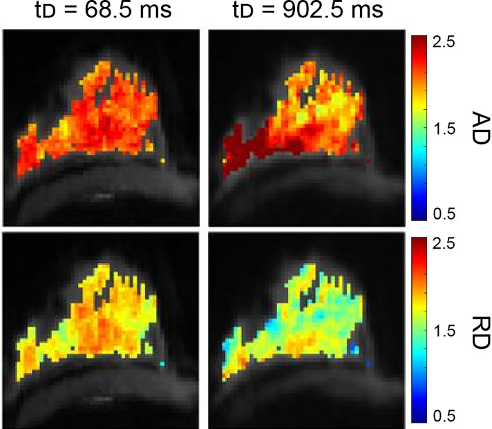
The role of diffusion weighted imaging (DWI) as a biomarker has been the subject of active investigation in the field of breast radiology. By quantifying the random motion of water within a voxel of tissue, DWI provides indirect metrics that reveal cellularity and architectural features. Studies show that data obtained from DWI may provide information related to the characterization, prognosis, and treatment response of breast cancer. The incorporation of DWI in breast imaging demonstrates its potential to serve as a non-invasive tool to help guide diagnosis and treatment. In this review, current technical literature of diffusion-weighted breast imaging will be discussed, in addition to clinical applications, advanced techniques, and emerging use in the field of radiomics.

Keywords: breast cancer; diagnostic breast imaging; diffusion tensor (DT) MRI; diffusion weighted (DW) breast MRI; imaging biomarker; non-gaussian diffusion; radiomics; restriction spectrum imaging.


Conflict of interest statement

RR-P: Human Longevity Inc: Consultant, Cortech Labs: Stock options, Curemetrix: Stock options, consultant. and GE: research agreement. The remaining authors declare that the research was conducted in the absence of any commercial or financial relationships that could be construed as a potential conflict of interest.

Figures

Figure 1
Figure 1
Simplified physical basis of advanced diffusion imaging. Water molecules moving at two different speeds are shown: fast-moving (free and hindered) which exist in extracellular space (blue), and slow-moving (restricted) molecules that are trapped intracellularly by the plasma membrane (orange). Note that exchange between the extra- and intracellular compartments also exists, dictated by membrane permeability. The schematic shows the dispersion of these water molecular diffusing across cellular compartments, at different timescales (Δ) of (A) 1msec, (B) 20 msec, and (C) 50 msec. (D) The root mean square (RMS) distance of water molecules experiencing hindered diffusion is linear with respect to the Δ (i.e. Gaussian diffusion, blue). In contrast, slow-moving water molecules in the intracellular compartment display Gaussian diffusion behavior (linear) at very short timescales (panel D, orange), dictated by the compartment’s intrinsic diffusivity (5). At intermediary timescales, molecules reach the plasma membrane boundary that restricts movement, indicated by the dotted black vertical line. Past this, the net squared displacement becomes sublinear with time and is dependent on the dimensions of the compartment. To note, at very long diffusion timescales (Δ>1s), restricted water diffusion becomes principally governed by the exchange rate between the intra- and extracellular compartments (5). (E) In DW-MRI, the measured signal (S) decays exponentially (in the case of Gaussian diffusion) with respect to b-value due to loss of spin coherence caused by dispersion of water molecules. Thus, the signal decay from water molecules experiencing hindered diffusion (blue) is faster than from water molecules experiencing restricted diffusion (orange). The measured diffusion signal at different b-value weighting (F–H) reflects the relative dephasing of water molecules in different tissue compartments. At short timescales (A, F), the measured signal, S, contains combined information from both hindered (SH) and restricted (SR) water signal. At progressively longer timescales (B, C, G, H), signal from hindered water dissipates more quickly than that from restricted water due to increased motion along the diffusion gradient axis, and the measured signal begins to arise predominantly from the restricted water signal (5). As shown in panel (E), restricted water will retain more signal at higher b-values than hindered water and, correspondingly, have a lower ADC than hindered water.
Figure 2
Figure 2
Example of conventional breast DWI at 3T, shown at b-values 0 and 800 s/mm2 and corresponding ADC map in a 49-year-old patient The lesion, indicated by the yellow arrow, has increased signal on b=800 s/mm2 images and displays lower ADC values compared to surrounding tissue, indicating a finding suspicious for malignancy. This lesion was found to be an invasive ductal carcinoma from pathology.
Figure 3
Figure 3
Example of SMS rs-EPI acquisition at b=800 s/mm2 in a 35-year-old healthy volunteer, wherein (B) two-fold (2×) SMS rs-EPI maintains comparable image quality as (A) conventional rs-EPI while reducing scan time in a 3T scanner. Panel (C) shows two simultaneously acquired slices used to generate a single-band equivalent image for the same patient at a different slice location (94). (Courtesy of Lukas Filli, MD, Zurich, Switzerland).
Figure 4
Figure 4
Reduced FOV EPI in a 63-year-old patient with invasive ductal carcinoma. T2-weighted, conventional DWI (b=0 s/mm2), full FOV EPI (b=0 s/mm2), and reduced FOV EPI (50% phase field of view) (b=0 s/mm2 acquired at 3T) images are shown. Reduction of percent phase encoding direction to 50% reduces geometric distortions caused by B0-inhomogeneity, especially in the nipple region (100).
Figure 5
Figure 5
Based on the results of the initial DW-DESS-Cones investigation in the breast at 3T (A), the diffusion-weighting and resolution of the method can be further increased (B) to better match contrast and resolution expectations for breast MRI (109). (Courtesy of Catherine Moran, PhD, Department of Radiology, Stanford University, Stanford, California, USA).
Figure 6
Figure 6
Effects of pseudo-diffusion on DWI signal. Signal curves in the presence of increasing IVIM effects deviate from the simple mono-exponential curve (fp=0, black line). The components have unique coefficients Dt = 0.001 s/mm2 and D* = 0.02 s/mm2 with relative proportions given by the pseudo-diffusion fraction fp (164). (Courtesy of Igor Vidić, PhD, previously at the Department of Physics, Norwegian University of Science and Technology, Trondheim, Norway).
Figure 7
Figure 7
Example of DKI analysis using b-values of 0, 500, 1000, and 2000 s/mm2, compared to conventional ADC images using b-values of 0 and 1000 s/mm2 and DCE-MRI peak intensity subtraction (1 min 30 s post-contrast). Invasive ductal carcinoma in a 67-year-old patient is indicated by the yellow arrow. The lesion displays higher mean kurtosis (Kapp) than surrounding healthy tissue. Images were acquired using a wide-bore 3T scanner, and mean kurtosis and diffusivity (Dapp) were calculated as previously demonstrated (181).
Figure 8
Figure 8
Stretched exponential modeling with b-values of 0, 500, 1000, 1500 and 2000 s/mm2 in a 73-year-old patient with invasive ductal carcinoma. Distributed diffusion coefficient (DDC) and alpha maps are overlaid on DWI b=0 s/mm2 images, acquired at 3T (56). (Courtesy of Shiteng Suo, PhD, and Jia Hua, MD, Department of Radiology, Ren Ji Hospital, School of Medicine, Shanghai Jiao Tong University, Shanghai, China).
Figure 9
Figure 9
Example of three-compartment RSI analysis in a 49-year-old patient with invasive ductal carcinoma. C1, C2, and C3 maps correspond to the slowest, intermediary, and fastest diffusion compartments, respectively. The lesion, indicated by the white arrow, is hypointense on C1 and C2 maps compared to surrounding healthy tissue, whereas there is little difference in the C3 compartment, which is suggested to correspond to vasculature. The product of C1 and C2 (C1C2) results in the greatest tumor conspicuity. DWI images were acquired at b = 0, 500, 1500, and 4000 s/mm2 on a 3T scanner, with 50% reduced FOV and without parallel imaging (206).
Figure 10
Figure 10
Example of STEAM analysis in a healthy volunteer. The protocol collected a prototype STEAM-DTI sequence with two b-values (0, 500 s/mm2) in six directions with parallel imaging in a 3T scanner. Axial diffusivity (AD, first row) and radial diffusivity (RD, second row) [µm2/ms] parametric maps at the shortest and longest diffusion time for healthy fibroglandular tissue are shown (212). (Courtesy of Jose Teruel, PhD, Department of Radiation Oncology, NYU Langone Medical Center, New York, New York, USA).
Figure 11
Figure 11
39-year-old female with invasive ductal carcinoma, imaged at 7T using a dedicated four-channel double-tuned 31P/1H breast coil. Images shown are b=0 s/mm2, corresponding ADC map, and DCE-MRI peak intensity subtraction. The mean ADC of the tumor shown is 0.71 ± 0.18 × 10⁻³ mm2/s. For DWI, a transverse DWI read-out segmented echo-planar imaging sequence was used. For DCE, patients were intravenously injected with a single dose of contrast agent (gadoterate meglumine) as a bolus, followed by a 20-mL saline flush after three of the 18 repetitions of the time-resolved angiographic imaging with stochastic trajectories sequence (26). (Courtesy of Katja Pinker-Domenig, MD, PhD, Department of Radiology, Memorial Sloan Kettering Cancer Center, New York, New York, USA).

Similar articles

Cited by

References

    1. Lehman CD, Schnall MD. Imaging in Breast Cancer: Magnetic Resonance Imaging. Breast Cancer Res (2005) 7(5):215. doi: 10.1186/bcr1309 - DOI - PMC - PubMed
    1. Kaiser WA, Zeitler E. MR Imaging of the Breast: Fast Imaging Sequences With and Without Gd-DTPA. Preliminary Observations. Radiology (1989) 170(3 Pt 1):681–6. doi: 10.1148/radiology.170.3.2916021 - DOI - PubMed
    1. Heywang SH, Fenzl G, Hahn D, Krischke I, Edmaier M, Eiermann W, et al. . MR Imaging of the Breast: Comparison With Mammography and Ultrasound. J Comput Assist Tomogr. (1986) 10(4):615–20. doi: 10.1097/00004728-198607000-00014 - DOI - PubMed
    1. Aminololama-Shakeri S, Lewin J, Appelton C, Lee CS, Giess CS, Ojeda-Fournier H, et al. . ACR Practice Parameter for the Performance of Contrast-Enhanced Magnetic Resonance Imaging (MRI) of the Breast. American College of Radiology; (2021). Available at: https://www.acr.org/-/media/ACR/Files/Practice-Parameters/mr-guided-brea....
    1. White NS, McDonald C, Farid N, Kuperman J, Karow D, Schenker-Ahmed NM, et al. . Diffusion-Weighted Imaging in Cancer: Physical Foundations and Applications of Restriction Spectrum Imaging. Cancer Res (2014) 74(17):4638–52. doi: 10.1158/0008-5472.CAN-13-3534 - DOI - PMC - PubMed