

Imaging pathological tau in atypical parkinsonisms: A review
- PMID: 35880206
- PMCID: PMC9307942
- DOI: 10.1016/j.prdoa.2022.100155
Imaging pathological tau in atypical parkinsonisms: A review
Abstract
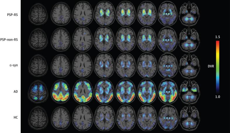
Atypical parkinsonisms (APs) are a group of diseases linked to tau pathology. These include progressive supranuclear palsy (PSP) and corticobasal degeneration (CBD). In the initial stages, these APs may have similar clinical manifestations to Parkinson's disease (PD) and other parkinsonisms: bradykinesia, postural instability, tremor, and cognitive decline. Because of this, one major hurdle is the accurate early diagnosis of APs. Recent advances in positron emission tomography (PET) radiotracer development have allowed for targeting pathological tau in Alzheimer's disease (AD). Currently, work is still in progress for identifying a first-in-class radiotracer for imaging tau in APs. In this review, we evaluate the literature on in vitro and in vivo testing of current tau PET radiotracers in APs. The tau PET tracers assessed include both first-generation tracers ([18F]AV-1451, [18F]FDDNP, [18F]THK derivatives, and [11C]PBB3) and second-generation tracers ([18F]PM-PBB3, [18F]PI-2620, [18F]RO-948, [18F]JNJ-067, [18F]MK-6240, and [18F]CBD-2115). Concerns regarding off-target binding to cerebral white matter and the basal ganglia are still prominent with first-generation tracers, but this seems to have been mediated in a handful of second-generation tracers, including [18F]PI-2620 and [18F]PM-PBB3. Additionally, these two tracers and [18F]MK-6240 show promising results for imaging PSP- and CBD-tau. Overall, [18F]AV-1451 is the most widely studied tracer but the mixed results regarding its efficacy for use in imaging AP-tau is a cause for concern moving forward. Instead, future work may benefit from focusing on the second-generation radiotracers which seem to have a higher specificity for AP-tau than those originally developed for imaging AD-tau.
Keywords: CBD; CBD, corticobasal degeneration; Neuroimaging; PET; PET, positron emission tomography; PSP; PSP, progressive supranuclear palsy; Parkinsonism; Parkinson’s disease; SPECT; SPECT, single-photon emission computerized tomography.
© 2022 The Authors. Published by Elsevier Ltd.
Conflict of interest statement
The authors declare that they have no known competing financial interests or personal relationships that could have appeared to influence the work reported in this paper.
Figures
Similar articles
-
Tau in Atypical Parkinsonisms: A Meta-Analysis of in Vivo PET Imaging Findings.Mov Disord Clin Pract. 2023 Sep 29;10(12):1725-1737. doi: 10.1002/mdc3.13885. eCollection 2023 Dec. Mov Disord Clin Pract. 2023. PMID: 38094644 Free PMC article. Review.
-
Imaging tau pathology in Parkinsonisms.NPJ Parkinsons Dis. 2017 Jun 29;3:22. doi: 10.1038/s41531-017-0023-3. eCollection 2017. NPJ Parkinsons Dis. 2017. PMID: 28685158 Free PMC article. Review.
-
Mechanistic Insights into the Binding of Different Positron Emission Tomography Tracers to Chronic Traumatic Encephalopathy Tau Protofibrils.ACS Chem Neurosci. 2023 Mar 31. doi: 10.1021/acschemneuro.3c00061. Online ahead of print. ACS Chem Neurosci. 2023. PMID: 37000128
-
Tau Positron Emission Tomography Imaging in Degenerative Parkinsonisms.J Mov Disord. 2018 Jan;11(1):1-12. doi: 10.14802/jmd.17071. Epub 2018 Jan 23. J Mov Disord. 2018. PMID: 29381890 Free PMC article. Review.
-
Oligomeric α-synuclein and tau aggregates in NDEVs differentiate Parkinson's disease from atypical parkinsonisms.Neurobiol Dis. 2023 Jan;176:105947. doi: 10.1016/j.nbd.2022.105947. Epub 2022 Dec 5. Neurobiol Dis. 2023. PMID: 36481435
Cited by
-
New Perspectives in Radiological and Radiopharmaceutical Hybrid Imaging in Progressive Supranuclear Palsy: A Systematic Review.Cells. 2023 Dec 6;12(24):2776. doi: 10.3390/cells12242776. Cells. 2023. PMID: 38132096 Free PMC article.
-
Tau in Atypical Parkinsonisms: A Meta-Analysis of in Vivo PET Imaging Findings.Mov Disord Clin Pract. 2023 Sep 29;10(12):1725-1737. doi: 10.1002/mdc3.13885. eCollection 2023 Dec. Mov Disord Clin Pract. 2023. PMID: 38094644 Free PMC article. Review.
-
Brain Evaluation by Dual PET/CT with [18F] FDOPA and [18F] FDG in Differential Diagnosis of Parkinsonian Syndromes.Brain Sci. 2024 Sep 18;14(9):930. doi: 10.3390/brainsci14090930. Brain Sci. 2024. PMID: 39335427 Free PMC article.
-
The role of Apolipoprotein E4 on cognitive impairment in Parkinson's disease and Parkinsonisms.Front Neurosci. 2025 Feb 20;19:1515374. doi: 10.3389/fnins.2025.1515374. eCollection 2025. Front Neurosci. 2025. PMID: 40052092 Free PMC article. Review.
-
Multinomial logistic regression algorithm for the classification of patients with parkinsonisms.EJNMMI Res. 2025 Mar 16;15(1):24. doi: 10.1186/s13550-025-01210-0. EJNMMI Res. 2025. PMID: 40091087 Free PMC article.
References
-
- Wenning G.K., Litvan I., Tolosa E. Milestones in atypical and secondary Parkinsonisms. Mov. Disord. 2011;26(6):1083–1095. - PubMed
-
- Greene P. Progressive Supranuclear Palsy, Corticobasal Degeneration, and Multiple System Atrophy. Continuum Lifelong Learn. Neurol. 2019;25(4):919–935. - PubMed
-
- Steele J.C., Richardson J.C., Olszewski J. Progressive supranuclear palsy. A heterogeneous degeneration involving the brain stem, basal ganglia and cerebellum with vertical gaze and pseudobulbar palsy, nuchal dystonia and dementia. Arch. Neurol. 1964;10(4):333–358. - PubMed
Publication types
LinkOut - more resources
Full Text Sources
Other Literature Sources
Research Materials
Miscellaneous
