. 2022 Nov;45(11):1683-1698.
doi: 10.1111/jfd.13691. Epub 2022 Jul 26.

Pathology and virulence of Edwardsiella tarda, Edwardsiella piscicida, and Edwardsiella anguillarum in channel (Ictalurus punctatus), blue (Ictalurus furcatus), and channel × blue hybrid catfish

Affiliations

Pathology and virulence of Edwardsiella tarda, Edwardsiella piscicida, and Edwardsiella anguillarum in channel (Ictalurus punctatus), blue (Ictalurus furcatus), and channel × blue hybrid catfish

Abigail R Armwood et al. J Fish Dis. 2022 Nov.

Abstract

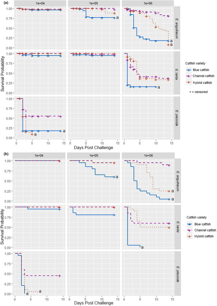
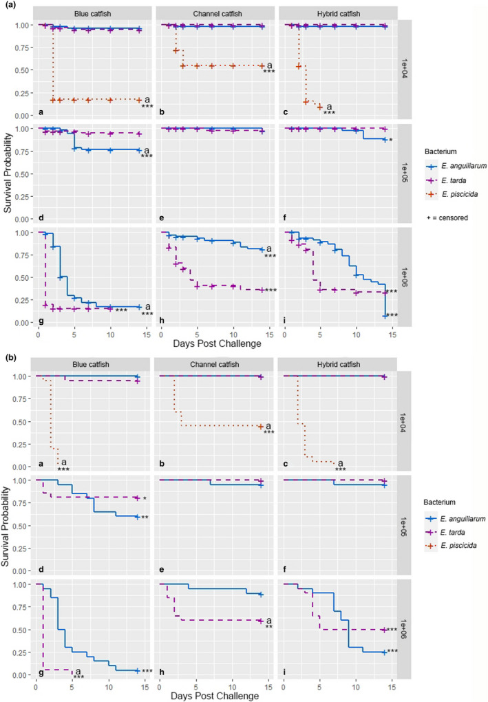
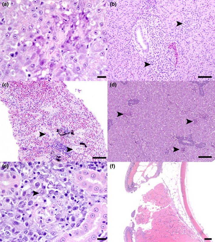
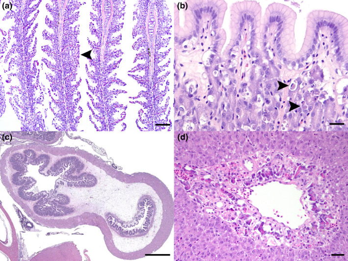
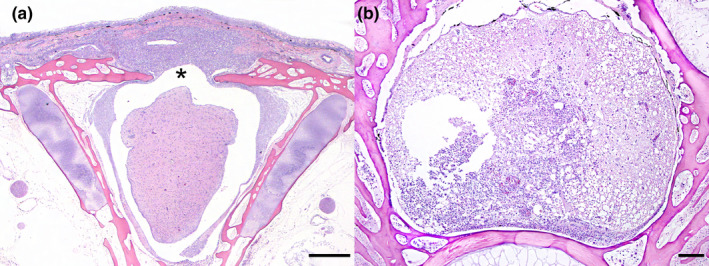
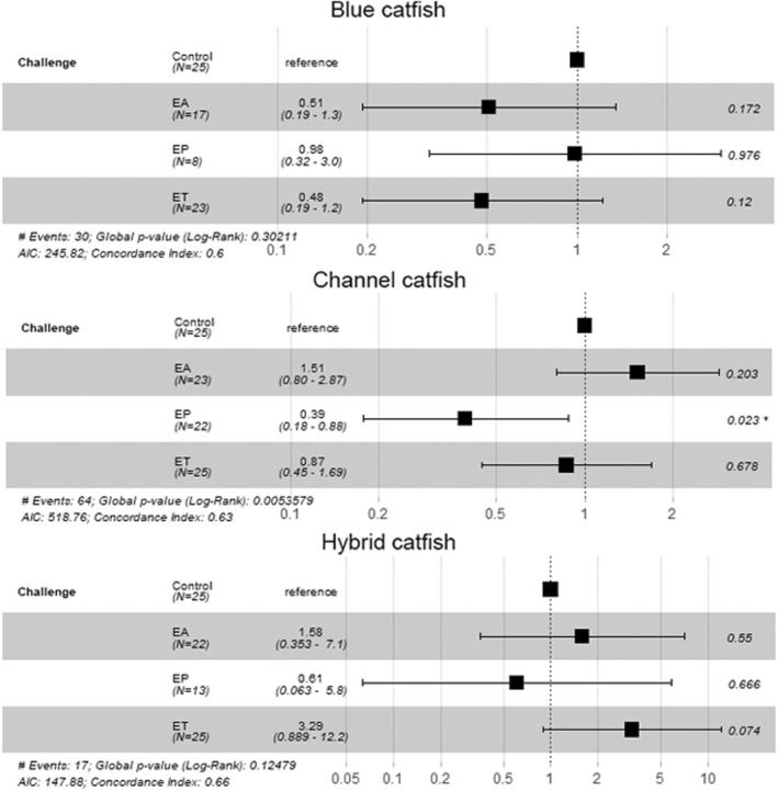
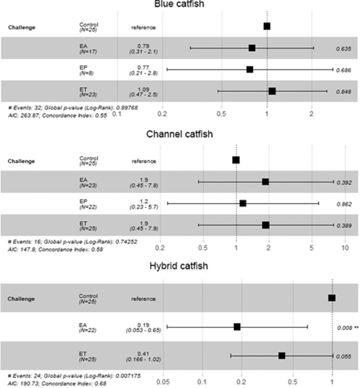
In the mid-2010s, Edwardsiella tarda was reaffiliated into three discrete taxa (E. anguillarum, E. piscicida, and E. tarda), obscuring previous descriptions of E. tarda-induced pathology in fish. To clarify ambiguity regarding the pathology of E. tarda, E. piscicida, and E. anguillarum infections in US farm-raised catfish, channel catfish (Ictalurus punctatus), blue catfish (I. furcatus), and channel × blue catfish hybrids were challenged with comparable doses of each bacterium. The most severe pathology and mortality occurred in fish challenged with E. piscicida, supporting previous reports of increased pathogenicity in commercially important ictalurids, while E. anguillarum and E. tarda warrant only minimal concern. Acute pathologic lesions among bacterial species were predominantly necrotizing and characteristic of gram-negative sepsis but became progressively granulomatous over time. After 100 days, survivors were exposed to the approximate median lethal doses of E. piscicida and E. ictaluri, revealing some cross-protective effects among E. piscicida, E. anguillarum, and E. ictaluri. In contrast, no fish that survived E. tarda challenge demonstrated any protection against E. piscicida or E. ictaluri. This work supports reports of increased susceptibility of channel, blue, and hybrid catfish to E. piscicida, while highlighting potential cross-protective affects among fish associated Edwardsiella spp.

Keywords: Edwardsiella anguillarum; Edwardsiella piscicida; Edwardsiella tarda; catfish; histopathology.


Conflict of interest statement

The author(s) declared no potential conflicts of interest.

Figures

FIGURE 1
FIGURE 1
(a) Kaplan–Meier survival curves with censoring from sampling tanks (n = 20 fish each tank; 3 tanks) for delivered doses of Edwardsiella anguillarum, Edwardsiella tarda, and Edwardsiella piscicida in channel, blue, and hybrid catfish. Controls (1.0 survival probability) are excluded from the figure. Survival probabilities for hybrid and blue catfish were compared with channel catfish using Cox proportional hazards models. Significant differences (p < .01) are indicated by “a”. No challenges were performed at the 1e + 05 or 1e + 06 levels for E. piscicida. Doses approximate colony forming units (CFU) per gram of fish. (b) Kaplan–Meier survival curves from non‐sampling tanks (n = 20 fish each tank; one tank) for delivered doses of Edwardsiella anguillarum, Edwardsiella tarda, and Edwardsiella piscicida in channel, blue, and hybrid catfish. Controls (1.0 survival probability) are excluded from the figure. Survival probabilities for hybrid and blue catfish were compared with channel catfish using Cox proportional hazards models. Significant differences (p < .05) are indicated by “a”. Data was transformed when one or more treatment's mortality equaled 0%. No challenges were performed at the 1e + 05 or 1e + 06 levels for E. piscicida. Doses approximate colony forming units (CFU) per gram of fish.
FIGURE 2
FIGURE 2
(a) Kaplan–Meier survival curves with censoring from sampling tanks (n = 20 fish each tank; three tanks) for channel, blue, and hybrid catfish at comparable delivered doses of Edwardsiella anguillarum, Edwardsiella tarda, and Edwardsiella piscicida. Controls (1.0 survival probability) are excluded from the figure. Bacteria were individually compared with controls using log‐tank tests (*p < .05; ***p < .001). (a–c) Kaplan–Meier survival curves for catfish exposed to E. tarda and E. anguillarum were compared with fish exposed to E. piscicida using Cox proportional hazards models. Significant differences (p < .05) are indicated by “a”. (d–i) Kaplan–Meier survival curves for catfish exposed to E. tarda were compared with fish exposed to E. anguillarum using Cox proportional hazards models. Significant differences (p < .05) are indicated by “a”. No challenges were performed at the 1e + 05 or 1e + 06 levels for E. piscicida. Doses approximate colony forming units (CFU) per gram of fish. (b) Kaplan–Meier survival curves from non‐sampling tanks (n = 20 fish each tank; 1 tank) for channel, blue, and hybrid catfish at comparable delivered doses of Edwardsiella anguillarum, Edwardsiella tarda, and Edwardsiella piscicida. Controls (1.0 survival probability) are excluded from the figure. Bacteria were individually compared with controls using log‐tank tests (*p < .05; ***p < .001). (a–c) Kaplan–Meier survival curves for catfish exposed to E. piscicida were compared individually to catfish exposed to E. tarda and E. anguillarum using log‐rank tests. Significant differences (p < .05) are indicated by “a”. (d–i) Kaplan–Meier survival curves for catfish exposed to E. tarda were compared with fish exposed to E. anguillarum using log‐rank tests. Significant differences (p < .05) are indicated by “a”. No challenges were performed at the 1e + 05 or 1e + 06 levels for E. piscicida. Doses approximate colony forming units (CFU) per gram of fish.
FIGURE 3
FIGURE 3
Gross lesions of Edwardsiella spp. infection. (a) Edwardsiella piscicida infection, channel catfish, 10 days post challenge (dpc). The skin overlying cranial fontanelle is moderately raised, light pink, and minimally ulcerated. (b) Edwardsiella tarda infection, channel catfish, 2 dpc. The lateral body wall is transmurally ulcerated caudal to the operculum. (c) E. tarda infection, hybrid catfish, 2 dpc. The transmural body wall ulceration exposes the underlying swim bladder and coelomic viscera. (d) E. anguillarum infection, channel catfish, 5 dpc. The coelomic cavity contains a moderate amount of light pink to yellow, serosanguinous, thin liquid.
FIGURE 4
FIGURE 4
Histopathology of Edwardsiella spp. necrotizing and inflammatory lesions. (a) Liver, hybrid catfish, E. piscicida low dose, 3 dpc. Acute necrotic areas characterized by hypereosinophilic cells with pyknotic nuclei, scattered cellular debris, and loss of cellular detail. Eosinophilic fibrin, abundant bacterial rods (arrowhead) and cellular debris are within the lesion center. Haematoxylin and eosin stain (H&E). Bar = 10 μm. (b) Liver, blue catfish, E. anguillarum middle dose, 14 dpc. Multifocal, well‐demarcated aggregates of macrophages (arrowheads) infiltrate the parenchyma and contain rare intracytoplasmic bacterial rods better visualized in figure (a) above. H&E. Bar = 50 μm (c) Spleen, channel catfish, E. tarda high dose, 1 dpc. Severely congested red pulp highlights necrotic splenic ellipsoids (arrowheads) containing abundant extracellular bacterial rods and karyorrhectic debris better visualized at higher magnification. H&E. Bar = 50 μm. (d) Liver, blue catfish, E. piscicida high dose, 3 dpc. Multiple random foci of necrosis (arrowheads) with swollen, hypereosinophilic hepatocytes containing pyknotic nuclei. H&E. Bar = 150 μm. (e) Posterior kidney, hybrid catfish, E. piscicida low dose, 3 dpc. Interstitial haematopoietic tissue is effaced by a focal area of necrosis, characterized by swollen hypoeosinophilic cells, loss of cellular detail, karyorrhectic debris, and abundant bacterial rods (arrowhead). H&E. Bar = 10 μm. (f) Lateral body wall, hybrid catfish, E. tarda high dose, 3 dpc. Ulceration and transmural necrosis within an extensive area of body wall. Necrotic and adjacent intact skeletal muscle is expanded by haemorrhage, macrophages, lymphocytes, karyorrhectic debris, and small numbers of bacterial rods. H&E. Bar = 200 μm.
FIGURE 5
FIGURE 5
Histopathology Edwardsiella spp. non‐inflammatory lesions. (a) Gills, hybrid catfish, E. anguillarum medium dose, 5 dpc. Epidermal hyperplasia multifocally expands lamellae and fills interlamellar troughs (arrowhead) accompanied by low numbers of mucous and inflammatory cells. H&E. Bar = 50 μm. (b) Stomach, channel catfish, E. anguillarum high dose, 2 dpc. Clear space surrounds apoptotic gastric glandular cells that are shrunken, rounded, and hypereosinophilic with pyknotic nuclei (arrowheads). H&E. Bar = 20 μm. (c) Stomach, channel catfish, E. anguillarum high dose, 2 dpc. The submucosa is diffusely expanded by abundant clear space characteristic of severe edema. H&E. Bar = 1 mm. (d) Intrahepatic exocrine pancreas, channel catfish, E. tarda high dose, 3 dpc. Pancreatic cells surrounding hepatic venules are often shrunken and surrounded by clear space with decreased cytoplasm, decreased zymogen granules, and/or condensed nuclei. H&E. Bar = 20 μm.
FIGURE 6
FIGURE 6
Central nervous system lesions in an Edwardsiella piscicida high‐dose challenged channel catfish 10 dpc. (a) Brain. The forebrain meninges are markedly expanded by sheets of granulomatous inflammation, predominated by macrophages, that extends through the cranial fontanel (asterisk) to infiltrate the dermis and elevate the overlying epidermis. The neuroparenchyma is minimally affected. H&E. Bar = 500 μm. (b) Spinal cord. Similar inflammatory infiltrates efface a focally extensive area of the spinal cord and ventral meninges, with marked vacuolation of the adjacent spinal tissue and dorsal displacement of the central canal. H&E. Bar = 100 μm.
FIGURE 7
FIGURE 7
Cox proportional hazard models for blue, channel, and hybrid catfish initially exposed to E. anguillarum (EA), E. piscicida (EP), or E. tarda (ET) and subsequently exposed to E. ictaluri via immersion bath or a sham challenge. Results indicate probability of mortality compared with naive controls. Risk ratio of 1 (dotted lines) indicate risk of mortality equal to naive controls. Treatments with whiskers lying fully to the left or right of the dotted line indicate a significant reduction or increase in risk of mortality, respectively. *p < .05. Concordance index is a goodness‐of‐fit metric with values >0.5 indicating a model with discriminatory power.
FIGURE 8
FIGURE 8
Cox proportional hazard ratios comparing blue, channel, and hybrid catfish initially exposed to E. anguillarum (EA), E. piscicida (EP), or E. tarda (ET) and subsequently exposed to E. piscicida via intracelomic injection or a sham challenge (**p < .01). Risk ratio of 1 (dotted lines) indicate risk of mortality equal to naive controls. Treatments with whiskers lying fully to the left or right of the dotted line indicate a significant reduction or increase in risk of mortality, respectively. E. piscicida in hybrids is excluded from the figure based on 0% mortality in hybrids previously challenged with E. piscicida but was significantly different (p < .01) than naive controls based on a log‐rank test. Concordance index is a goodness‐of‐fit metric with values >0.5 indicating a model with discriminatory power.

Similar articles

Cited by

References

    1. Aarattuthodiyil, S. , Griffin, M. J. , Greenway, T. E. , Khoo, L. H. , Byars, T. S. , Lewis, M. , Steadman, J. , & Wise, D. J. (2020). An orally delivered, live‐attenuated Edwardsiella ictaluri vaccine efficiently protects channel catfish fingerlings against multiple Edwardsiella ictaluri field isolates. Journal of the World Aquaculture Society, 51(6), 1354–1372. 10.1111/jwas.12693 - DOI
    1. Abayneh, T. , Colquhoun, D. , & Sørum, H. (2013). Edwardsiella piscicida sp. nov., a novel species pathogenic to fish. Journal of Applied Microbiology, 114, 644–654. 10.1111/jam.12080 - DOI - PubMed
    1. Abayneh, T. , Colquhoun, D. J. , & Sørum, H. (2012). Multi‐locus sequence analysis (MLSA) of Edwardsiella tarda isolates from fish. Veterinary Microbiology, 158(3–4), 367–375. 10.1016/j.vetmic.2012.03.006 - DOI - PubMed
    1. Armwood, A. R. , Camus, A. C. , López‐Porras, A. , Ware, C. , Griffin, M. J. , & Soto, E. (2019). Pathologic changes in cultured Nile tilapia (Oreochromis niloticus) associated with an outbreak of Edwardsiella anguillarum . Journal of Fish Diseases, 42, 1463–1469. 10.1111/jfd.13058 - DOI - PubMed
    1. Baumgartner, W. A. , Ford, L. , & Hanson, L. (2017). Lesions caused by virulent Aeromonas hydrophila in farmed catfish (Ictalurus punctatus and I. punctatus× I. furcatus) in Mississippi. Journal of Veterinary Diagnostic Investigation, 29, 747–751. 10.1177/1040638717708584 - DOI - PubMed

Supplementary concepts

LinkOut - more resources