. 2022 Jul 26:11:e77156.
doi: 10.7554/eLife.77156.

Lung evolution in vertebrates and the water-to-land transition

Affiliations

Lung evolution in vertebrates and the water-to-land transition

Camila Cupello et al. Elife. .

Abstract

A crucial evolutionary change in vertebrate history was the Palaeozoic (Devonian 419-359 million years ago) water-to-land transition, allowed by key morphological and physiological modifications including the acquisition of lungs. Nonetheless, the origin and early evolution of vertebrate lungs remain highly controversial, particularly whether the ancestral state was paired or unpaired. Due to the rarity of fossil soft tissue preservation, lung evolution can only be traced based on the extant phylogenetic bracket. Here we investigate, for the first time, lung morphology in extensive developmental series of key living lunged osteichthyans using synchrotron x-ray microtomography and histology. Our results shed light on the primitive state of vertebrate lungs as unpaired, evolving to be truly paired in the lineage towards the tetrapods. The water-to-land transition confronted profound physiological challenges and paired lungs were decisive for increasing the surface area and the pulmonary compliance and volume, especially during the air-breathing on land.

Keywords: actinopterygii; developmental biology; evolutionary biology; osteichthyes; sarcopterygii; tetrapod.

Plain language summary

All life on Earth started out under water. However, around 400 million years ago some vertebrates, such as fish, started developing limbs and other characteristics that allowed them to explore life on land. One of the most pivotal features to evolve was the lungs, which gave vertebrates the ability to breathe above water. Most land-living vertebrates, including humans, have two lungs which sit on either side of their chest. The lungs extract oxygen from the atmosphere and transfer it to the bloodstream in exchange for carbon dioxide which then gets exhaled out in to the atmosphere. How this important organ first evolved is a hotly debated topic. This is largely because lung tissue does not preserve well in fossils, making it difficult to trace how the lungs of vertebrates changed over the course of evolution. To overcome this barrier, Cupello et al. compared the lungs of living species which are crucial to understand the early stages of the water-to-land transition. This included four species of lunged bony fish which breathe air at the water surface, and a four-legged salamander that lives on land. Cupello et al. used a range of techniques to examine how the lungs of the bony fish and salamander changed shape during development. The results suggested that the lungs of vertebrates started out as a single organ, which became truly paired later in evolution once vertebrates started developing limbs. This anatomical shift increased the surface area available for exchanging oxygen and carbon dioxide so that vertebrates could breathe more easily on land. These findings provide new insights in to how the lung evolved into the paired structure found in most vertebrates alive today. It likely that this transition allowed vertebrates to fully adapt to breathing above water, which may explain why this event only happened once over the course of evolution.


Conflict of interest statement

CC, TH, NT, YY, PG, SI, RM, TS, AK, MH, KU, MO, PB No competing interests declared

Figures

Figure 1.
Figure 1.. Three-dimensional reconstructions of the pulmonary complex of Polypterus senegalus.
(A) Early embryo (9.3mm) total length (TL) in right lateral view, (B) isolated lung of the early embryo in dorsal view, (C) juvenile (45mm TL) in right lateral view, (D) isolated lung of the juvenile in dorsal view, (E) close-up of (D) highlighting the lung in ventral view and pointing out the region of the independent and secondary connection of the left sac to the right one by a lateral opening. Yellow, foregut including the stomach; red, lung. Black arrow in (A) pointing to the lung. Arrowheads in (B) pointing to the lung connection to the foregut and in (D) pointing to the pneumatic duct connection to the foregut. Black arrow in (E) pointing to the independent connection. Ls, left sac; rs, right sac; ulb, unpaired lung bud. Scale bars, 5.0mm (A); 0.075mm (B); 5.0mm (C, D); 1.0mm (E).
Figure 1—figure supplement 1.
Figure 1—figure supplement 1.. Sections of synchrotron x-ray microtomography of a juvenile of Polypterus senegalus (23 mm total length; TL).
(A) Unpaired lung origin. (B) Right sac arising from the foregut. (C) Left sac arising from an independent and lateral connection to the right sac. (D) Right and left sacs. Yellow, foregut; red, lung. Orange arrow, opened connection between foregut and lung. Fg, foregut; ls, left sac; rs, right sac. Scale bars, 0.5 mm (A–D).
Figure 1—figure supplement 2.
Figure 1—figure supplement 2.. Three-dimensional reconstructions of the pulmonary complex of Polypterus senegalus.
(A) Virtual section of the juvenile (45 mm total length; TL) in anterior view, evidencing the esophagus and the lung in 3D. (B) Isolated right lung of the juvenile in lateral view, evidencing the independent and secondary connection of the left sac to the right one by a lateral opening. Yellow, foregut including the stomach; red, right sac; blue, left sac, dashed line, independent and secondary connection of the left sac to the right one. Scale bars, 1.0 mm (A, B).
Figure 2.
Figure 2.. Coronal sections of the unpaired lung in the living actinopterygian fish Polypterus senegalus.
(A) No lung bud in 8.0mm total length (TL) specimen, (B) origin of an unpaired lung bud in 8.5mm TL specimen, (C) unpaired lung bud in 9.1mm TL specimen, (D) first register of an independent and lateral second lung bud in 12mm TL specimen, (E, F) independent and lateral second lung bud arising from the principal tube in 15.5mm TL and 18mm TL specimens. (A, C, E–F) Histological thin-sections. (B, D) Sections of synchrotron x-ray microtomography of the early embryo. Black and white head arrows pointing to the lumen of the unpaired lung buds; arrows pointing to the undifferentiated cells surrounding the glottis. Fg, foregut; ilb, independent lateral bud; rb, right bud; ulb, unpaired lung bud. Scale bars, 0.2mm (A, E); 0.1mm (B, D, F); 0.05mm (C).
Figure 3.
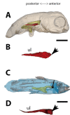
Figure 3.. Three-dimensional reconstructions of the pulmonary complex of Latimeria chalumnae.
(A) Early embryo of Latimeria chalumane (45mm total length; TL) in right lateral view (Cupello et al., 2015), (B) isolated unpaired lung of the early embryo in dorsal view, (C) adult specimen of Latimeria chalumnae (1300mm TL) in right lateral view (Cupello et al., 2015), (D) isolated unpaired lung of the adult specimen in dorsal view. Yellow, foregut including the stomach; red, lung. Arrowheads in (B) and (D) pointing to the lung connection to the foregut. Black arrow in (C) pointing to the lung. Ul, unpaired lung bud in (B) and unpaired lung in (D). Scale bars, 5.0mm (A); 5.0mm (B); 200.0mm (C); 40mm (D). Modified from Cupello et al., 2015.
Figure 4.
Figure 4.. Three-dimensional reconstructions of the pulmonary complex of two species of lungfishes.
(A) Early embryo of Neoceratodus forsteri (13.5mm total length; TL) in right lateral view, (B) isolated unpaired lung of the early embryo in dorsal view, (C) adult specimen of Neoceratodus forsteri (200mm TL) in right lateral view, (D) isolated unpaired lung of the adult specimen in dorsal view, (E) close-up of the lung unpaired connection to the foregut in (D), (F) larva of Lepidosiren paradoxa (46mm TL) in lateral view, (G) isolated lung of the larval specimen in dorsal view, (H) close-up of the lung unpaired connection to the foregut in (G), (I) juvenile of Lepidosiren paradoxa young adult (68mm TL) in lateral view, (J) isolated lung of the juvenile specimen in dorsal view, (K) close-up of the lung unpaired connection to the foregut in (J).Yellow, foregut including the stomach; red, lung. Black arrow in (A) pointing to the lung. Arrowheads in (B), pointing to the lung connection to the foregut and in (D), (G) and (J) pointing the pneumatic duct connection to the foregut. Ls, left sac; rs, right sac; ul, unpaired lung; ulb, unpaired lung bud. Scale bars, 2.5mm (A); 0.1mm (B); 20mm (C); 10mm (D, I); 7.2 mm (J); 5.0mm (F, G).
Figure 4—figure supplement 1.
Figure 4—figure supplement 1.. Sections of synchrotron x-ray microtomography of a larva of Neoceratodus forsteri (19 mm total length; TL).
(A) Unpaired lung origin. (B) Unique sac arising from the foregut. (C, D) Unique sac developing. Yellow, foregut; red, lung. Orange arrow, opened connection between foregut and lung. fg, foregut; us, unique sac. Scale bars, 0.5 mm (A–D).
Figure 4—figure supplement 2.
Figure 4—figure supplement 2.. Sections of synchrotron x-ray microtomography of a juvenile of Lepidosiren paradoxa (68mm total length; TL).
(A) Unpaired lung origin. (B) Right sac arising from the foregut. (C) Left sac arising from an independent and lateral connection to the right sac. (D) Right and left sacs. Yellow, foregut; red, lung. Orange arrow, opened connection between foregut and lung. Fg, foregut; ls, left sac; rs, right sac. Scale bars, 0.5mm (A–D).
Figure 4—figure supplement 3.
Figure 4—figure supplement 3.. Dissection of the lung of an adult Lepidosiren paradoxa (400mm total length; TL).
Red arrow, lung. Black arrow, ventral insertion of the right sac. Ls, left sac; rs, right sac. Scale bars, 50mm (A, B); 10mm (C).
Figure 5.
Figure 5.. Three-dimensional reconstructions of the pulmonary complex of Salamandra salamandra.
(A) Early larva of Salamandra salamandra (35.5mm total length; TL) in right lateral view, (B) isolated paired lung of the larva embryo in dorsal view, (C) juvenile of Salamandra salamandra (81.85mm TL) in right lateral view, (D) isolated paired lung of the juvenile specimen in dorsal view. Yellow, foregut including the stomach; red, lung. Arrowheads in (B) and (D) pointing to the trachea connection to the foregut. Ll, left lung; rl, right lung. Scale bars, 5.0mm (A); 3.125mm (B); 10mm (C); 6.25cm (D).
Figure 5—figure supplement 1.
Figure 5—figure supplement 1.. Sections of synchrotron x-ray microtomography of a larva of Salamandra salamandra (42.8mm total length; TL).
(A, B) Trachea arising. (C, D) Fist order bronchioles. (E) Right and left lungs arising simultaneously and symmetrically. Yellow, foregut; red, lung. Orange arrow, opened connection from the foregut. Br, braonchile; fg, foregut; ll, left lung; rl, right lung; tr, trachea. Scale bars, 0.5mm (A–D).
Figure 6.
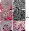
Figure 6.. Comparison of sections showing the difference in lung origin and connection between unpaired (A–H) and true paired lungs (I, J).
(A, B) Virtual section of Polypterus senegalus (12mm total length; TL), (C) histological thin section of Latimeria chalumnae (127cm) (Cupello et al., 2017a), (D) virtual section of L. chalumnae (40mm TL; modified from Cupello et al., 2017a), (E, F) virtual section of Neoceratodus forsteri (16mm TL), (G, H) virtual section of Lepidosiren paradoxa (46mm TL), (I, J) virtual section of Salamandra salamandra (35.5mm TL). Yellow, foregut including the stomach; red, lung. Orange arrows, opened connection between the foregut and the lung. Fg, foregut; ll, left lung; ls, left sac; rb, right bud; ilb, independent lung bud; rl, right lung; rs, right sac; ulb, unpaired lung bud. Scale bars, 0.25mm (A, B); 3.0mm (C); 1.0mm (D); 0.1mm (E, F); 0.5mm (G, H); 1.25mm (I, J).
Figure 7.
Figure 7.. Schematic figure reconstructing the evolutionary history of vertebrate lungs.
All living actinopterygian and sarcopterygian fishes have unpaired lungs. True paired lungs are a synapomorphy of tetrapods. Dashed circle in Cladistia lung pointing to the secondary and independent opening to a left sac, at the lung level. Modified from Liem, 1988. This figure was made with free silhouettes from PhyloPic.

References

    1. Amemiya CT, Alföldi J, Lee AP, Fan S, Philippe H, Maccallum I, Braasch I, Manousaki T, Schneider I, Rohner N, Organ C, Chalopin D, Smith JJ, Robinson M, Dorrington RA, Gerdol M, Aken B, Biscotti MA, Barucca M, Baurain D, Berlin AM, Blatch GL, Buonocore F, Burmester T, Campbell MS, Canapa A, Cannon JP, Christoffels A, De Moro G, Edkins AL, Fan L, Fausto AM, Feiner N, Forconi M, Gamieldien J, Gnerre S, Gnirke A, Goldstone JV, Haerty W, Hahn ME, Hesse U, Hoffmann S, Johnson J, Karchner SI, Kuraku S, Lara M, Levin JZ, Litman GW, Mauceli E, Miyake T, Mueller MG, Nelson DR, Nitsche A, Olmo E, Ota T, Pallavicini A, Panji S, Picone B, Ponting CP, Prohaska SJ, Przybylski D, Saha NR, Ravi V, Ribeiro FJ, Sauka-Spengler T, Scapigliati G, Searle SMJ, Sharpe T, Simakov O, Stadler PF, Stegeman JJ, Sumiyama K, Tabbaa D, Tafer H, Turner-Maier J, van Heusden P, White S, Williams L, Yandell M, Brinkmann H, Volff JN, Tabin CJ, Shubin N, Schartl M, Jaffe DB, Postlethwait JH, Venkatesh B, Di Palma F, Lander ES, Meyer A, Lindblad-Toh K. The African coelacanth genome provides insights into tetrapod evolution. Nature. 2013;496:311–316. doi: 10.1038/nature12027. - DOI - PMC - PubMed
    1. Arsenault M, Desbiens S, Janvier P, Kerr J. In: Recent Advances in the Origin and Early Radiation of Vertebrates. Arratia G, Wilson MVH, Cloutier R, editors. Munich, Germany: Verlag Dr. Friedrich Pfeil; 2004. New data on the soft tissues and external morphology of the antiarch Bothriolepis canadensis (Whiteaves, 1880) from the Upper Devonian of Miguasha, Québec; pp. 439–454.
    1. Bartsch P, Gemballa S, Piotrowski T. The embryonic and larval development of Polypterus senegalus cuvier, 1829: its staging with reference to external and skeletal features, behaviour and locomotory habits. Acta Zoologica. 1997;78:309–328. doi: 10.1111/j.1463-6395.1997.tb01014.x. - DOI
    1. Béchard I, Arsenault F, Cloutier R, Kerr J. The Devonian placoderm fish Bothriolepis canadensis revisited with three-dimensional digital imagery. Palaeontologia Electronica. 2014;17:1–19. doi: 10.26879/417. - DOI
    1. Bi X, Wang K, Yang L, Pan H, Jiang H, Wei Q, Fang M, Yu H, Zhu C, Cai Y, He Y, Gan X, Zeng H, Yu D, Zhu Y, Jiang H, Qiu Q, Yang H, Zhang YE, Wang W, Zhu M, He S, Zhang G. Tracing the genetic footprints of vertebrate landing in non-teleost ray-finned fishes. Cell. 2021;184:1377–1391. doi: 10.1016/j.cell.2021.01.046. - DOI - PubMed

Publication types