. 2022 Aug 11;65(15):10251-10284.
doi: 10.1021/acs.jmedchem.2c00552. Epub 2022 Jul 26.

Discovery of an Orally Bioavailable and Selective PKMYT1 Inhibitor, RP-6306

Affiliations

Discovery of an Orally Bioavailable and Selective PKMYT1 Inhibitor, RP-6306

Janek Szychowski et al. J Med Chem. .

Abstract

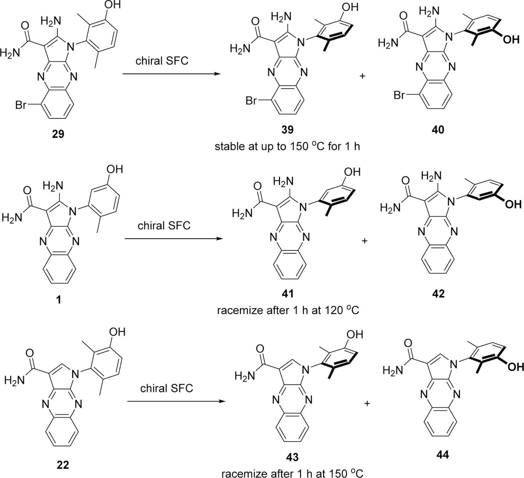
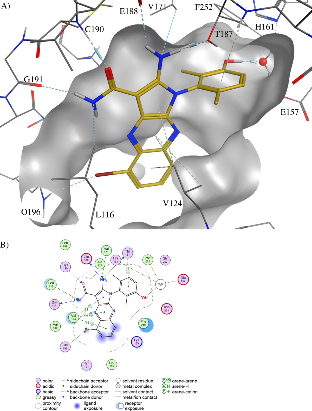
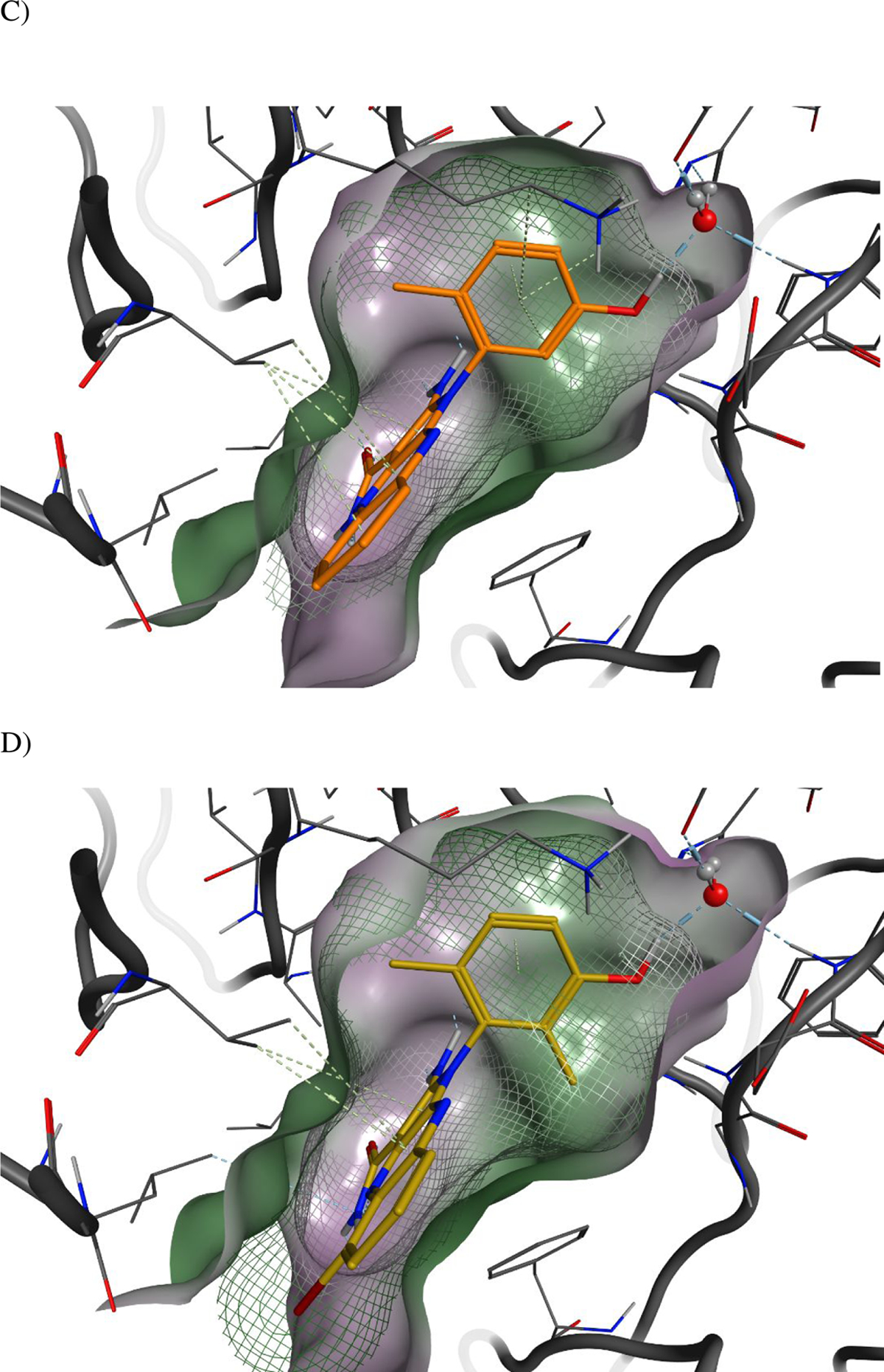
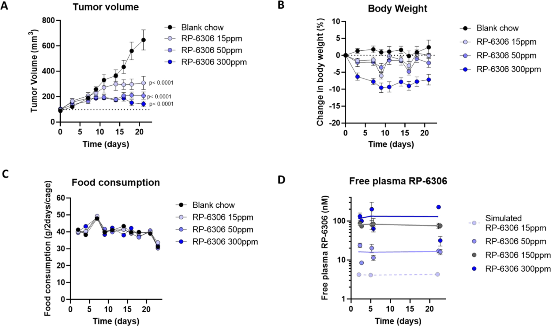
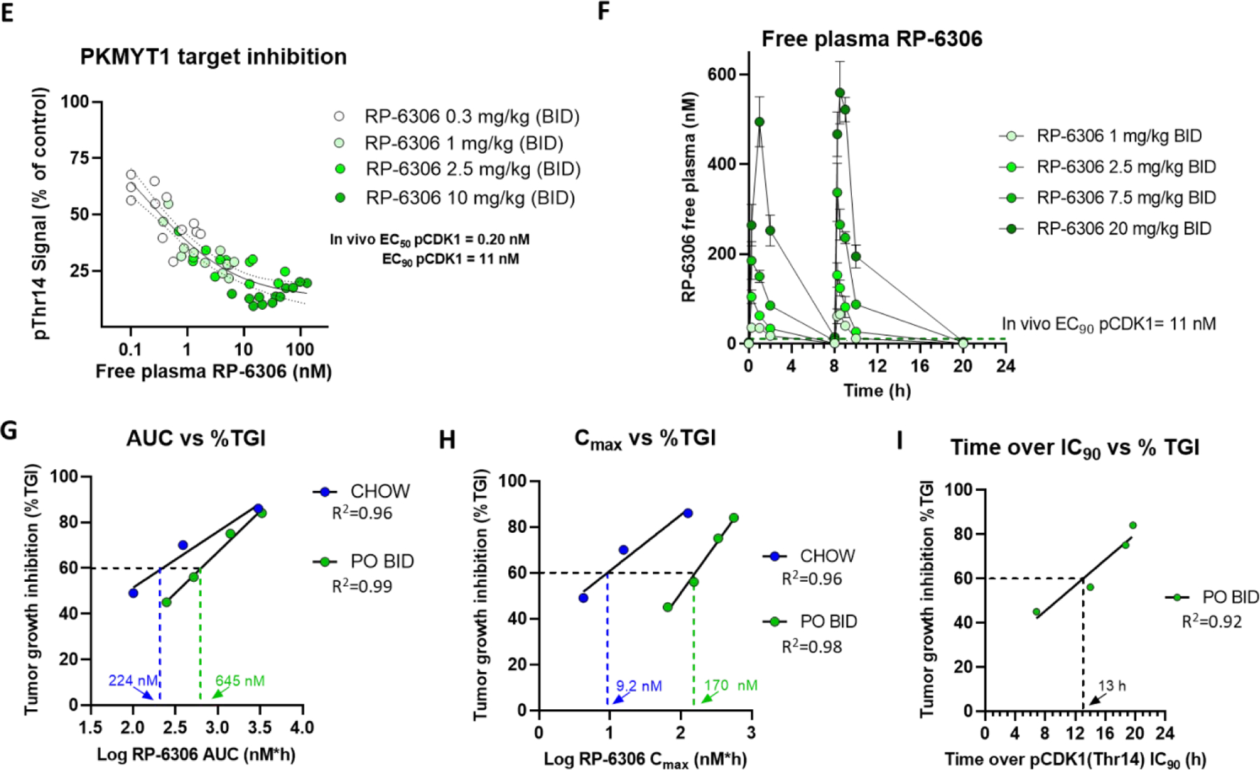
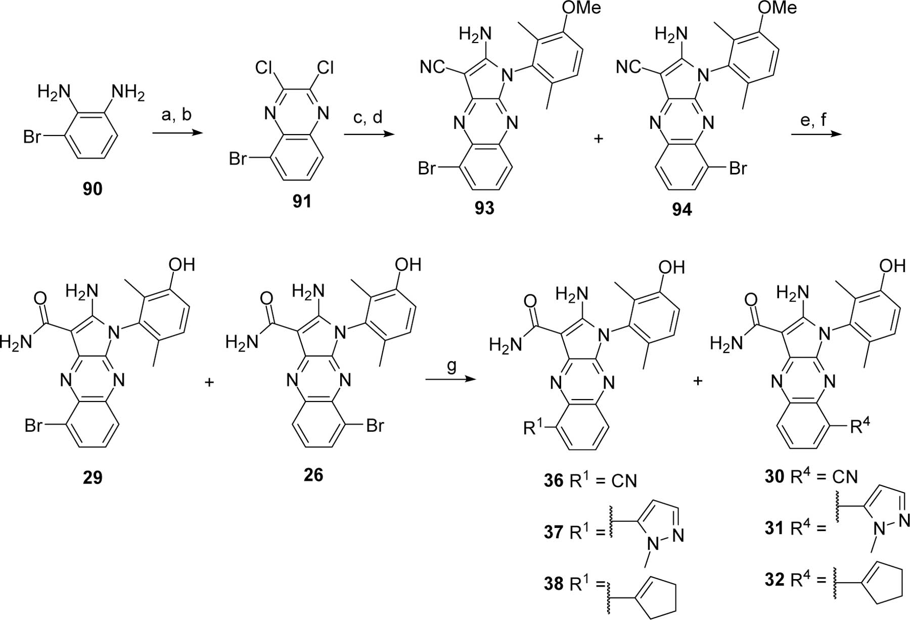
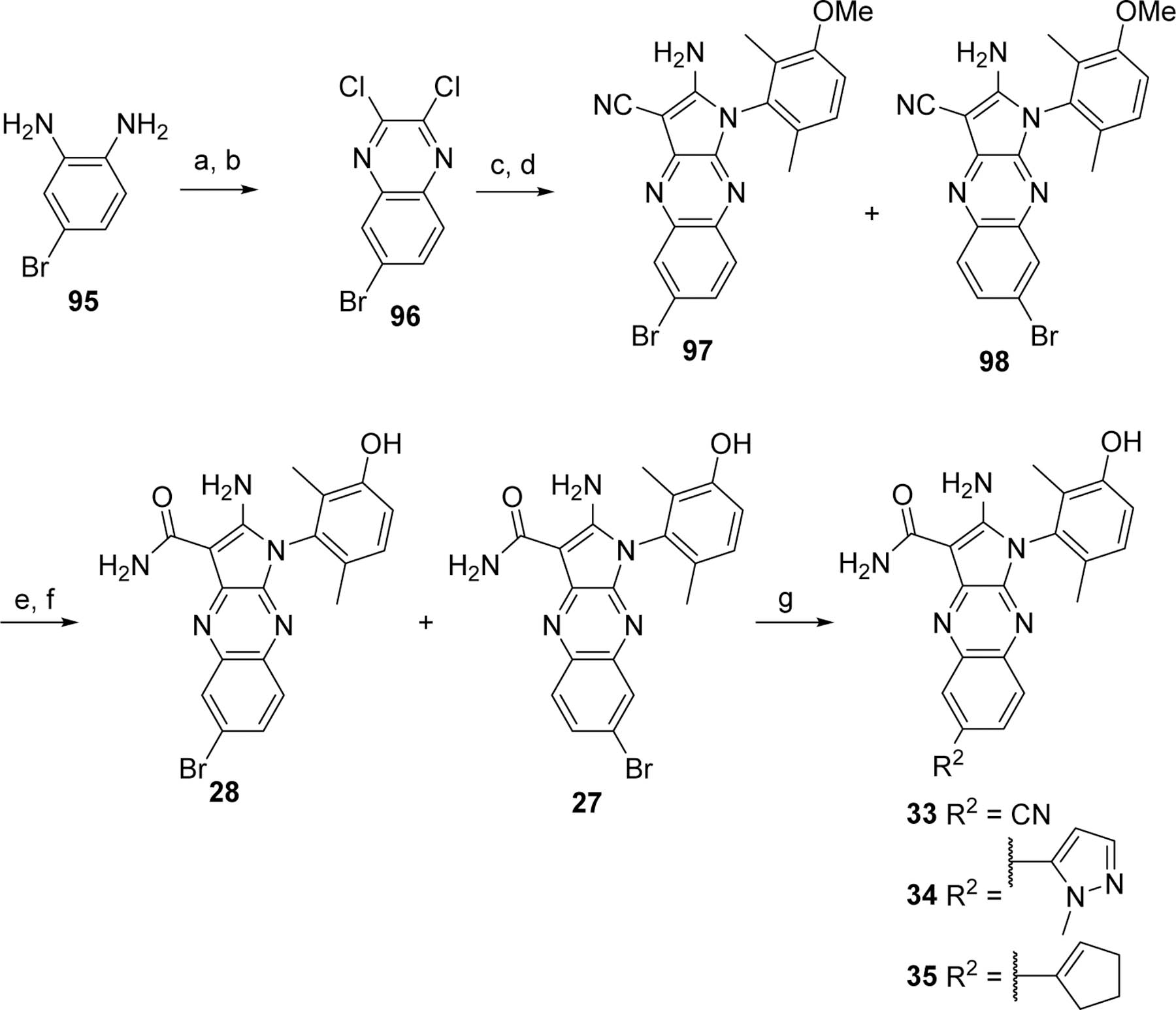
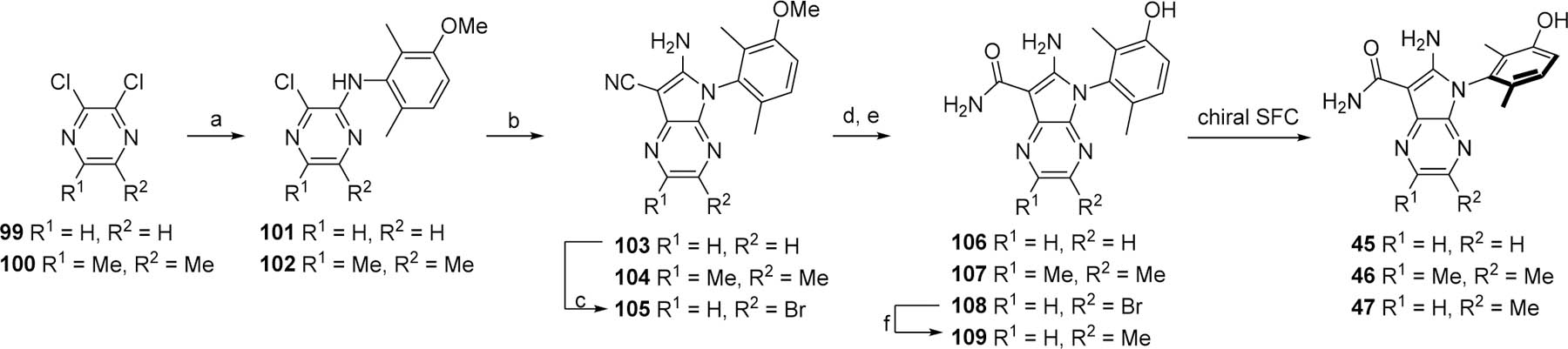
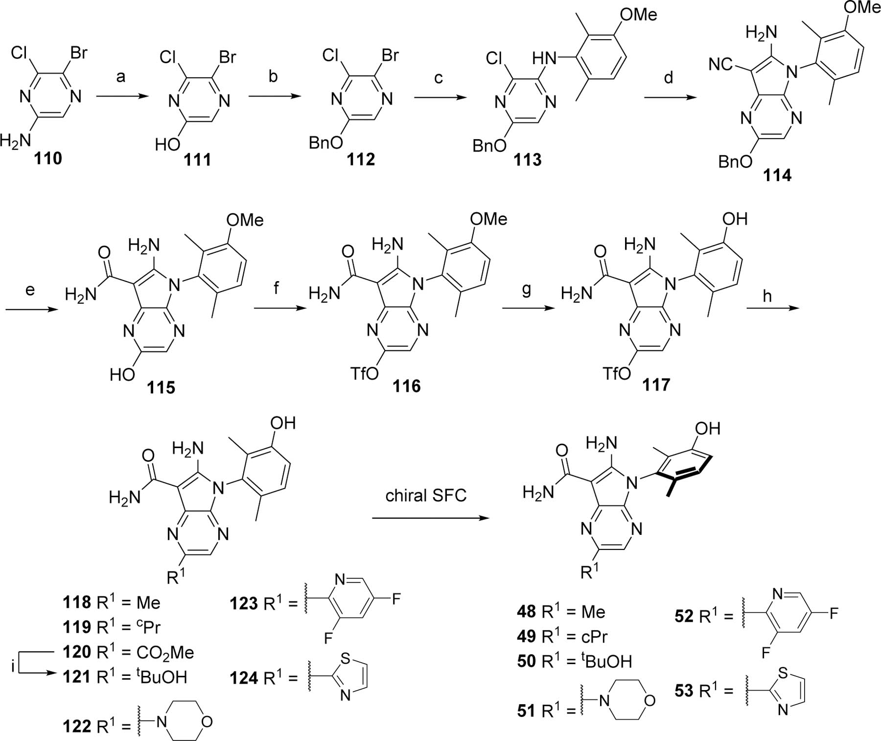
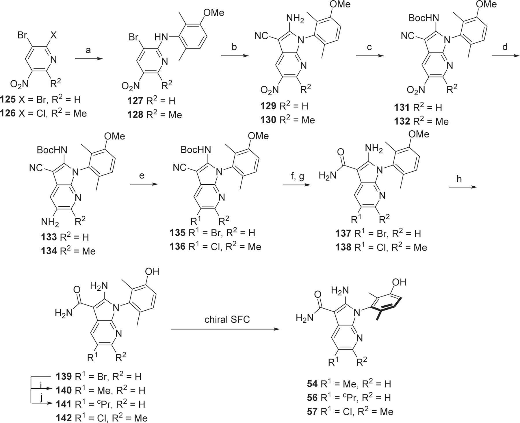
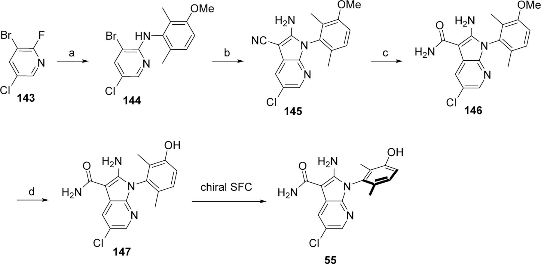
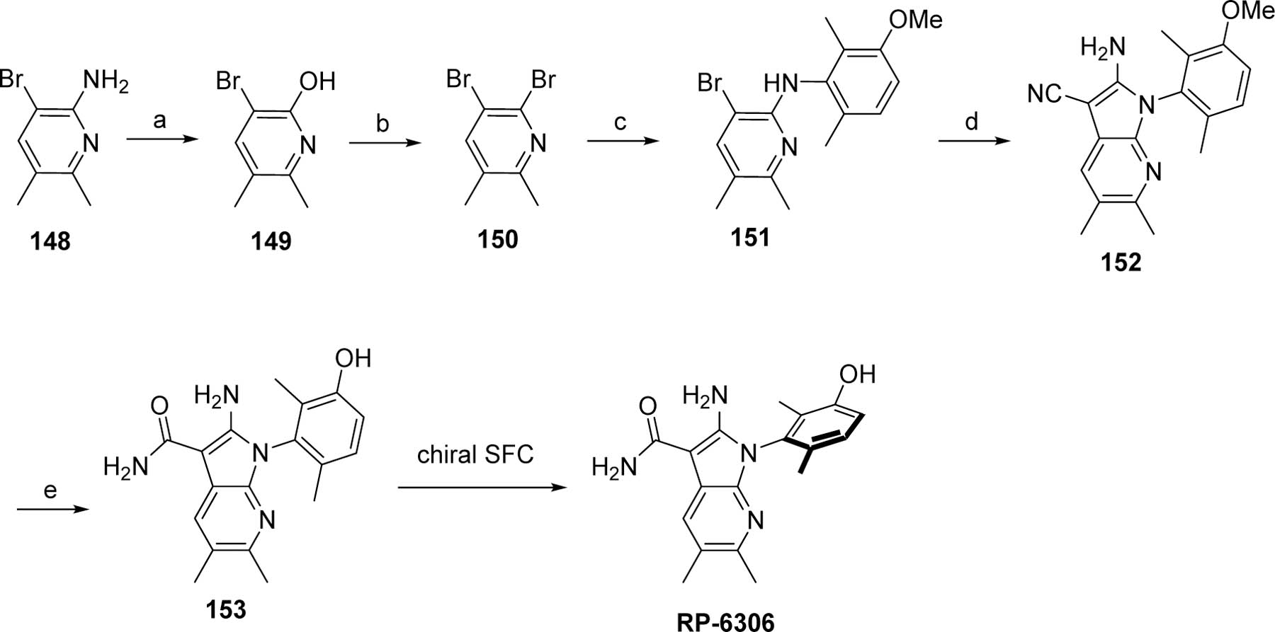
PKMYT1 is a regulator of CDK1 phosphorylation and is a compelling therapeutic target for the treatment of certain types of DNA damage response cancers due to its established synthetic lethal relationship with CCNE1 amplification. To date, no selective inhibitors have been reported for this kinase that would allow for investigation of the pharmacological role of PKMYT1. To address this need compound 1 was identified as a weak PKMYT1 inhibitor. Introduction of a dimethylphenol increased potency on PKMYT1. These dimethylphenol analogs were found to exist as atropisomers that could be separated and profiled as single enantiomers. Structure-based drug design enabled optimization of cell-based potency. Parallel optimization of ADME properties led to the identification of potent and selective inhibitors of PKMYT1. RP-6306 inhibits CCNE1-amplified tumor cell growth in several preclinical xenograft models. The first-in-class clinical candidate RP-6306 is currently being evaluated in Phase 1 clinical trials for treatment of various solid tumors.


Conflict of interest statement

Conflict of Interest Disclosure

J. S.; R. P.; E. D.; B. L.; F. V.; M.-E. L.; J. F.; A. L. P.; S. Y. Y.; A. R.; C. G. M.; N. M. D.; R. S.; C. G.; A. B.-F.; R. K.; V. B.; M. G. and W. C. B. are employees of Repare Therapeutics. F. S. is a founder and consultant of Repare Therapeutics. G. M.; V. P.; P. M.; J.-F. T.; M. D.; D. M.; S. O.; P. B.; P. T.; I. K. and Y. M. declare no competing financial interest.

Figures

Figure 1.
Figure 1.
Separation of representative atropisomers.
Figure 2.
Figure 2.
Kinativ Colo-20 cell lysate kinase binding profile of RP-6306 at 1.2 μM.
Figure 3.
Figure 3.
The binding mode of 39 to PKMYT1. A) Ribbons representation of 39 bound to PKMYT1 (crystal structure PDB ID 8D6D). 39 is shown as sticks with golden carbon atoms, and part of the solvent-accessible surface of the binding pocket is shown in light gray. Oxygen is rendered in red, nitrogen in blue, carbon in yellow, bromine in brown, and polar hydrogens in white. Favorable interactions are highlighted with dashed lines with stronger hydrogen bonds additionally highlighted with cylinders (as calculated by the Molecular Operating Environment, MOE, from the Chemical Computing Group, Inc.). A bridging water molecule is shown in ball-and-stick representation. B) 2D plot of the interactions that compound 39 forms with PKMYT1, as calculated by MOE. C) Surface-surface complementarity of the 6-monomethyl phenol of analog 41 bound to PKMYT1 (crystal structure PDB ID 8D6F). The solvent-accessible surface of the protein is shown as a solid surface, with polar regions in purple and hydrophobic patches in green. The solvent-accessible surface of the ligand is displayed as a mesh. D) Surface-surface complementarity of the 2,6-dimethyl phenol motif of analog 39 bound to PKMYT1. The solvent-accessible surfaces of the protein and ligand are represented, using the same convention as in panel C. E) The carboxamide-side pyrazine’s nitrogen is sandwiched between the side-chains of hydrophobic residues. The solvent-accessible surfaces of the protein and ligand are represented, using the same convention as in panel C.
Figure 3.
Figure 3.
The binding mode of 39 to PKMYT1. A) Ribbons representation of 39 bound to PKMYT1 (crystal structure PDB ID 8D6D). 39 is shown as sticks with golden carbon atoms, and part of the solvent-accessible surface of the binding pocket is shown in light gray. Oxygen is rendered in red, nitrogen in blue, carbon in yellow, bromine in brown, and polar hydrogens in white. Favorable interactions are highlighted with dashed lines with stronger hydrogen bonds additionally highlighted with cylinders (as calculated by the Molecular Operating Environment, MOE, from the Chemical Computing Group, Inc.). A bridging water molecule is shown in ball-and-stick representation. B) 2D plot of the interactions that compound 39 forms with PKMYT1, as calculated by MOE. C) Surface-surface complementarity of the 6-monomethyl phenol of analog 41 bound to PKMYT1 (crystal structure PDB ID 8D6F). The solvent-accessible surface of the protein is shown as a solid surface, with polar regions in purple and hydrophobic patches in green. The solvent-accessible surface of the ligand is displayed as a mesh. D) Surface-surface complementarity of the 2,6-dimethyl phenol motif of analog 39 bound to PKMYT1. The solvent-accessible surfaces of the protein and ligand are represented, using the same convention as in panel C. E) The carboxamide-side pyrazine’s nitrogen is sandwiched between the side-chains of hydrophobic residues. The solvent-accessible surfaces of the protein and ligand are represented, using the same convention as in panel C.
Figure 4.
Figure 4.
The binding mode of RP-6306 to PKMYT1. A) RP-6306 (crystal structure PDB ID 8D6E) is shown as sticks with pink carbon atoms, and part of the solvent-accessible surface of the binding pocket is rendered in light gray. Oxygen is rendered in red, nitrogen in blue, carbon in pink, bromine in brown, and polar hydrogens in white. Favorable interactions are highlighted with dashed lines with stronger hydrogen bounds additionally highlighted with cylinders (as calculated by MOE). Water molecules are shown as balls-and-sticks. B) 2D plot of the interactions that compound RP-6306 forms with PKMYT1, as calculated by MOE.
Figure 5.
Figure 5.. RP-6306 free plasma exposure and in vivo efficacy in the OVCAR3 CCNE1 amplified xenograft model.
A) Tumor xenograft volume and B) change in body weight in OVCAR3-bearing mice treated with RP-6306 formulated in chow for 21 days. Results are expressed as mean tumor volume ± SEM, N=8 mice / group. Statistical significance relative to vehicle control was established by One-Way ANOVA followed by Fisher’s LSD test (GraphPad Prism v8). C) The two-day mean chow consumption in mice receiving blank chow or chow mixed with RP-6306 at the indicated concentrations D) Measured free plasma levels of RP-6306 in chow formulation at the indicated doses measured at 6:30am and 4:30pm on Days 2, 5 and 22. The 15 ppm dose was simulated, assuming linearity from the 50 ppm dose. E) The proportion of OVCAR3 tumor pCDKThr14 signal relative to vehicle treated mice for each dose at 2, 6 and 10 h post PO dosing; mean ± SEM (N=4/group/time point). The tumor pCDK1(Thr14) EC50 was determined by a non-linear dose-response model (GraphPad Prism v9.30). F) Pharmacokinetics of RP-6306 administered PO BID at the indicated doses. G-I) The relationship between measured tumor growth inhibition (TGI) and free plasma RP-6306 exposure (AUC) G), Cmax H) or time over pCDK1(Thr14) EC90 I) at each chow (A, D) and BID dose (F and Gallo et al.) evaluated in efficacy studies.
Scheme 1.
Scheme 1.
Preparation of analogs for phenol ring SARs Reagents and conditions. a. malononitrile, NaH, DME; b. ArNH2, NMP; c. ArNH2, KOtBu, THF; d. malononitrile, NaH, Pd(PPh3)4, dioxane; e. H2SO4; f. BBr3 for methoxy deprotection or H2, Pd/C for NO2 reduction.
Scheme 2.
Scheme 2.
Preparation of substituted carboxamide analogs. Reagents and conditions. a. 81 NaOtBu, Pd2(dba)3, XantPhos, toluene; b. R3NH(CO)CH2CN, KOtBu, THF or Cs2CO3, DMF; c. HCl, dioxane.
Scheme 3.
Scheme 3.
Preparation of analogs with pyrrole NH2 replacements. Reagents and conditions. a. tBuONO, THF; b. tBuONO, CuCl, ACN; c. H2SO4; d. BBr3, CH2Cl2; e. BCl3, TBAI, CH2Cl2; f. Ghaffar-Parkins catalyst, EtOH, H2O.
Scheme 4.
Scheme 4.
Preparation of N-regioisomers 24 and 25. Reagents and conditions. a. 81, NaOtBu, Pd2(dba)3, Xantphos, toluene; b. malononitrile, KOtBu, DME; c. malononitrile, NaH, Pd(PPh3)4; d. HCl, dioxane; e. H2SO4.
Scheme 5.
Scheme 5.
Preparation and derivatization of the bromo regioisomers 26 and 29 Reagents and conditions. a. diethyl oxalate; b. SOCl2, DMF cat; c. malononitrile, NaH, DME; d. 92, NMP; e. H2SO4; f. BBr3, CH2Cl2; g. RB(OR)2, Pd(dppf)Cl2.CH2Cl2, K2CO3, DMF or CuCN, DMF.
Scheme 6.
Scheme 6.
Preparation and derivatization of the bromo regioisomers 27 and 28. Reagents and conditions. a. diethyl oxalate, reflux; b. SOCl2, DMF cat.; c. Malononitrile, NaH, DME; d. 92, NMP; e. H2SO4; f. BBr3, CH2Cl2; g. RB(OR)2, Pd(dppf)Cl2.CH2Cl2, K2CO3, DMF or CuCN, DMF.
Scheme 7.
Scheme 7.
Preparation of bicyclic pyrrolopyrazine analogs 45–47. Reagents and conditions. a. Malononitrile, NaH, THF; b. 92, NMP or 92 KOtBu, Pd-PEPPSI-SIPr, NMP; c. NBS, DMF; d. H2SO4; e. BBr3, CH2Cl2; f. MeMgBr, ZnCl2, THF, then Pd(PPh3)4, THF.
Scheme 8.
Scheme 8.
Preparation of bicyclic pyrrolopyrazine analogs 49–53. Reagents and conditions. a. NaNO2, H2SO4; b. BnBr, Ag2CO3, toluene; c. 92, Pd2(dba)3, Xantphos, toluene; d. Malononitrile, NaH, Pd(PPh3)4, DME; e. H2SO4; f. PhNTf2, Cs2CO3, DMF; g. BBr3, CH2Cl2; h. OTf derivatization; i. MeMgCl, THF.
Scheme 9.
Scheme 9.
Preparation of 7-azaindole analogs 54 and 56–57. Reagents and conditions. a. 92, 2,6-lutidine, NMP; b. Malononitrile, NaH, Pd(dppf)Cl2·CH2Cl2, DME; c. Boc2O, Et3N, 4-DMAP, THF, then ethylenediamine; d. H2, Pd/C, DCM, MeOH; e. tBuONO, CuX2, ACN, DMF; f. HCl, EtOH or 160 °C, NMP; g. LiOH, H2O2, EtOH, H2O; h. BBr3, DCM; i. Me2Zn, Pd(PtBu3)2, THF; j. cPrZnBr, Pd(PtBu3)2, THF.
Scheme 10.
Scheme 10.
Preparation of analog 55. Reagents and conditions. a. 92, LiHMDS, THF; b. Malononitrile, NaH, Pd(dppf)Cl2·CH2Cl2, DME; c. LiOH, H2O2, H2O, EtOH or H2SO4. d. BBr3, DCM.
Scheme 11.
Scheme 11.
Alternative preparation of RP-6306. Reagents and conditions. a. NaNO2, H2SO4, H2O; b. POBr3, DMF, toluene; c. 92, Pd2(dba)3, Xantphos, Cs2CO3; d. Malononitrile, NaOtBu, Pd(dppf)Cl2·CH2Cl2, DME; e. H2SO4, MeSO3H, H2O, then DL-methionine.

References

    1. Sonntag R; Giebeler N; Nevzorova YA; Bangen J-M; Fahrenkamp D; Lambertz D; Haas U; Hu W; Gassler N; Cubero FJ; Müller-Newen G; Abdallah AT; Weiskirchen R; Ticconi F; Costa IG; Barbacid M; Trautwein C; Liedtke. C. Cyclin E1 and cyclin-dependent kinase 2 are critical for initiation, but not for progression of hepatocellular carcinoma. Proc. Natl. Acad. Sci 2018, 115, 9282–9287 - PMC - PubMed
    1. Teixeira LK; Reed SI Cyclin E deregulation and genomic instability. Adv. Exp. Med. Biol 2017, 1042, 527–547. - PubMed
    1. Schmidt M; Rohe A; Platzer C; Najjar A; Erdmann F; Sippl W Regulation of G2/M transition by inhibition of WEE1 and PKMYT1 kinases. Molecules, 2017, 22, 2045. - PMC - PubMed
    1. Xu H; George E; Kinose Y; Kim H; Shah JB; Peake JD; Ferman B; Medvedev S; Murtha T; Barger CJ; Devins KM; D’Andrea K; Wubbenhorst B; Schwartz LE; Hwang WT; Mills GB; Nathanson KL; Karpf AR; Drapkin R; Brown EJ; Simpkins F CCNE1 copy number is a biomarker for response to combination WEE1-ATR inhibition in ovarian and endometrial cancer models. Cell Rep. Med 2021, 2, 100394. - PMC - PubMed
    1. Chow JP; Poon RY The CDK1 inhibitory kinase MYT1 in DNA damage checkpoint recovery. Oncogene 2013, 32, 4778–4788. - PubMed

Publication types

LinkOut - more resources