Effects of moderate-continuous and high-intensity interval aerobic training on cardiac function of spontaneously hypertensive rats
- PMID: 35880885
- PMCID: PMC9597206
- DOI: 10.1177/15353702221110823
Effects of moderate-continuous and high-intensity interval aerobic training on cardiac function of spontaneously hypertensive rats
Abstract
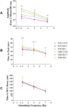
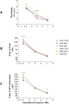
The aim of this study was to verify the effects of moderate-intensity continuous (MICT) and high-intensity interval (HIIT) aerobic training on cardiac morphology and function and the mechanical properties of single cardiomyocytes in spontaneously hypertensive rats (SHR) in the compensated phase of hypertension. Sixteen-week-old male SHR and normotensive Wistar (WIS) rats were allocated to six groups of six animals each: SHR CONT or WIS CONT (control); SHR MICT or WIS MICT (underwent MICT, 30 min/day, five days per week for eight weeks); and SHR HIIT or WIS HIIT (underwent HIIT, 30 min/day, five days per week for eight weeks). Total exercise time until fatigue and maximum running speed were determined using a maximal running test before and after the experimental period. Systolic (SAP), diastolic (DAP), and mean (MAP) blood pressures were measured using tail plethysmography before and after the experimental period. Echocardiographic evaluations were performed at the end of the experimental period. The rats were euthanized after in vivo assessments, and left ventricular myocytes were isolated to evaluate global intracellular Ca2+ transient ([Ca2+]i) and contractile function. Cellular measurements were performed at basal temperature (~37°C) at 3, 5, and 7 Hz. The results showed that both training programs increased total exercise time until fatigue and, consequently, maximum running speed. In hypertensive rats, MICT decreased SAP, DAP, MAP, interventricular septal thickness during systole and diastole, and the contraction amplitude at 5 Hz. HIIT increased heart weight and left ventricular wall thickness during systole and diastole and reduced SAP, MAP, and the time to peak [Ca2+]i at all pacing frequencies. In conclusion, both aerobic training protocols promoted beneficial adaptations to cardiac morphology, function, and mechanical properties of single cardiomyocytes in SHR.
Keywords: Cardiomyocytes; aerobic training; calcium transient; cardiac function; hypertension; spontaneously hypertensive rats.
Conflict of interest statement
The author(s) declared no potential conflicts of interest with respect to the research, authorship, and/or publication of this article.
Figures


Similar articles
-
High-intensity intermittent training is as effective as moderate continuous training, and not deleterious, in cardiomyocyte remodeling of hypertensive rats.J Appl Physiol (1985). 2019 Apr 1;126(4):903-915. doi: 10.1152/japplphysiol.00131.2018. Epub 2019 Jan 31. J Appl Physiol (1985). 2019. PMID: 30702976
-
The benefits of endurance training in cardiomyocyte function in hypertensive rats are reversed within four weeks of detraining.J Mol Cell Cardiol. 2013 Apr;57:119-28. doi: 10.1016/j.yjmcc.2013.01.013. Epub 2013 Jan 30. J Mol Cell Cardiol. 2013. PMID: 23376037
-
Moderate Exercise in Spontaneously Hypertensive Rats Is Unable to Activate the Expression of Genes Linked to Mitochondrial Dynamics and Biogenesis in Cardiomyocytes.Front Endocrinol (Lausanne). 2020 Aug 19;11:546. doi: 10.3389/fendo.2020.00546. eCollection 2020. Front Endocrinol (Lausanne). 2020. PMID: 32973679 Free PMC article.
-
Physical Exercise and Regulation of Intracellular Calcium in Cardiomyocytes of Hypertensive Rats.Arq Bras Cardiol. 2018 Aug;111(2):172-179. doi: 10.5935/abc.20180113. Epub 2018 Jul 2. Arq Bras Cardiol. 2018. PMID: 29972415 Free PMC article.
-
Effectiveness of High-Intensity Interval Training Versus Moderate-Intensity Continuous Training in Hypertensive Patients: a Systematic Review and Meta-Analysis.Curr Hypertens Rep. 2020 Mar 3;22(3):26. doi: 10.1007/s11906-020-1030-z. Curr Hypertens Rep. 2020. PMID: 32125550
Cited by
-
Animal Models of Exercise and Cardiometabolic Disease.Circ Res. 2025 Jul 7;137(2):139-162. doi: 10.1161/CIRCRESAHA.124.325704. Epub 2025 Jul 3. Circ Res. 2025. PMID: 40608858 Review.
References
-
- McCrossan ZA, Billeter R, White E. Transmural changes in size, contractile and electrical properties of SHR left ventricular myocytes during compensated hypertrophy. Cardiovasc Res 2004;63:283–92 - PubMed
-
- Fowler MR, Naz JR, Graham MD, Bru-Mercier G, Harrison SM, Orchard CH. Decreased Ca2+ extrusion via Na+/Ca2+ exchange in epicardial left ventricular myocytes during compensated hypertrophy. Am J Physiol Heart Circ Physiol 2005;288:H2431–1248 - PubMed
-
- Bernardo BC, Weeks KL, Pretorius L, McMullen JR. Molecular distinction between physiological and pathological cardiac hypertrophy: experimental findings and therapeutic strategies. Pharmacol Ther 2010;128:191–227 - PubMed
-
- Roman-Campos D, Carneiro-Júnior MA, Prímola-Gomes TN, Silva KA, Quintão-Júnior JF, Gondim AN, Duarte HL, Cruz JS, Natali AJ. Chronic exercise partially restores the transmural heterogeneity of action potential duration in left ventricular myocytes of spontaneous hypertensive rats. Clin Exp Pharmacol Physiol 2012;39:155–7 - PubMed
Publication types
MeSH terms
LinkOut - more resources
Full Text Sources
Medical
Miscellaneous

