. 2022 Aug 31;10(4):e0119922.
doi: 10.1128/spectrum.01199-22. Epub 2022 Jul 26.

Inhibition of Biofilm Formation and Virulence Factors of Cariogenic Oral Pathogen Streptococcus mutans by Shikimic Acid

Affiliations

Inhibition of Biofilm Formation and Virulence Factors of Cariogenic Oral Pathogen Streptococcus mutans by Shikimic Acid

Zhong Zhang et al. Microbiol Spectr. .

Abstract

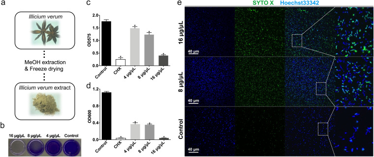
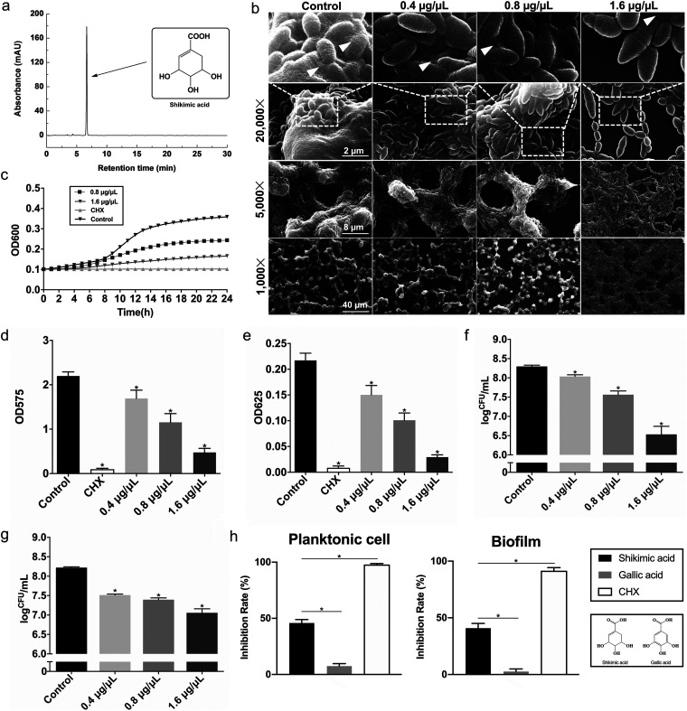
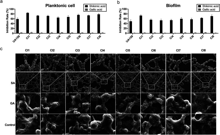
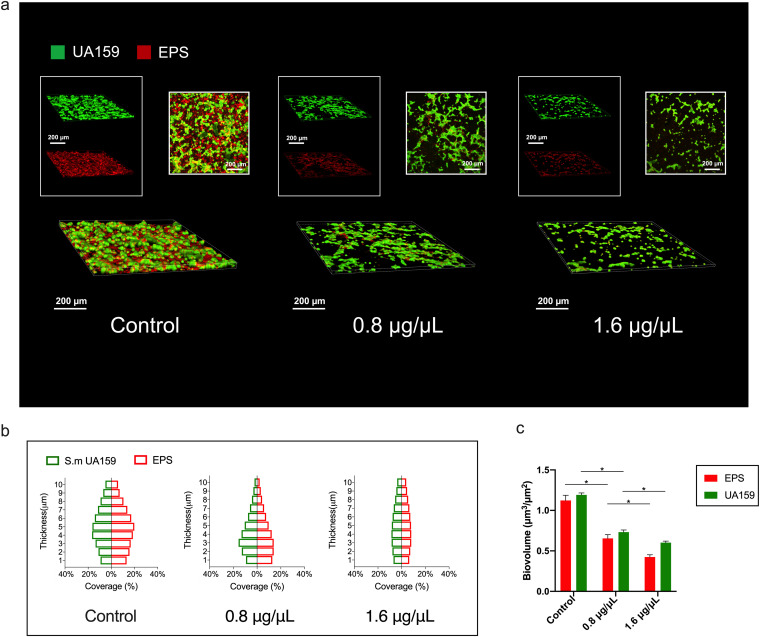
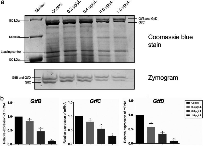
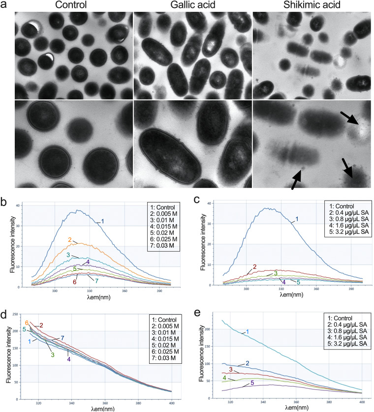
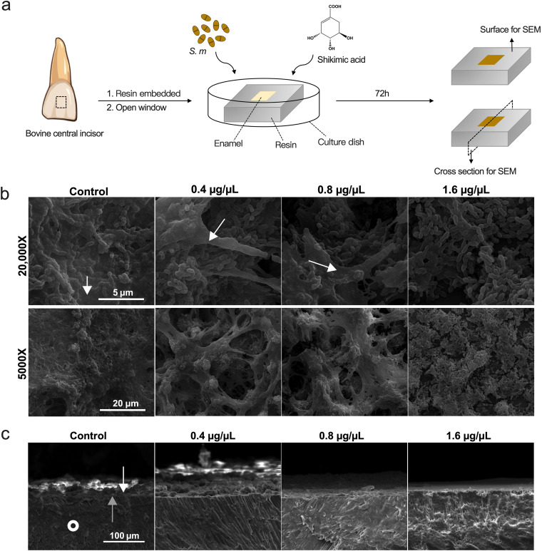
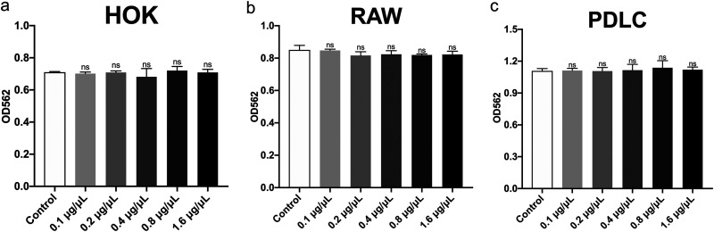
Streptococcus mutans is known as an important oral pathogen causing dental caries, a widespread oral infectious disease. S. mutans synthesize exopolysaccharide (EPS) using glucosyltransferases (Gtfs), resulting in biofilm formation on the tooth surface. Bacterial cells in the biofilms become strongly resistant to a harsh environment, such as antibiotics and host defense mechanisms, making biofilm-based infections difficult to eliminate. Discovering novel antibiofilm agents, especially from natural products, helps to develop effective strategies against this kind of diseases. The present study investigated the inhibitory effect of shikimic acid (SA), one abundant compound derived from Illicium verum extract, on the biofilm formation of S. mutans. We found SA can reduce the EPS synthesized by this oral pathogen and modulate the transcription of biofilm formation related genes, leading to fewer bacterial cells in its biofilm. SA also interacted with cell membrane and membrane proteins, causing damage to bacterial cells. Ex vivo testing of biofilm formation on bovine teeth showed SA strongly decreased the number of S. mutans cells and the number of EPS accumulated on dental enamel surfaces. Moreover, SA exhibits almost no toxicity to human oral cells evaluated by in vitro biocompatibility assay. In conclusion, shikimic acid exhibits remarkable antibiofilm activity against S. mutans and has the potential to be further developed as a novel anticaries agent. IMPORTANCE Natural products are an important and cost-effective source for screening antimicrobial agents. Here, we identified one compound, shikimic acid, from Illicium verum extract, exhibiting antimicrobial activity against S. mutans proliferation. It also inhibits biofilm formation of this bacteria through decreasing Gtf expression and EPS synthesis. Furthermore, this compound exhibits no significant cytotoxicity at its MIC against S. mutans, providing evidence for its clinical application.

Keywords: anti-biofilm agent; dental biofilm; glucosyltransferase; natural compound; shikimic acid.


Conflict of interest statement

The authors declare no conflict of interest.

Figures

FIG 1
FIG 1
Illicium verum extract exhibits antimicrobial activity against S. mutans. (a) The brief description of Illicium verum extract preparation. (b–c) Effect of Illicium verum extract on S. mutans biofilm formation. Extract was added into bacterial suspension and cocultured for 24 h to assess biofilm formation. (c) Crystal violet staining of S. mutans biofilm treated with Illicium verum extract or chlorhexidine (CHX). (d) Effect of Illicium verum extract on planktonic S. mutans. Bacterial cells were cocultured with drugs for 24 h to assess antimicrobial activity. The concentration of 0.2% (wt/vol) chlorhexidine (CHX) was used as a positive control in the antimicrobial assays (c and d). (e) Live and dead staining of planktonic S. mutans cells treated with Illicium verum extract. SYTO X marks the dead cells, while Hoechst33342 marks all bacterial cells.
FIG 2
FIG 2
Shikimic acid inhibits S. mutans proliferation and biofilm formation. The concentration of 0.2% (wt/vol) chlorhexidine (CHX) was used as a positive control in the antimicrobial assays (c, d, e, and i). (a) HPLC-mass spectrometry identification of shikimic acid as the most abundant compound in Illicium verum extract. (b) SEM images of S. mutans UA159 24-h biofilms on glass coverslips. Images were taken at 1,000, 5,000, and 20,000× magnification, respectively. White triangles indicate the EPS produced by S. mutans. (c) A typical growth curve of S. mutans under the treatment of shikimic acid. The concentrations of 0.8 and 1.6 μg/μL of shikimic acid were incubated with S. mutans for 24 h. (d) Effect of shikimic acid on S. mutans biofilm formation assessed by crystal violet staining. (e) Quantitative measurement of water-insoluble EPS by anthrone assay. (f) Effect of shikimic acid on S. mutans biofilm formation assessed by measuring the average number of CFU in biofilms. (g) Effect of shikimic acid on S. mutans mature biofilms by the determination of the average number of CFU in biofilms. The S. mutans biofilms were cultured for 24 h without shikimic acid treatment, and the data were collected 24 h after the addition of shikimic acid. (h) Molecular structures of shikimic acid and one of its derivatives, gallic acid. (i) The antimicrobial activity of 0.8 μg/μL shikimic acid or gallic acid on S. mutans planktonic cells or biofilms. Values represent the means standard deviations from three independent experiments. *, P < 0.05 compared with the untreated control.
FIG 3
FIG 3
Antimicrobial activity of shikimic acid on S. mutans clinical isolates. (a) The inhibitory effect of 0.8 μg/μL shikimic acid or gallic acid on planktonic cell growth of S. mutans clinical isolates. (b) The inhibitory effect of 0.8 μg/μL shikimic acid or gallic acid on biofilm formation of S. mutans clinical isolates. (c) SEM images of the 24-h biofilms of S. mutans clinical isolates on glass coverslips. The bacteria were treated with 1.6 μg/μL shikimic acid or gallic acid. Values represent the means standard deviations from three independent experiments. *, P < 0.05 compared with the untreated control.
FIG 4
FIG 4
Shikimic acid decreases the amounts of bacteria and EPS in S. mutans biofilms. (a) Three-dimensional visualization and double-labeling imaging of S. mutans biofilm formed on glass coverslips. The green fluorescence (SYTO9) marks the live bacteria, while the red fluorescence (Dextran 647) marks the EPS synthesized by S. mutans. (b) The distribution of bacteria and EPS inside S. mutans biofilms. (c) Biovolume of bacteria and EPS. *, P < 0.05 between compared groups.
FIG 5
FIG 5
Shikimic acid decreases the transcription level of Gtf-encoding genes. (a) Enzymatic activity of Gtfs measured by zymogram assay. Gtfs were separated by SDS-PAGE. One gel was stained by Coomassie blue (upper gel), and the other gel was coincubated with sucrose and dextran T70 (lower gel). Glucan bands synthesized by GtfB, GtfD, and GtfC were marked in zymography, respectively. (b) Relative expression of gtfB, gtfC, and gtfD measured by qRT-PCR. S. mutans UA159 16S rRNA was used as the internal control. Values represent the means standard deviations from three independent experiments. *, P < 0.05 compared with the control group.
FIG 6
FIG 6
Shikimic acid interacts with the subcellular structures of S. mutans. (a) Transmission electron micrographs of S. mutans UA159. The bacteria were treated with 0.8 μg/μL shikimic acid or gallic acid. The black arrows point to the damage of membrane. (b) Effect of KI on fluorescence intensity of the membrane proteins of S. mutans at λex 258 nm. (c) Effect of shikimic acid on fluorescence intensity of the membrane proteins of S. mutans at λex 258 nm. (d) Effect of KI on fluorescence intensity of the membrane proteins of S. mutans at λex 296 nm. (e) Effect of shikimic acid on fluorescence intensity of the membrane proteins of S. mutans at λex 296 nm.
FIG 7
FIG 7
Shikimic acid inhibits S. mutans cells and EPS accumulation on dental enamel. (a) Brief description of ex vivo biofilm formation assay. (b) Surface images of S. mutans biofilm taken by SEM. EPS are indicated by the white arrows. (c) Cross-section images of S. mutans biofilm taken by SEM. S. mutans were treated with different concentrations of SA for 72 h and formed biofilms on bovine dental enamel surface. The white arrows point to the bacteria on the enamel surface, while the gray arrows point to the boundary between the cross section and the surface of enamel. White circles point to the morphology of enamel in the cross section.
FIG 8
FIG 8
Biocompatibility of shikimic acid with several types of cells evaluated by CCK-8 assay. (a) Cytotoxicity of SA on human oral keratinocytes (HOKs). (b) Cytotoxicity of SA on RAW264.7. (c) Cytotoxicity of SA on human periodontal ligament cells (PDLCs). Values represent the means standard deviations from three independent experiments. *, P < 0.05 compared with the untreated control. ns, not statistically significant compared with the untreated control.

Similar articles

Cited by

References

    1. Pitts NB, Zero DT, Marsh PD, Ekstrand K, Weintraub JA, Ramos-Gomez F, Tagami J, Twetman S, Tsakos G, Ismail A. 2017. Dental caries. Nat Rev Dis Primers 3:17030. doi:10.1038/nrdp.2017.30. - DOI - PubMed
    1. Krzyściak W, Jurczak A, Kościelniak D, Bystrowska B, Skalniak A. 2014. The virulence of Streptococcus mutans and the ability to form biofilms. Eur J Clin Microbiol Infect Dis 33:499–515. doi:10.1007/s10096-013-1993-7. - DOI - PMC - PubMed
    1. Kreth J, Zhang Y, Herzberg MC. 2008. Streptococcal antagonism in oral biofilms: Streptococcus sanguinis and Streptococcus gordonii interference with Streptococcus mutans. J Bacteriol 190:4632–4640. doi:10.1128/JB.00276-08. - DOI - PMC - PubMed
    1. Kolenbrander PE, Palmer RJ, Periasamy S, Jakubovics NS. 2010. Oral multispecies biofilm development and the key role of cell–cell distance. Nat Rev Microbiol 8:471–480. doi:10.1038/nrmicro2381. - DOI - PubMed
    1. Wang C, Hou J, van der Mei HC, Busscher HJ, Ren Y. 2019. Emergent properties in Streptococcus mutans biofilms are controlled through adhesion force sensing by initial colonizers. mBio 10:e01908-19. doi:10.1128/mBio.01908-19. - DOI - PMC - PubMed

Publication types