. 2022 Oct 5;12(10):2454-2473.
doi: 10.1158/2159-8290.CD-21-1690.

Reprogrammed Schwann Cells Organize into Dynamic Tracks that Promote Pancreatic Cancer Invasion

Affiliations

Reprogrammed Schwann Cells Organize into Dynamic Tracks that Promote Pancreatic Cancer Invasion

Sylvie Deborde et al. Cancer Discov. .

Abstract

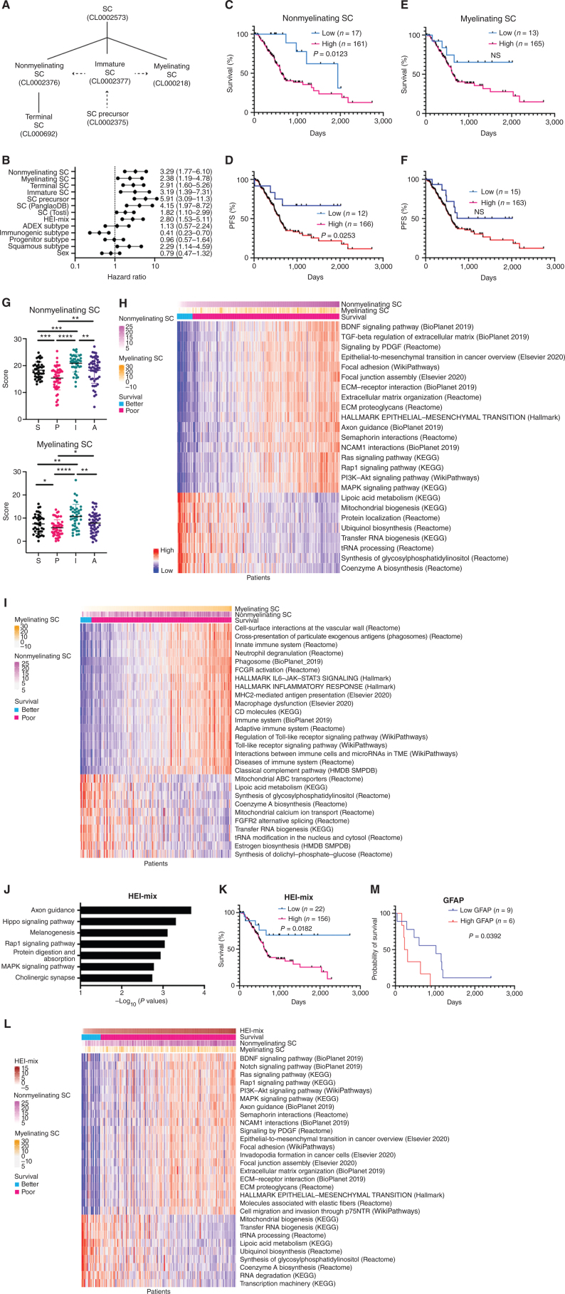
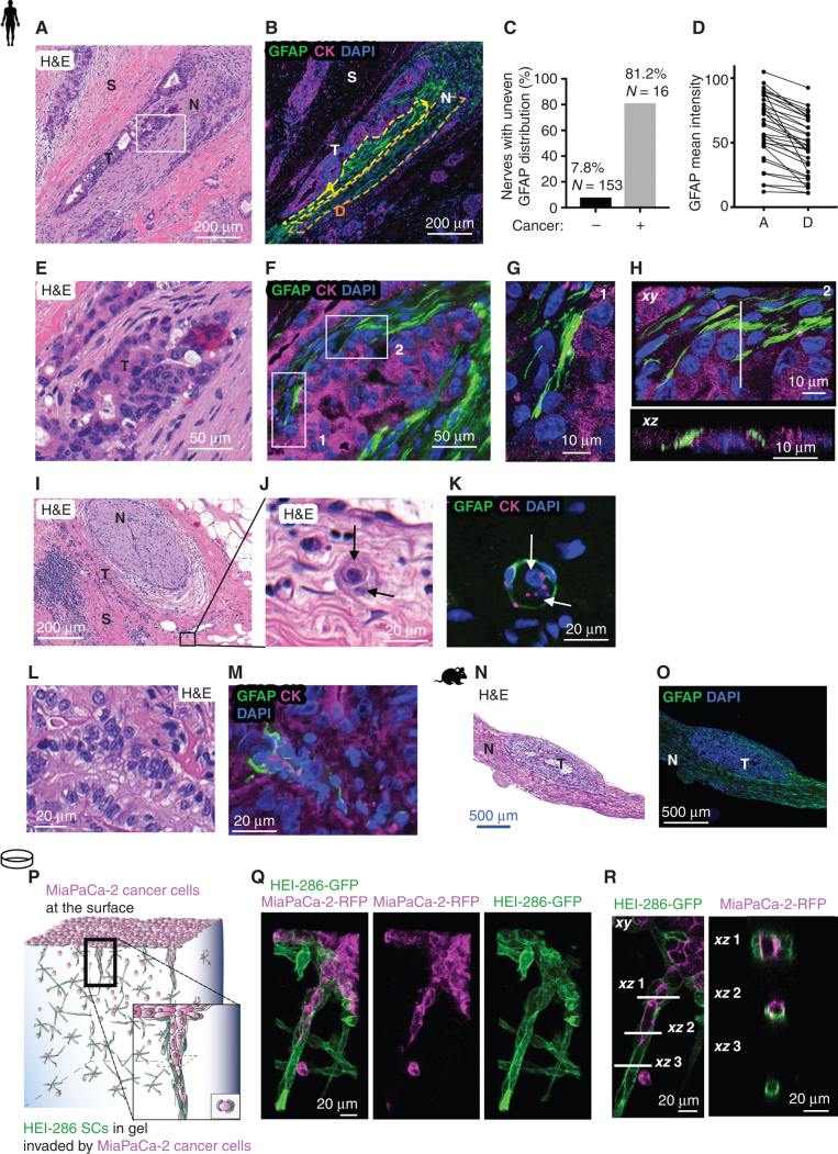
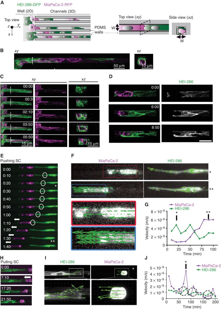
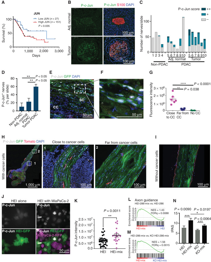
Nerves are a component of the tumor microenvironment contributing to cancer progression, but the role of cells from nerves in facilitating cancer invasion remains poorly understood. Here we show that Schwann cells (SC) activated by cancer cells collectively function as tumor-activated Schwann cell tracks (TAST) that promote cancer cell migration and invasion. Nonmyelinating SCs form TASTs and have cell gene expression signatures that correlate with diminished survival in patients with pancreatic ductal adenocarcinoma. In TASTs, dynamic SCs form tracks that serve as cancer pathways and apply forces on cancer cells to enhance cancer motility. These SCs are activated by c-Jun, analogous to their reprogramming during nerve repair. This study reveals a mechanism of cancer cell invasion that co-opts a wound repair process and exploits the ability of SCs to collectively organize into tracks. These findings establish a novel paradigm of how cancer cells spread and reveal therapeutic opportunities.

Significance: How the tumor microenvironment participates in pancreatic cancer progression is not fully understood. Here, we show that SCs are activated by cancer cells and collectively organize into tracks that dynamically enable cancer invasion in a c-Jun-dependent manner. See related commentary by Amit and Maitra, p. 2240. This article is highlighted in the In This Issue feature, p. 2221.


Figures

Figure 1. Nonmyelinating SC signature scores correlate with diminished survival in patients with pancreatic adenocarcinoma and with pathways related to cancer invasion. A, Hierarchical organization of SCs from Tabula-Sapiens (full lines) with dashed arrows indicating transitions in the SC lineage (22). B, Forest plot with hazard ratio and 95% CI. C–F, Kaplan–Meier curves of overall survival (C, E) and PFS (D, F) with high or low scores for signatures of nonmyelinating SC (C, D), and myelinating SC (E, F) in 178 TCGA PAAD patients. G, Nonmyelinating and myelinating SC signature scores in PDAC subtypes (S: squamous, P: progenitor, I: immunogenic, A: ADEX). H and I, Heatmap of gene sets correlating with high and low scores for nonmyelinating SC signature (H) and myelinating SC signature (I) in TCGA PAAD patients. Columns represent TCGA PAAD samples that have been rank ordered by the top row signature. Arrows indicate survival cutoff used in B and D with low survival patients at the left of the arrow. J, Top 7 enriched pathways in HEI-286 cocultured with MiaPaCa-2 as compared with HEI-286 SCs alone (EnrichR, human KEGG 2019 data set). K, Kaplan–Meier curve of overall survival with high or low scores for the cancer-exposed HEI-286 SC (HEI-mix) signature in 178 TCGA PAAD patients. L, Heatmap of gene sets correlating with high and low scores for cancer-exposed HEI-286 SC (HEI-mix) signature in TCGA PAAD patients. Columns represent TCGA PAAD samples that have been rank ordered by the top row signature. M, Kaplan–Meier of overall survival of PDAC patients with high or low GFAP expression in SCs determined histologically.
Figure 1.
Nonmyelinating SC signature scores correlate with diminished survival in patients with pancreatic adenocarcinoma and with pathways related to cancer invasion. A, Hierarchical organization of SCs from Tabula Sapiens (full lines), with dashed arrows indicating transitions in the SC lineage (22). B, Forest plot with hazard ratio and 95% confidence interval. ADEX, aberrantly differentiated endocrine exocrine. C–F, Kaplan–Meier curves of overall survival (C, E) and progression-free survival (PFS; D, F) with high or low scores for signatures of nonmyelinating SCs (C, D) and myelinating SCs (E, F) in 178 TCGA pancreatic adenocarcinoma (PAAD) patients. NS, not signficant. G, Nonmyelinating and myelinating SC signature scores in PDAC subtypes (A, ADEX; I, immunogenic; P, progenitor; S, squamous). *, P < 0.05; **, P < 0.01; ***, P < 0.001; ****, P < 0.0001. H, Heat map of gene sets correlating with high and low scores for nonmyelinating SC signature in TCGA PAAD patients. I, Heat map of gene sets correlating with high and low scores for myelinating SC signature in TCGA PAAD patients. Columns represent TCGA PAAD samples that have been rank ordered by the top row signature. ECM, extracellular matrix; HMDB, Human Metabolic Database; KEGG, Kyoto Encyclopedia of Genes and Genomes; SMPDB, Small Molecule Pathway Database. J, Top seven enriched pathways in HEI-286 cocultured with MiaPaCa-2 as compared with HEI-286 SCs alone (EnrichR, human KEGG 2019 data set). K, Kaplan–Meier curve of overall survival with high or low scores for the cancer-exposed HEI-286 SC (HEI-mix) signature in 178 TCGA PAAD patients. L, Heat map of gene sets correlating with high and low scores for cancer-exposed HEI-286 SC (HEI-mix) signature in TCGA PAAD patients. Columns represent TCGA PAAD samples that have been rank ordered by the top row signature. M, Kaplan–Meier of overall survival of PDAC patients with high or low GFAP expression in SCs determined histologically.
Figure 2. SCs wrap and align cancer cells. A, Hematoxylin and eosin (H&E) section of a human PDAC specimen with PNI. Scale bar, 200 μm. N, nerve; S, stroma; T, tumor. B, GFAP (green) and cytokeratin (CK; magenta) staining in a section adjacent to A showing cancer cells surrounded by GFAP+ SCs in a nerve. The yellow and orange dotted regions of the nerve are adjacent (A) or distal (D) to the cancer cells. Scale bar, 200 μm. C, Quantification of nerves with an uneven GFAP distribution comparing when cancer is visibly present or absent. D, Quantification of GFAP mean intensity in adjacent and distal regions of nerves (paired t test, P < 0.0001). E, Enlargement of rectangle in A. Scale bar, 50 μm. F, GFAP and CK staining of an adjacent section of E. Scale bar, 50 μm. G, Confocal images corresponding to rectangle 1 in F. Scale bar, 10 μm. H, Confocal images corresponding to rectangle 2 in F. Top image is an xy maximum projection, and the bottom image is an xz image corresponding to the dotted line. Scale bars, 10 μm. I, H&E section of a human PDAC specimen with a nerve (N) and tumor cells (T) in the neighboring stroma (S). Scale bar, 200 μm. J, Enlargement of I. Arrows indicate two cancer cells. Scale bar, 20 μm. K, GFAP and CK staining of a section adjacent to J showing GFAP+ SCs wrapping two cancer cells (arrows). Scale bar, 20 μm. L and M, H&E staining and adjacent GFAP and CK staining of a human PDAC specimen showing GFAP+ SCs around aligned cancer cells. Scale bars, 20 μm. N and O, H&E staining and adjacent GFAP staining of a longitudinal section of a murine sciatic nerve injected with cancer cells. Scale bars, 500 μm. P, Schematic representation of HEI-286 SCs and MiaPaCa-2 cancer cells in a 3D Matrigel invasion assay. The MiaPaCa-2-RFP cancer cells are placed on the surface of a Matrigel chamber in which HEI-286 SCs have grown. Cancer cells invade the gel as a chain of cancer cells surrounded by HEI-286 SC tracks. Q, Maximum projection of confocal images showing a chain of cancer cells lined up within a tubular structure of HEI-286 SCs in Matrigel. Scale bar, 20 μm. R, Single focal planes of confocal images showing MiaPaCa-2 cancer cells and HEI-286 SCs organized into columns. Longitudinal (left) and transverse (right) images correspond to the indicated positions. Scale bars, 20 μm.
Figure 2.
SCs wrap and align cancer cells. A, Hematoxylin and eosin (H&E) section of a human PDAC specimen with PNI. Scale bar, 200 μm. N, nerve; S, stroma; T, tumor. B, GFAP (green) and cytokeratin (CK; magenta) staining in a section adjacent to A showing cancer cells surrounded by GFAP+ SCs in a nerve. The yellow and orange dotted regions of the nerve are adjacent (A) or distal (D) to the cancer cells. Scale bar, 200 μm. C, Quantification of nerves with an uneven GFAP distribution comparing when cancer is visibly present or absent. D, Quantification of GFAP mean intensity in adjacent and distal regions of nerves (paired t test, P < 0.0001). E, Enlargement of rectangle in A. Scale bar, 50 μm. F, GFAP and CK staining of an adjacent section of E. Scale bar, 50 μm. G, Confocal images corresponding to rectangle 1 in F. Scale bar, 10 μm. H, Confocal images corresponding to rectangle 2 in F. Top image is an xy maximum projection, and the bottom image is an xz image corresponding to the dotted line. Scale bars, 10 μm. I, H&E section of a human PDAC specimen with a nerve (N) and tumor cells (T) in the neighboring stroma (S). Scale bar, 200 μm. J, Enlargement of I. Arrows indicate two cancer cells. Scale bar, 20 μm. K, GFAP and CK staining of a section adjacent to J showing GFAP+ SCs wrapping two cancer cells (arrows). Scale bar, 20 μm. L and M, H&E staining and adjacent GFAP and CK staining of a human PDAC specimen showing GFAP+ SCs around aligned cancer cells. Scale bars, 20 μm. N and O, H&E staining and adjacent GFAP staining of a longitudinal section of a murine sciatic nerve injected with cancer cells. Scale bars, 500 μm. P, Schematic representation of HEI-286 SCs and MiaPaCa-2 cancer cells in a 3D Matrigel invasion assay. The MiaPaCa-2-RFP cancer cells are placed on the surface of a Matrigel chamber in which HEI-286 SCs have grown. Cancer cells invade the gel as a chain of cancer cells surrounded by HEI-286 SC tracks. Q, Maximum projection of confocal images showing a chain of cancer cells lined up within a tubular structure of HEI-286 SCs in Matrigel. Scale bar, 20 μm. R, Single focal planes of confocal images showing MiaPaCa-2 cancer cells and HEI-286 SCs organized into columns. Longitudinal (left) and transverse (right) images correspond to the indicated positions. Scale bars, 20 μm.
Figure 3. SCs form dynamic tracks for cancer cells. A, Schematic of microchannels with HEI-286 SCs (green) and MiaPaCa-2 cells (magenta). Both cell types are seeded in the adjacent well (left, 2D) and enter microchannels (3D) where they make contact with each other. PDMS, polydimethylsiloxane. B, Confocal images of HEI-286 SCs and MiaPaCa-2 within microchannels in longitudinal (xy) and transverse (xz) sections. Scale bars, 50 μm. C, Confocal images of time-lapse movies showing a cancer cell moving in a microchannel lined by HEI-286 SCs. Time is h:min. Scale bars, 15 μm. D, Confocal images of time-lapse movies showing an HEI-286 SC wrapping around a cancer cell. Scale bar, 50 μm. E, Fluorescent images of time-lapse movie showing an HEI-286 SC pushing a cancer cell. Arrows indicate cancer cell displacement. Circles indicate intracellular movement within the SC (* and ** are time points shown in E). F, Fluorescent images of an HEI-286 SC and cancer cell from E at two time points (* and **) overlaid with vectors obtained by PIV analysis indicating vector direction. G, Quantification of the mean instantaneous velocity of the HEI-286 SC and cancer cell in E, F, and G. Corresponding time points are indicated by * and **. H, Fluorescent images of time-lapse movie showing HEI-286 SCs pulling a cancer cell. Arrows indicate cancer cell displacement. I, Fluorescent images of the HEI-286 SCs and cancer cells from H at one time point (*) overlaid with vectors obtained by PIV analysis. J, Quantification of the mean instantaneous velocity. *, Corresponding time point in I. Dotted circles indicate periods with synchronized increases in velocity for both cancer cells and SCs.
Figure 3.
SCs form dynamic tracks for cancer cells. A, Schematic of microchannels with HEI-286 SCs (green) and MiaPaCa-2 cells (magenta). Both cell types are seeded in the adjacent well (left, 2D) and enter microchannels (3D) where they make contact with each other. PDMS, polydimethylsiloxane. B, Confocal images of HEI-286 SCs and MiaPaCa-2 within microchannels in longitudinal (xy) and transverse (xz) sections. Scale bars, 50 μm. C, Confocal images of time-lapse movies showing a cancer cell moving in a microchannel lined by HEI-286 SCs. Time is h:min. Scale bars, 15 μm. D, Confocal images of time-lapse movies showing an HEI-286 SC wrapping around a cancer cell. Scale bar, 50 μm. E, Fluorescent images of time-lapse movie showing an HEI-286 SC pushing a cancer cell. Arrows indicate cancer cell displacement. Circles indicate intracellular movement within the SC (* and ** are time points shown in E). F, Fluorescent images of an HEI-286 SC and cancer cell from E at two time points (* and **) overlaid with vectors obtained by PIV analysis indicating vector direction. G, Quantification of the mean instantaneous velocity of the HEI-286 SC and cancer cell in E, F, and G. Corresponding time points are indicated by * and **. H, Fluorescent images of time-lapse movie showing HEI-286 SCs pulling a cancer cell. Arrows indicate cancer cell displacement. I, Fluorescent images of the HEI-286 SCs and cancer cells from H at one time point (*) overlaid with vectors obtained by PIV analysis. J, Quantification of the mean instantaneous velocity. *, Corresponding time point in I. Dotted circles indicate periods with synchronized increases in velocity for both cancer cells and SCs.
Figure 4. Cancer cells induce SC c-Jun activation and reprogramming. A, Kaplan–Meier curve of overall survival with high or low JUN expression in TCGA PAAD patients. B, P-c-Jun staining in S100-labeled nerves from PDAC specimens that are close to tumor as compared with nerves from adjacent (Adj.) normal tissue. Scale bars, 20 μm. C and D, Assessment of nerves from within pancreatic specimens of non-PDAC pathology, PDAC, and the normal tissues adjacent to PDAC. C, Quantification of the number of nerves per slide with no (-), low (+)-, or high (++)-intensity P-c-Jun staining. Each x-axis number represents a patient. D, Percentage of P-c-Jun–positive nerves (scored + or ++) in the pancreatic specimens of non-PDAC pathology, PDAC, and the normal tissues adjacent to PDAC (non-PDAC, n = 4; PDAC and adjacent normal, n = 6 each; mean ± SEM). E and F, Immunofluorescence of P-c-Jun (white) in a Panc02-injected murine sciatic nerve expressing GFP+ SCs. P-c-Jun is expressed in the green SCs but not in the Panc02 cancer cells (CC). Scale bars, 50 μm. G, Quantification of P-c-Jun fluorescence intensity in SCs of uninjected nerves and in SCs close to or far from injected Panc-02 tumor cells (near tumor: n = 6; far from tumor: n = 4; PBS-injected: n = 7; mean ± SEM, representative of three independent experiments). Fluorescence intensity is the mean intensity measured per area of nerve covered by green SCs. H, P-c-Jun staining in a sciatic nerve injected with Panc-02. (1) Region adjacent to the tumor. (2) Region far from the tumor. Scale bars, 1,000 and 100 μm. I, P-c-Jun staining in a normal nerve without cancer cells. Scale bar, 100 μm. J, Immunofluorescence of P-c-Jun in HEI-286 GFP SCs grown alone or grown mixed with MiaPaCa-2-RFP. Top images show P-c-Jun staining alone in HEI-286 SCs alone (left) and cocultured HEI-286 mixed with MiaPaCa-2 (right). Bottom overlay images allow identification of HEI-286 SCs (green) and MiaPaCa-2 (magenta). Scale bar, 50 μm. K, Quantification of P-c-Jun fluorescence intensity in HEI-286 SCs grown alone or mixed with MiaPaCa-2. Mean fluorescence intensity was measured per HEI-286 SC (n > 15 cells/group, mean ± SEM, representative of three independent experiments). L and M, GSEA assessing axon guidance genes in cancer cocultured HEI-286 SCs compared with HEI-286 SCs alone (L and in cancer cocultured HEI-286 compared with cancer cocultured c-Jun KO HEI-286 SCs (M). NES, normalized enrichment score. N, IPAS scores for the axon guidance genes (KEGG 2019).
Figure 4.
Cancer cells induce SC c-Jun activation and reprogramming. A, Kaplan–Meier curve of overall survival with high or low JUN expression in TCGA PAAD patients. B, P-c-Jun staining in S100-labeled nerves from PDAC specimens that are close to tumor as compared with nerves from adjacent (Adj.) normal tissue. Scale bars, 20 μm. C and D, Assessment of nerves from within pancreatic specimens of non-PDAC pathology, PDAC, and the normal tissues adjacent to PDAC. C, Quantification of the number of nerves per slide with no (-), low (+)-, or high (++)-intensity P-c-Jun staining. Each x-axis number represents a patient. D, Percentage of P-c-Jun–positive nerves (scored + or ++) in the pancreatic specimens of non-PDAC pathology, PDAC, and the normal tissues adjacent to PDAC (non-PDAC, n = 4; PDAC and adjacent normal, n = 6 each; mean ± SEM). E and F, Immunofluorescence of P-c-Jun (white) in a Panc02-injected murine sciatic nerve expressing GFP+ SCs. P-c-Jun is expressed in the green SCs but not in the Panc02 cancer cells (CC). Scale bars, 50 μm. G, Quantification of P-c-Jun fluorescence intensity in SCs of uninjected nerves and in SCs close to or far from injected Panc-02 tumor cells (near tumor: n = 6; far from tumor: n = 4; PBS-injected: n = 7; mean ± SEM, representative of three independent experiments). Fluorescence intensity is the mean intensity measured per area of nerve covered by green SCs. H, P-c-Jun staining in a sciatic nerve injected with Panc-02. (1) Region adjacent to the tumor. (2) Region far from the tumor. Scale bars, 1,000 and 100 μm. I, P-c-Jun staining in a normal nerve without cancer cells. Scale bar, 100 μm. J, Immunofluorescence of P-c-Jun in HEI-286 GFP SCs grown alone or grown mixed with MiaPaCa-2-RFP. Top images show P-c-Jun staining alone in HEI-286 SCs alone (left) and cocultured HEI-286 mixed with MiaPaCa-2 (right). Bottom overlay images allow identification of HEI-286 SCs (green) and MiaPaCa-2 (magenta). Scale bar, 50 μm. K, Quantification of P-c-Jun fluorescence intensity in HEI-286 SCs grown alone or mixed with MiaPaCa-2. Mean fluorescence intensity was measured per HEI-286 SC (n > 15 cells/group, mean ± SEM, representative of three independent experiments). L and M, GSEA assessing axon guidance genes in cancer cocultured HEI-286 SCs compared with HEI-286 SCs alone (L and in cancer cocultured HEI-286 compared with cancer cocultured c-Jun KO HEI-286 SCs (M). NES, normalized enrichment score. N, IPAS scores for the axon guidance genes (KEGG 2019).
Figure 5. SC c-Jun facilitates cancer migration along SC tracks in microchannels. A, Confocal images of HEI-286 with MiaPaCa-2 (top) and c-Jun KO HEI-286 with MiaPaCa-2 (bottom) within microchannels showing longitudinal (xy) and transverse (xz) sections. Scale bars, 10 μm. B, Quantification of percentage of HEI-286 SCs wrapping around MiaPaCa-2 cells for control or c-Jun KO HEI-286 SCs (n = 5–7 recordings per group with a total of 23 to 27 cells/group, mean ± SEM). C, Quantification of distance migrated by cancer cells in microchannels of different widths and occupied by HEI-286 SCs (n = 9–20 cells per channel size). D, Tracks of cancer cells in microchannels occupied by control and c-Jun KO HEI-286 SCs. Average representation of 21 MiaPaCa-2 cells in each group (F test with F = 930.1; P < 0.0001). E, Quantification of cancer cell speed (absolute values) in microchannels occupied by control versus c-Jun KO HEI-286 SCs (n = 21 cells in each group). F, Quantification of MiaPaCa-2 cell directional persistence (absolute values) in microchannels occupied by control versus c-Jun KO HEI-286 SCs (n = 21 cells in each group). G, Fluorescent images of time-lapse movies showing the behavior of a MiaPaCa-2 after HEI-286 SC contact. A cancer cell passes by a control HEI-286 SC, whereas another is blocked by a c-Jun KO HEI-286 SC. Scale bars, 15 μm. H, Quantification of G showing the percentage of cancer cells (CC) passing by an HEI-286 SC (n = 3 experiments per group with at least 11 cells/group in each experiment, mean ± SEM). I, Images showing stiffness maps of cocultured HEI-286 SCs versus c-Jun KO HEI-286 SCs. Scale bar, 15 μm. J, Quantification of stiffness of HEI-286 SCs versus c-Jun KO HEI-286 SCs, measured by AFM (n = 12–13 cells/group, mean ± SEM, representative of two independent experiments). K, Images showing actin staining in cocultured HEI-286 SCs versus c-Jun KO HEI-286 SCs. Scale bar, 20 μm.
Figure 5.
SC c-Jun facilitates cancer migration along SC tracks in microchannels. A, Confocal images of HEI-286 with MiaPaCa-2 (top) and c-Jun KO HEI-286 with MiaPaCa-2 (bottom) within microchannels showing longitudinal (xy) and transverse (xz) sections. Scale bars, 10 μm. B, Quantification of percentage of HEI-286 SCs wrapping around MiaPaCa-2 cells for control or c-Jun KO HEI-286 SCs (n = 5–7 recordings per group with a total of 23 to 27 cells/group, mean ± SEM). C, Quantification of distance migrated by cancer cells in microchannels of different widths and occupied by HEI-286 SCs (n = 9–20 cells per channel size). D, Tracks of cancer cells in microchannels occupied by control and c-Jun KO HEI-286 SCs. Average representation of 21 MiaPaCa-2 cells in each group (F test with F = 930.1; P < 0.0001). E, Quantification of cancer cell speed (absolute values) in microchannels occupied by control versus c-Jun KO HEI-286 SCs (n = 21 cells in each group). F, Quantification of MiaPaCa-2 cell directional persistence (absolute values) in microchannels occupied by control versus c-Jun KO HEI-286 SCs (n = 21 cells in each group). G, Fluorescent images of time-lapse movies showing the behavior of a MiaPaCa-2 after HEI-286 SC contact. A cancer cell passes by a control HEI-286 SC, whereas another is blocked by a c-Jun KO HEI-286 SC. Scale bars, 15 μm. H, Quantification of G showing the percentage of cancer cells (CC) passing by an HEI-286 SC (n = 3 experiments per group with at least 11 cells/group in each experiment, mean ± SEM). I, Images showing stiffness maps of cocultured HEI-286 SCs versus c-Jun KO HEI-286 SCs. Scale bar, 15 μm. J, Quantification of stiffness of HEI-286 SCs versus c-Jun KO HEI-286 SCs, measured by AFM (n = 12–13 cells/group, mean ± SEM, representative of two independent experiments). K, Images showing actin staining in cocultured HEI-286 SCs versus c-Jun KO HEI-286 SCs. Scale bar, 20 μm.
Figure 6. c-Jun coordinates SC collective organization and enhances cancer cell invasion in 3D Matrigel through track formation. A, Confocal images of control and c-Jun KO HEI-GFP SCs in Matrigel showing a lack of SC organization in c-Jun KO HEI-286 SCs. Scale bars, 30 μm. B and C, Quantification of the length of the SC structures created in Matrigel from one cell after 80 hours for control and c-Jun KO cells (control n = 10, c-Jun KO n = 6, mean ± SEM; B) and JNK inhibitor–treated cells (JNK inh; control n = 14, JNK inh n = 30, mean ± SEM; C). D, Quantification of cancer cell invasion into a 3D Matrigel chamber in the presence of control versus c-Jun KO HEI-286 SCs grown in the Matrigel (n = 8–12 measurements/condition, mean ± SEM). E, Maximum projection view of confocal images of MiaPaCa-2 (magenta) invasion in the presence of HEI-286-SCs (green) growing in Matrigel. Scale bar, 50 μm. F, Confocal image enlarged from E and schematic showing a chain of cancer cells (CC) aligned within a tubular, linear structure of control SCs in Matrigel. Scale bar, 50 μm. G, Confocal image enlarged from E and schematic showing branched structures created by aligned SCs in Matrigel. Scale bar, 50 μm. H, Maximum projection view of confocal images of MiaPaCa-2 invasion in the presence of c-Jun KO HEI-286 SCs growing in Matrigel. Scale bar, 50 μm. I, Confocal image enlarged from F and schematic showing a disorganized cluster of c-Jun KO HEI-286 SCs lacking organization. Scale bar, 50 μm.
Figure 6.
c-Jun coordinates SC collective organization and enhances cancer cell invasion in 3D Matrigel through track formation. A, Confocal images of control and c-Jun KO HEI-GFP SCs in Matrigel showing a lack of SC organization in c-Jun KO HEI-286 SCs. Scale bars, 30 μm. B and C, Quantification of the length of the SC structures created in Matrigel from one cell after 80 hours for control and c-Jun KO cells (control n = 10, c-Jun KO n = 6, mean ± SEM; B) and JNK inhibitor–treated cells (JNK inh; control n = 14, JNK inh n = 30, mean ± SEM; C). D, Quantification of cancer cell invasion into a 3D Matrigel chamber in the presence of control versus c-Jun KO HEI-286 SCs grown in the Matrigel (n = 8–12 measurements/condition, mean ± SEM). E, Maximum projection view of confocal images of MiaPaCa-2 (magenta) invasion in the presence of HEI-286-SCs (green) growing in Matrigel. Scale bar, 50 μm. F, Confocal image enlarged from E and schematic showing a chain of cancer cells (CC) aligned within a tubular, linear structure of control SCs in Matrigel. Scale bar, 50 μm. G, Confocal image enlarged from E and schematic showing branched structures created by aligned SCs in Matrigel. Scale bar, 50 μm. H, Maximum projection view of confocal images of MiaPaCa-2 invasion in the presence of c-Jun KO HEI-286 SCs growing in Matrigel. Scale bar, 50 μm. I, Confocal image enlarged from F and schematic showing a disorganized cluster of c-Jun KO HEI-286 SCs lacking organization. Scale bar, 50 μm.
Figure 7. c-Jun–reprogrammed SCs promote cancer invasion in vivo. A, Histologic analysis of injected murine sciatic nerves in P0-CRE− (WT) and P0-CRE+ (c-Jun KO) c-Junfl/fl mice. Representative samples of cancer invasion detected by H&E staining. Boxes show the invasion of areas away from the site of injection. Scale bars, 2,000 μm. Box scale bars, 500 μm. B, Quantification of distance of nerve invasion by Panc02 cells (WT n = 7, c-Jun KO n = 11, mean ± SEM) and KPC cells (WT n = 13, c-Jun KO n = 11, mean ± SEM). C, Representative images of murine hind limbs after Panc02 cancer cell injections show less paralysis in c-Jun KO SC mice. D, Quantification of the maximum width of hind limb paw in P0-CRE− (WT) versus P0-CRE+ (c-Jun KO) c-Junfl/fl mice 10 days after cancer injection (n = 8 mice/condition, mean ± SEM). E, Quantification of sciatic nerve function in P0-CRE− (WT) versus P0-CRE+ (c-Jun KO) c-Junfl/fl mice (n = 8 mice/condition, mean ± SEM). F, Effect of SP600125 on sciatic nerve invasion. Quantification of the length of nerve invasion in mice injected with Panc02 and SP600125 [JNK inhibitor (JNKi)] or Panc02 and DMSO (n = 11 mice/treatment, mean ± SEM). G, Representative H&E staining images of sciatic nerves injected with Panc02 and SP600125 versus Panc02 and DMSO. Top images are from proximal nerve regions at the spinal cord. No cancer is seen within the SP600125-injected nerve. Bottom images are from the site of injection, distal to the spinal cord. CC, cancer cells. Scale bars, 200 μm. H, Effect of SP600125 on flank tumor growth. Quantification of tumor volume in mice coinjected with Panc02 and SP600125 and with Panc02 and DMSO (n = 5 mice/treatment, mean ± SEM). NS, not significant. I, Survival analysis of WT and c-Jun KO mice injected with Panc02 in the sciatic nerve (WT n = 9, c-Jun KO = 11, P = 0.0003).
Figure 7.
c-Jun–reprogrammed SCs promote cancer invasion in vivo.A, Histologic analysis of injected murine sciatic nerves in P0-CRE (WT) and P0-CRE+ (c-Jun KO) c-Junfl/fl mice. Representative samples of cancer invasion detected by H&E staining. Boxes show the invasion of areas away from the site of injection. Scale bars, 2,000 μm. Box scale bars, 500 μm. B, Quantification of distance of nerve invasion by Panc02 cells (WT n = 7, c-Jun KO n = 11, mean ± SEM) and KPC cells (WT n = 13, c-Jun KO n = 11, mean ± SEM). C, Representative images of murine hind limbs after Panc02 cancer cell injections show less paralysis in c-Jun KO SC mice. D, Quantification of the maximum width of hind limb paw in P0-CRE (WT) versus P0-CRE+ (c-Jun KO) c-Junfl/fl mice 10 days after cancer injection (n = 8 mice/condition, mean ± SEM). E, Quantification of sciatic nerve function in P0-CRE (WT) versus P0-CRE+ (c-Jun KO) c-Junfl/fl mice (n = 8 mice/condition, mean ± SEM). F, Effect of SP600125 on sciatic nerve invasion. Quantification of the length of nerve invasion in mice injected with Panc02 and SP600125 [JNK inhibitor (JNKi)] or Panc02 and DMSO (n = 11 mice/treatment, mean ± SEM). G, Representative H&E staining images of sciatic nerves injected with Panc02 and SP600125 versus Panc02 and DMSO. Top images are from proximal nerve regions at the spinal cord. No cancer is seen within the SP600125-injected nerve. Bottom images are from the site of injection, distal to the spinal cord. CC, cancer cells. Scale bars, 200 μm. H, Effect of SP600125 on flank tumor growth. Quantification of tumor volume in mice coinjected with Panc02 and SP600125 and with Panc02 and DMSO (n = 5 mice/treatment, mean ± SEM). NS, not significant. I, Survival analysis of WT and c-Jun KO mice injected with Panc02 in the sciatic nerve (WT n = 9, c-Jun KO = 11, P = 0.0003).

Comment in

References

    1. Chambers AF, Groom AC, MacDonald IC. Dissemination and growth of cancer cells in metastatic sites. Nat Rev Cancer 2002;2:563–72. - PubMed
    1. Friedl P, Wolf K. Tumour-cell invasion and migration: diversity and escape mechanisms. Nat Rev Cancer 2003;3:362–74. - PubMed
    1. Kai F, Drain AP, Weaver VM. The extracellular matrix modulates the metastatic journey. Dev Cell 2019;49:332–46. - PMC - PubMed
    1. Winkler J, Abisoye-Ogunniyan A, Metcalf KJ, Werb Z. Concepts of extracellular matrix remodelling in tumour progression and metastasis. Nat Commun 2020;11:5120. - PMC - PubMed
    1. van Helvert S, Storm C, Friedl P. Mechanoreciprocity in cell migration. Nat Cell Biol 2018;20:8–20. - PMC - PubMed

Publication types

MeSH terms