. 2022 Sep 13;55(9):1645-1662.e7.
doi: 10.1016/j.immuni.2022.06.021. Epub 2022 Jul 25.

Keratinocyte-derived defensins activate neutrophil-specific receptors Mrgpra2a/b to prevent skin dysbiosis and bacterial infection

Affiliations

Keratinocyte-derived defensins activate neutrophil-specific receptors Mrgpra2a/b to prevent skin dysbiosis and bacterial infection

Xintong Dong et al. Immunity. .

Abstract

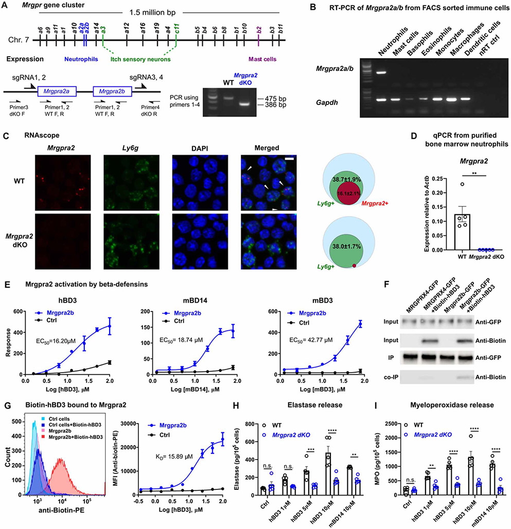
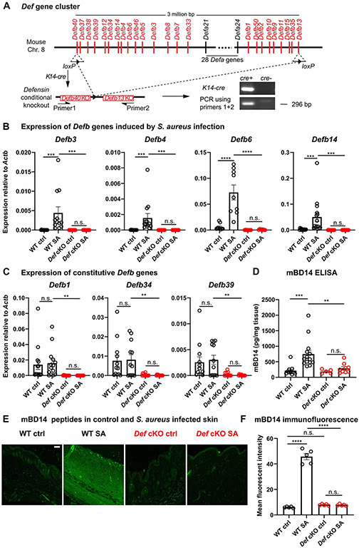
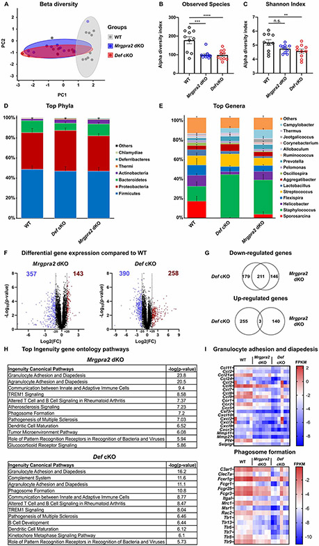
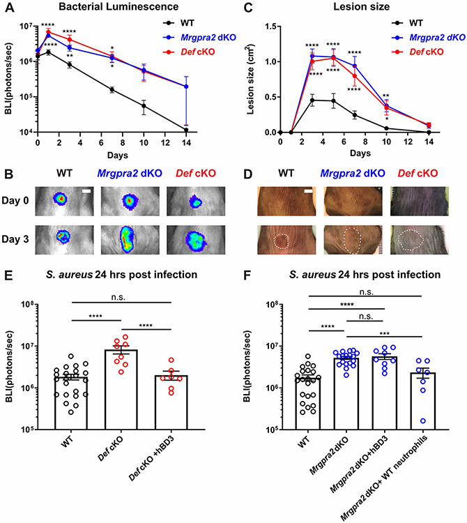
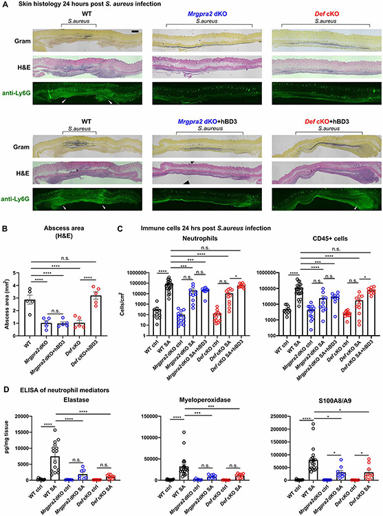
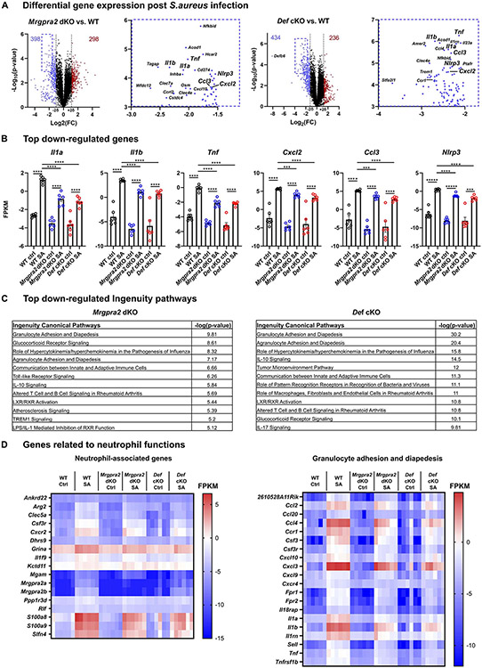
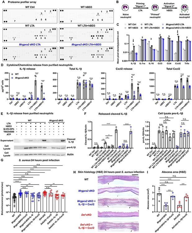
Healthy skin maintains a diverse microbiome and a potent immune system to fight off infections. Here, we discovered that the epithelial-cell-derived antimicrobial peptides defensins activated orphan G-protein-coupled receptors (GPCRs) Mrgpra2a/b on neutrophils. This signaling axis was required for effective neutrophil-mediated skin immunity and microbiome homeostasis. We generated mutant mouse lines lacking the entire Defensin (Def) gene cluster in keratinocytes or Mrgpra2a/b. Def and Mrgpra2 mutant animals both exhibited skin dysbiosis, with reduced microbial diversity and expansion of Staphylococcus species. Defensins and Mrgpra2 were critical for combating S. aureus infections and the formation of neutrophil abscesses, a hallmark of antibacterial immunity. Activation of Mrgpra2 by defensin triggered neutrophil release of IL-1β and CXCL2 which are vital for proper amplification and propagation of the antibacterial immune response. This study demonstrated the importance of epithelial-neutrophil signaling via the defensin-Mrgpra2 axis in maintaining healthy skin ecology and promoting antibacterial host defense.

Keywords: CXCL2; GPCR; IL-1β; Mrgpr; antimicrobial peptide; defensin; innate immunity; neutrophil; skin dysbiosis; skin microbiome.


Conflict of interest statement

Declaration of interests Xinzhong Dong is a co-founder and a scientific advisory board member of Escient Pharmaceuticals, a company focused on developing small molecules targeting MRGPRs. N.K.A. has received previous grant support from Pfizer and Boehringer Ingelheim and was a paid consultant for Janssen Pharmaceuticals. L.S.M. is currently a full-time paid employee of Janssen Research & Development, the pharmaceutical companies of Johnson & Johnson, and owns Johnson & Johnson stock. L.S.M. performed all work at his prior affiliation at Johns Hopkins University School of Medicine and he has received prior grant support from AstraZeneca, Pfizer, Boehringer Ingelheim, Regeneron Pharmaceuticals, and Moderna Therapeutics; was a paid consultant for Armirall and Janssen Research and Development; was on the scientific advisory board of Integrated Biotherapeutics; and is a shareholder of Noveome Biotherapeutics, which are all developing therapeutics against infections (including S. aureus and other pathogens) and/or inflammatory conditions. B.S.K. has served as a consultant for AbbVie, ABRAX Japan, Almirall, Cara Therapeutics, Maruho, Menlo Therapeutics, Pfizer, and Third Rock Ventures. He has also participated on the advisory board for Almirall, Boehringer Ingelheim, Cara Therapeutics, Kiniksa Pharmaceuticals, Menlo Therapeutics, Regeneron Pharmaceuticals, Sanofi Genzyme, and Trevi Therapeutics. He is also Founder, Chief Scientific Officer, and stockholder of Nuogen Pharma, Inc. He is stockholder of Locus Biosciences. C.V. is a paid consultant and stockholder of AstraZeneca.

Figures

Figure 1.
Figure 1.. Mrgpra2a/b were neutrophil-specific receptors for defensi
(A) Schematic illustration of the Mrgpr gene cluster on mouse Chromosome 7 and cellular expression specificities of a few well-studied members of the gene family. Mrgpra2a and a2b were deleted by CRISPR/cas9 using four gRNAs flanking the two genes, removing the entire genomic region. PCR genotyping using primers 1-4 as indicated in the diagram detects a WT band of 475 base pairs and an Mrgpra2 dKO mutant band of 386 base pairs. (B) RT-PCR gel showing expression of Mrgpra2 and housekeeping gene Gapdh in FACS-sorted neutrophils, mast cells, basophils, eosinophils, monocytes, macrophages, dendritic cells, and the no reverse transcriptase control (nRT Ctrl) of the neutrophil sample. (C) Representative RNAscope in situ hybridization images of bone marrow cells labeled using probes for Mrgpra2 (red), Ly6g (green), and DAPI (blue). Arrowheads point to Mrgpra2+; Ly6g+ neutrophils. Scale bar=5 μm. Venn diagrams depict percentages of Ly6g+ (green) and Mrgpra2+ (red) cells in all bone marrow cells (blue). n=3 (D) qPCR of mRNA from purified bone marrow neutrophils showed that Mrgpra2 were expressed highly in WT neutrophils but deleted from Mrgpra2 dKO neutrophils. n=5 (E) HEK cells expressing Mrgpra2b activation by hBD3 (EC50=16.2μM), mBD14 (EC50=18.74μM) and mBD3 (EC50=42.77μM) as determined by FLIPR intracellular calcium mobilization assay. n=4-9 (F) Western blot showing biotin-hBD3 being co-immunoprecipitated (co-IP) by Mrgpra2b-GFP. MRGPRX4-GFP (negative control) and Mrgpra2b-GFP were pulled down using anti-GFP beads, biotin-hBD3 was co-precipitated only with Mrgpra2b-GFP but not MRGPRX4-GFP. (G) Dissociation constant (KD=15.89μM) between biotin-hBD3 and Mrgpra2b determined by flow cytometry. Left, representative flow cytometry histogram showing control HEK cells (blue) and Mrgpra2b-GFP-expressing cells (red) incubated with (darker shades) and without (lighter shades) biotin-hBD3 (100μM). Right, quantification of mean fluorescent intensity (MFI) of PE-anti-biotin showing the amount of biotin-hBD3 bound to the cells at each concentration. n=6 (H-I) hBD3 and mBD14 triggered WT neutrophils to release elastase (H) and myeloperoxidase (I) (black). Neutrophils purified from Mrgpra2 dKO animals did not respond to hBD3 stimuli (blue). n=5 Results are presented as mean ± SEM from at least three independent experiments. **p < 0.01, ***p < 0.001, ****p < 0.0001, n.s. not significant by two-tailed unpaired Student’s t test (D) or two-way ANOVA (H-I). See also Figure S1.
Figure 2.
Figure 2.. Generation of Defensin cluster conditional knockout mice
(A) Schematic illustration of the Def gene cluster on mouse Chromosome 8. Two loxP sites were inserted into exons of Defb40 and Defb13 on the two ends of the gene cluster. K14-cre-mediated recombination successfully removed the entire 3 million base pair genomic region from keratinocytes. Genotyping PCR using primers flanking the cluster detected a band corresponding to partial sequence in Defb40, one recombined loxP site, and partial sequence in Defb13. (B-C) mRNA quantification of infection-induced Defb3 (n=12), Defb4 (n=13), Defb6 (n=9) and Defb14 (n=15) (B) and constitutively expressed Defb1 (n=14), Defb34 (n=12) and Defb39 (n=12) (C) in WT (black) and Def cKO (red) control (Ctrl) skin and 24 hours post S. aureus infection (SA) by qPCR. The amounts of mRNA for each gene were normalized to housekeeping gene Actb. (D) Quantification of mBD14 peptide in control and S. aureus infected skin of WT and Def cKO animals by ELISA. n=5-14 (E) Representative images of mBD14 immunofluorescence of WT control skin, WT skin infected with S. aureus, Def cKO control skin and Def cKO skin infected with S. aureus. mBD14 was low in uninfected WT skin but was robustly induced 24 hours after infection by S. aureus. In Def cKO animals, infection failed to induce mBD14 production. Scale bar=100μm (F) Quantification of mean fluorescent intensity of anti-mBD14 immunofluorescence across the thickness of the skin tissue as shown in (E). n=5 Results are presented as mean ± SEM from at least three independent experiments. **p < 0.01, ***p < 0.001, ****p < 0.0001 n.s. not significant by one-way ANOVA. See also Figure S2.
Figure 3.
Figure 3.. Def and Mrgpra2 mutant animals had altered skin microbiomes
(A-E) 16S sequencing analysis of microbial communities on the skin of WT, Mrgpra2 dKO and Def cKO mice. n=10 (A) Principal coordinates analysis showed that the clustering of Mrgpra2 dKO (blue) and Def cKO (red) microbial communities overlapped with each other but were distinct from WT (grey). (B) Total numbers of bacterial species observed in WT (black), Mrgpra2 dKO (blue), and Def cKO (red) skin swab samples. (C) Shannon indices of WT (black), Mrgpra2 dKO (blue), and Def cKO (red) skin swab samples. (D-E) Stacked bar charts depicting the relative abundance of top phyla (D) and top genera (E) in WT, Mrgpra2 dKO and Def cKO skin microbiota. (F-I) RNA-seq analysis of gene expression in naïve skin of WT, Mrgpra2 dKO, and Def cKO mice. n=6 (F) Volcano plots illustrating genes with significantly down- (blue) or up-regulated (red) expression in the Mrgpra2 dKO (left) or Def cKO (right) naïve skin compared to WT. (G) Top: Venn diagram showing overlap of down-regulated gene sets between Mrgpra2 dKO and Def cKO. Bottom: Very few overlap in the up-regulated gene sets. (H) Top altered Ingenuity gene ontology pathways in Mrgpra2 dKO (top) and Def cKO (bottom) naïve skin compared to WT. (I) Heat maps depicting fragments per kilobase per million mapped reads (FPKM) counts of genes related to granulocyte adhesion and diapedesis (top) and phagosome formation (bottom). n=6 Results are presented as mean ± SEM from one experiment. *p < 0.05, **p < 0.01, ***p < 0.001, n.s. not significant by one-way ANOVA. See also Figure S3, S4.
Figure 4.
Figure 4.. Mrgpra2 and defensins were critical for anti-S. aureus immunity
(A) Bacterial luminescence (mean total flux [photons per second]) of WT (black, n=22), Mrgpra2 dKO (blue, n=13) and Def cKO (red, n=8) back skin intradermally infected with S. aureus. (B) Representative images of in vivo bioluminescence. Scale bar=0.5cm (C) Mean total infected area (cm2) of WT (black), Mrgpra2 dKO (blue) and Def cKO (red) back skin infected with S. aureus. (D) Representative photographs of S. aureus-infected skin. Dotted lines indicate the dermonecrotic area caused by the infection. Scale bar=0.5cm (E) hBD3 injection 6 hours post S. aureus infection was sufficient to rescue the anti-bacterial defect of Def cKO mice. n=7-22 (F) Anti-bacterial defect of Mrgpra2 dKO animals could not be rescued by hBD3 injection but was fully rescued by adoptive transfer of purified WT neutrophils. n=7-22 Results are presented as mean ± SEM from at least three independent experiments. *p < 0.05, **p < 0.01, ***p < 0.001, ****p < 0.0001, n.s. not significant by two-way ANOVA (A and C) and one-way ANOVA (E and F). In (A) and (C), comparisons were made between each mutant and WT. See also Figure S5.
Figure 5.
Figure 5.. Mrgpra2 dKO and Def cKO animals showed severe defects in neutrophil abscess formation following S. aureus infection
(A) Top row: Histology staining of WT, Def cKO, and Mrgpra2 dKO mouse skin 24 hours post S. aureus infection using Gram stain (top), H&E (middle), and immunofluorescent staining using a monoclonal antibody against Ly6G (bottom) to show the locations of neutrophils. Black brackets indicate the bacteria band. White arrowheads point to neutrophil abscesses. Scale bar=1mm Bottom row: hBD3 injection rescued abscess formation in Def cKO but not Mrgpra2 dKO mutant animals. (B) Quantification of abscess areas based on H&E staining shown in (A). n=5 (C) Flow cytometry analyses of total CD45+ cells and neutrophils in the skin 24 hours post S. aureus infection. Both Def cKO and Mrgpra2 dKO animals showed reduced recruitment of neutrophils. hBD3 injection rescued neutrophil recruitment of Def but not Mrgpra2 mutants. n=9-23 (D) ELISA quantifications of elastase, myeloperoxidase (MPO), and calprotectin (S100A8/A9 dimer) in the skin of WT, Def cKO and Mrgpra2 dKO mice 24 hours post S. aureus infection. n=6-21 Results are presented as mean ± SEM from at least three independent experiments. *p < 0.05, ***p < 0.001, ****p < 0.0001, n.s. not significant by one-way ANOVA. See also Figure S5.
Figure 6.
Figure 6.. RNA-seq analysis of WT, Mrgpra2 dKO and Def cKO skin 24 hours post-S. aureus infection
(A) Volcano plots illustrating genes with significantly down- (blue) or up-regulated (red) expression in the Mrgpra2 dKO (left) or Def cKO (right) infected skin compared to WT. Boxed areas in the upper-left corners were magnified to show the most significantly down-regulated genes in detail. (B) FPKMs of key pro-inflammatory genes in WT, Mrgpra2 dKO and Def cKO naïve and S. aureus-infected skin. n=6 (C) Top altered Ingenuity gene ontology pathways in Mrgpra2 dKO (top) and Def cKO (bottom) S. aureus infected skin. (D) Heat maps depicting FPKM of neutrophil-associated genes and genes related to granulocyte adhesion and diapedesis. Results are presented as mean ± SEM from one experiment. ***p < 0.001, ****p < 0.0001, *****p < 0.00001, n.s. not significant by one-way ANOVA. See also Figure S6.
Figure 7.
Figure 7.. Defensin-Mrgpra2 interaction triggered neutrophil release of IL-1β and Cxcl2
(A) Proteome profiler array analysis of culture medium of bone marrow neutrophils purified from WT (black) or Mrgpra2 dKO (blue) animals stimulated by control medium, hBD3 only, S. aureus lipoteichoic acid (LTA), or LTA+hBD3. Numbers: 1=IL-1β, 2=IL-1ra, 3=IL-16, 4=Ccl3, 5=Ccl4, 6=Cxcl2, 7=Tnfα. Quantifications are shown in (C). (B) Schematic illustration of experimental design. Neutrophils are first primed with LTA (“signal 1”), then activated by hBD3 (“signal 2”). (C) Quantification of mean pixel intensity of proteome profiler arrays shown in (A). Arrows point to IL-1β (1 in A) and Cxcl2 (6 in A), whose releases were enhanced by hBD3. Pixel intensities were normalized to positive controls on the same array. (D) ELISA quantification of IL-1β and Cxcl2 release from neutrophils. WT neutrophils pretreated with LTA released IL-1β and Cxcl2 in response to hBD3 or mBD14 stimuli (black). Neutrophils purified from Mrgpra2 dKO animals failed to respond to hBD3 (blue). Total IL-1β and Cxcl2 was calculated as the sum of secreted and intracellular amounts. hBD3 or mBD14 alone did not induce the synthesis of IL-1β or Cxcl2. n=5 (E) Western blot showing the release of mature IL-1β (17kDa) released into the supernatant by WT neutrophils when stimulated with hBD3 (5μM). IL-1β release was inhibited by pan-caspase inhibitor Q-VD-OPh and Gq inhibitor YM-254890 and was abolished in the Mrgpra2 mutant neutrophils. ATP (5mM) was used as positive control. (F) Quantification of western blots of released IL-1β (17kDa) and cellular pro-IL-1β (31kDa) normalized to Actin. n=3 (G) Bacteria bioluminescence 24 hour post S. aureus infection. Cxcl2 alone did not rescue Mrgpra2 or Def KO phenotypes, whereas IL-1β or IL-1β+Cxcl2 together rescued the anti-S. aureus defect of Mrgpra2 dKO and Def cKO mice. n=8-36 (H) H&E staining showing that injecting IL-1β and Cxcl2 restored abscess formation in Mrgpra2 dKO (blue) and Def cKO (red) animals 24 hours post S. aureus infection. Arrow heads point to neutrophil abscesses. Scale bar=1mm (I) Quantification of abscess areas based on H&E staining shown in (H). n=5 Results are presented as mean ± SEM from at least three independent experiments. *p < 0.05, **p < 0.01, ***p < 0.001, ****p < 0.0001, n.s. not significant by one-way ANOVA. See also Figure S7.

Comment in

References

    1. Abdul Hamid AI, Cara A, Diot A, Laurent F, Josse J, and Gueirard P (2021). Differential Early in vivo Dynamics and Functionality of Recruited Polymorphonuclear Neutrophils After Infection by Planktonic or Biofilm Staphylococcus aureus. Frontiers in Microbiology 12. - PMC - PubMed
    1. Adolph TE, Tomczak MF, Niederreiter L, Ko H-J, Böck J, Martinez-Naves E, Glickman JN, Tschurtschenthaler M, Hartwig J, Hosomi S, et al. (2013). Paneth cells as a site of origin for intestinal inflammation. Nature 503, 272–276. - PMC - PubMed
    1. Ahrens K, Schunck M, Podda GF, Meingassner J, Stuetz A, Schröder JM, Harder J, and Proksch E (2011). Mechanical and metabolic injury to the skin barrier leads to increased expression of murine β-defensin-1, −3, and −14. The Journal of investigative dermatology 131, 443–452. - PubMed
    1. Ali RS, Falconer A, Ikram M, Bissett CE, Cerio R, and Quinn AG (2001). Expression of the peptide antibiotics human beta defensin-1 and human beta defensin-2 in normal human skin. The Journal of investigative dermatology 117, 106–111. - PubMed
    1. Amid C, Rehaume LM, Brown KL, Gilbert JGR, Dougan G, Hancock REW, and Harrow JL (2009). Manual annotation and analysis of the defensin gene cluster in the C57BL/6J mouse reference genome. BMC Genomics 10, 606. - PMC - PubMed

Publication types