. 2022 Jul 26;7(1):44.
doi: 10.1038/s41525-022-00315-y.

Processed pseudogene insertion in GLB1 causes Morquio B disease by altering intronic splicing regulatory landscape

Affiliations

Processed pseudogene insertion in GLB1 causes Morquio B disease by altering intronic splicing regulatory landscape

Igor Bychkov et al. NPJ Genom Med. .

Erratum in

Abstract

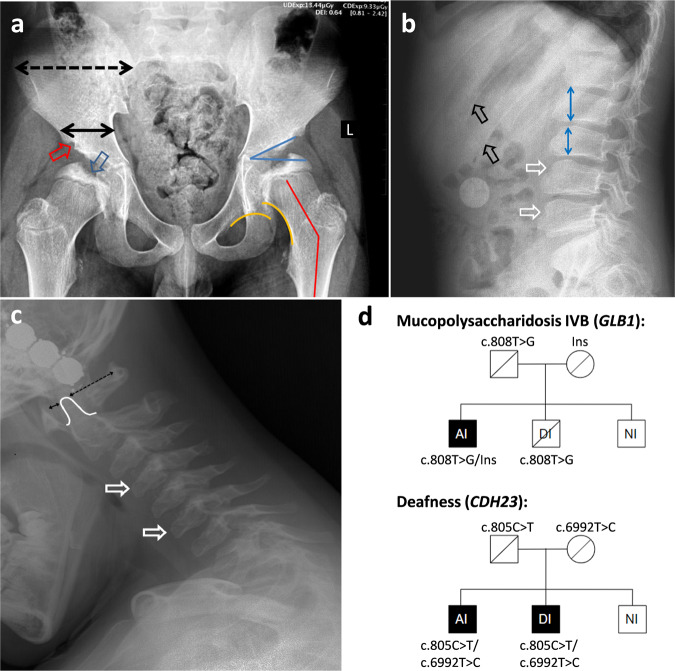
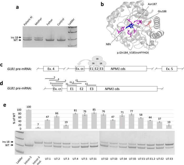
Morquio B disease (MBD) is an ultra-rare lysosomal storage disease, which represents the relatively mild form of GLB1-associated disorders. In this article, we present the unique case of "pure" MBD associated with an insertion of the mobile genetic element from the class of retrotransposons. Using whole-genome sequencing (WGS), we identified an integration of the processed pseudogene NPM1 deep in the intron 5 of GLB1. The patient's mRNA analysis and the detailed functional analysis revealed the underlying molecular genetic mechanism of pathogenesis, which is an alteration of the GLB1 normal splicing. By co-expression of minigenes and antisense splice-modulating oligonucleotides (ASMOs), we demonstrated that pseudogene-derived splicing regulatory motifs contributed to an activation of the cryptic exon located 36 bp upstream of the integration site. Blocking the cryptic exon with ASMOs incorporated in the modified U7 small nuclear RNA (modU7snRNA) almost completely restored the wild-type splicing in the model cell line, that could be further extended toward the personalized genetic therapy. To our knowledge, this is the second reported case of the processed pseudogene insertion for monogenic disorders. Our data emphasizes the unique role of WGS in identification of such rare and probably underrepresented in literature types of disease-associated genetic variants.


Conflict of interest statement

The authors declare no competing interests.

Figures

Fig. 1
Fig. 1. Patient’s summary.
a The anteroposterior radiograph of pelvis and hips: “mickey mouse” pelvis—wide iliac wing (dotted black line) and narrow iliac body (solid black line); deficient ossification of acetabular edge (red arrow) and femoral head (blue arrow); dysplastic acetabulum (acetabular angle 30°—blue lines); coxa valga (neck-shaft angle 150°—red lines); disrupted Shenton line (yellow lines). b Radiograph of the lumbar spine (lateral view in flexion): anterior wedging and beaking of the lumbar vertebrae (white arrows); anisospondyly—irregular height of the vertebral bodies in cranio-caudal direction (blue lines); widening of the ribs (black arrows). c Radiograph of the cervical spine (lateral view): anterior wedging and beaking of the cervical vertebrae (white arrows); mild hypoplasia of the C2 dens (white line); mild anterior atlanto-axial instability (black lines). d Segregation analysis of the variants, associated with MPS IVB and nonsyndromic deafness in the patient’s family. Ins—insertion of the processed pseudogene. Painted icons—affected patients, crossed icons—heterozygous asymptomatic carriers.
Fig. 2
Fig. 2. Results of functional analysis of the PP insertion.
a Results of the blood mRNA analysis demonstrated the presence of an additional GLB1 mRNA isoform with inclusion of the 18 bp fragment of the GLB1 intron 5 in patient and his mother, which is the heterozygous carrier of the PP insertion at the DNA level. b Fragment of the patient’s mutated β-GAL with the insertion of six amino acids (the 3D model was created by SWISS-MODEL based on the PDB:3WEZ template), located in the close proximity to the protein’s active site and two amino acids upstream of the catalytic residue Asn187. Catalytic residues are colored in pink, insertion is red and N8V ligand is blue. c The scheme of the GLB1 intron 5 fragment at the pre-mRNA level. The cryptic 18 bp exon (Ex. cr.) is located 36 bp upstream of the PP insertion (NPM1 cds) and was supposed to be activated by the PP-derived splicing enhancer motifs (E1-3). Splicing enhancers are enriched in binding sites of serine-rich proteins (SR), which promote the inclusion of the exons by interacting with spliceosome. d Location of studied ASMOs. Three clusters of splicing enhancer motifs were predicted by HExoSplice and were chosen as targets for ASMOs together with the cryptic exon (additional information is in Supplementary Figs. 1 and 2). e Fragment analysis and representative polyacrylamide gel electrophoresis visualization of PCR products obtained from minigene experiments. HEK293T cells were transfected with minigenes containing the fragment of the WT intron 5 and the mutated one (with PP insertion) alone (columns 2 and 3) or co-transfected with the mutated minigene and plasmids expressing modU7snRNAs (columns 4–17). Columns 4–8 (U7.1-5) correspond to modU7snRNAs targeting cryptic exon. Columns 9–13 (U7.S1–S5) correspond to the same modU7snRNAs but tailed with the splicing silencer hnRNPA1 motif. Columns 14–17 (U7.E1-E3) correspond to the modU7snRNAs targeting PP-derived splicing enhancer motifs. Error bars represent the standard deviation of biological replicates. The uncropped blots are presented in Supplementary Fig. 3. All blots derive from the same experiment and were processed in parallel.

References

    1. Morreau H, et al. Alternative splicing of β-galactosidase mRNA generates the classic lysosomal enzyme and a β-galactosidase-related protein. J. Biol. Chem. 1989;264:20655–20663. doi: 10.1016/S0021-9258(19)47114-7. - DOI - PubMed
    1. Callahan JW. Molecular basis of GM1 gangliosidosis and Morquio disease, type B. Structure–function studies of lysosomal β-galactosidase and the non-lysosomal β-galactosidase-like protein. Biochimica et. Biophysica Acta (BBA)-Mol. Basis Dis. 1999;1455:85–103. doi: 10.1016/S0925-4439(99)00075-7. - DOI - PubMed
    1. Stockler-Ipsiroglu S, et al. Morquio-like dysostosis multiplex presenting with neuronopathic features is a distinct GLB1-related phenotype. JIMD Rep. 2021;60:23–31. doi: 10.1002/jmd2.12211. - DOI - PMC - PubMed
    1. Abumansour IS, et al. Morquio‐B disease: clinical and genetic characteristics of a distinct GLB1‐related dysostosis multiplex. JIMD Rep. 2020;51:30–44. doi: 10.1002/jmd2.12065. - DOI - PMC - PubMed
    1. Latour YL, et al. Human GLB1 knockout cerebral organoids: a model system for testing AAV9-mediated GLB1 gene therapy for reducing GM1 ganglioside storage in GM1 gangliosidosis. Mol. Genet. Metab. Rep. 2019;21:100513. doi: 10.1016/j.ymgmr.2019.100513. - DOI - PMC - PubMed